自动乳腺全容积成像技术在BI-RADS4类结节再评估中的诊断价值
2021-09-08温祺祺郑晨笛
温祺祺 郑晨笛
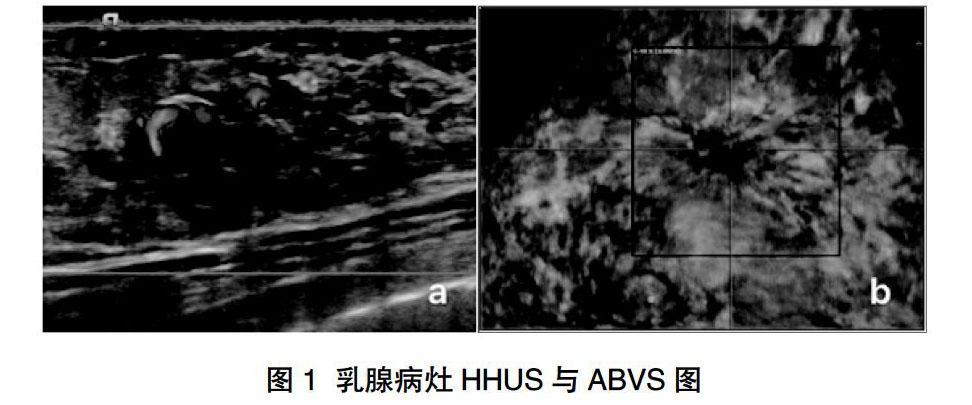
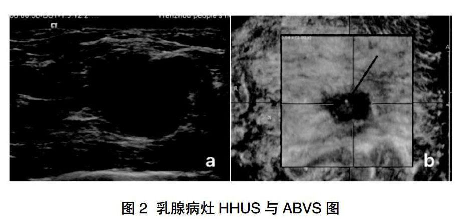
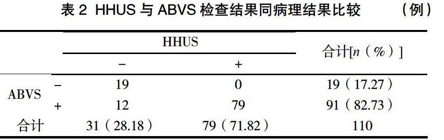
摘 要:目的 探讨自动乳腺全容积成像(ABVS)技术对乳腺影像学报告及数据系统(BI-RADS)4类结节二次评估分类的价值。方法 选取2015年6月~2019年12月温州市人民医院进行乳腺常规手持超声(HHUS)检查被诊断为BI-RADS 4类的110例患者,再做ABVS检查,根据获取的三维图像,针对增加的冠状切面信息,对结节进行再次BI-RADS分类,并与病理结果对照分析,分别计算ABVS与HHUS的各类结节恶性概率及诊断符合率。结果 110个乳腺结节,经病理检查证实其中79个为良性病灶,31个为恶性病灶。ABVS与HHUS对诊断4类结节的恶性概率分别为:4A类9.46%(7/74)vs 23.33%(21/90),4B类50.00%(11/22)vs 16.67%(2/12),4C类87.50%(7/8)vs 100.00%(8/8)。ABVS对4类病灶的恶性概率比HHUS更符合BI-RADS分类标准,若将BI-RADS 4A与4B作为良恶性截断点,ABVS对诊断乳腺癌的准确性高于HHUS(82.73% vs 71.82%),差异有统计学意义(P<0.05)。结论 ABVS检查提供更全面的诊断信息,有助于提高对BI-RADS 4类结节的诊断准确性,可以作為常规超声检查的一个技术补充。
关键词:自动乳腺全容积成像;常规超声;乳腺结节
Abstract: Objective To explore the values of the automated breast volume scanner(ABVS) in reassessment of breast tumors with BI-RADS 4. Methods A total of 110 patients with breast tumors of BI-RADS 4 grade by handheld ultrasound(HHUS) in our hospital from June 2015 to December 2019 were selected, and then ABVS was performed to obtain the three-dimensional volume data. And all the tumors were reassessed according to the newly added coronal information, and compared with postoperative pathology. The probability of malignant and diagnostic accordance rate of ABVS and HHUS were calculated. Results In 110 patients with breast tumors, 79 cases were benign, 31 cases were malignancy, all proved by pathology. The malignant probability of ABVS and HHUS for diagnosing 4 types of nodules was 9.46%(7/74) vs 23.33%(21/90) for class 4A, 50.00%(11/22) vs 16.67%(2/12) for Class 4B, 87.50%(7/8) vs 100.00% (8/8) for Class 4C, respectively. The malignant probability of ABVS and HHUS for diagnasing 4 tyres Malignancy probability in breast tumors of BI-RADS 4 were accord with criterion using ABVS than HHUS. Breast tumors of BI-RADS 4A were assessed with HHUS and were reassessed with ABVS, the coincidence rate in diagnosis of ABVS were higher than those of HHUS(82.73% vs 71.82%, P<0.05). Conclusions ABVS can provide more information and help to improve the diagnostic accuracy of BI-RADS 4 breast tumors,as a supplement for HHUS.
Keywords: Automated breast volume scanner; Ultrasonography; Breast tumors
乳腺癌是我国女性最常见的恶性肿瘤之一,但早期乳腺癌(临床分期为0、1期的患者)治愈率可达90%。因此,早发现、早治疗是应对乳腺恶性肿瘤的最有效方法。乳腺超声被广泛运用于诊断各类乳腺疾病,在乳腺肿瘤筛查方面具有重大作用,自动乳腺全容积成像(automated breast volume scanner,ABVS)的出现是对常规超声检查技术的提升,是一种最佳的可覆盖全乳腺组织的超声检查技术。ABVS通过自动扫描来获取乳腺的三维图像,可多层面、多角度地显示病灶。其规范化的扫描,保留乳腺全容积数据,可重复性好,有利于长期随访观察病灶,并大大减少因操作者经验不足而造成的漏诊。美国放射学会于1992年制定了乳腺影像学报告及数……
