Pingchuan formula (平喘方) improves allergic asthma in mice through inhibiting nuclear factor-kappa B/mitogen-activated protein kinase signaling pathway
2021-08-09WUMingyunYUJianerBAILiXUEZhengJIANGShenhuaLILiqingPIAOXiangXUWanchaoWANGJianiSHENQian
WU Mingyun,YU Jianer,BAI Li,XUE Zheng,JIANG Shenhua,LI Liqing,PIAO Xiang,XU Wanchao,WANG Jiani,SHEN Qian
WU Mingyun,JIANG Shenhua,XU Wanchao,WANG Jiani,Department of Pediatrics,Shanghai Municipal Hospital of Traditional Chinese Medicine,Shanghai 200071,China;Shanghai University of Traditional Chinese Medicine,Shanghai 201203,China
YU Jianer,BAI Li,XUE Zheng,LI Liqing,PIAO Xiang,Department of Pediatrics,Shanghai Municipal Hospital of Traditional Chinese Medicine,Shanghai 200071,China
SHEN Qian,Shanghai University of Traditional Chinese Medicine,Shanghai 201203,China;Department of Nephrology,Putuo Hospital Affiliated to Shanghai University of Traditional Chinese Medicine,Shanghai 200062,China
Abstract OBJECTIVE:To examine the role and decipher the mechanism of Pingchuan formula(平喘方,PCF)in treating allergic asthma.METHODS:The mice were treated with saline,dexamethasone (DXM) and PCF for 1 week after the asthma model was established and their respiratory function including respiratory resistance(RI),pulmonary dynamic compliance (Cdyn) and maximum voluntary ventilation (MVV) were measured.In addition,cellular changes in bronchoalveolar lavage fluid(BALF)and pathological changes in lung biopsy as well as the expression level ofα-smooth muscle actin (α-SMA),transforming growth factor-beta1 (TGF-β1) in BALF and interleukin-5 (IL-5),interleukin-13 (IL-13),tumor necrosis factor-α(TNF-α),interferon-γ (IFN-γ),nuclear factor-kappa B-p65 (NF-κBp65),inhibitor-α of nuclear transcription factor κB (IκBα),p38 mitogen-activated protein kinase (p38MAPK),c-jun n-terminal kinase(JNK) and its phosphorylated proteins in lung tissue were also examined and compared among different groups.RESULTS:Our data suggested that the respiratory functions were significantly improved and the pathological changes ameliorated in the DXM group and the PCF group compared to the model group.Both DXM and PCF effectively decreased the number of eosinophils,lymphocytes,and neutrophils in BAL as well as the secretion ofα-SMA and TGF-β1,IL-5,IL-13,while increased the expression of TNF-α and IFN-γ.Furthermore,our study indicated that the NF-κBp65,IκBα,p38MAPK and JNK pathways were inhibited under the treatment of PCF.CONCLUSION:Our data indicated that PCF can attenuate the inflammatory response in asthma through inhibiting the NF-κB/MAPK signaling pathway.This study not only supported the use of PCF in allergic asthma in clinic but also shed light upon afurtherunderstandingofthediseasepathogenesis.
Keywords:asthma;NF-kappa B;p38 mitogen-activated protein kinases;signal transduction;Pingchuan formula
INTRODUCTION
Bronchial asthma is a chronic airway inflammation characterized by deteriorating respiratory function and symptoms including wheezing,shortness of breath,chest tightness and coughing etc.It has been suggested that allergic asthma is one of the most common subtype of bronchial asthma and accounts for more than 50% of adult asthma and 80% of childhood asthma.1Under the view of Traditional Chinese Medicine(TCM),asthma falls into the category of "Wheezing Dyspnea" which pathogenesis mainly involves persistent phlegm associated obstruction and constriction of the airway after being triggered by external afflictions,dietary factors,emotional changes,fatigue and other factors that can result disequilibrium in lungQicirculation.2Pingchuan formula (平喘方,PCF) was first invented by Professor Yu Jianer,the fourth generation successor of Xu Family of Pediatrics from Haipai Chinese Medicine.Previous studies confirmed that PCF can regulate Th1 (helper T cell T1)/Th2 (helper T cell T2) balance to resolve airway inflammation in athma.3However,the specific mechanism has not been well studied.
The mitogen-activated protein kinase (MAPK) signal transduction pathway is an important pathway involved in the pathogenesis and progression of bronchial asthma.On the other hand,the nuclear-factor-κB(NF-κB) family is a group of nuclear transcriptional regulators and widely distributed in mammalian cells.Studies have confirmed that the NF-κB signaling pathway is involved in dendritic cell phenotypic expression,4lymphocytes activation and differentiation,5cytokine secretion and other aspects in the inflammatory process of bronchial asthma.6,7In this study,we aimed to explore the effects of PCF on allergic asthma with a special interest in NF-κB/MAPK signaling pathway and identify its possible mechanism of action to provide better reference for its clinical.
MATERIALS AND METHODS
Animals
Forty naive BALB/c mice with 4-6 weeks old,weighing 18-22 g were provided by Shanghai Xipuer-Beikai Experimental Animal Co,Ltd.(license No.SCXK(Shanghai,China) 2013-C016) and raised at 20 ℃-24 ℃temperatures and 50%-70% humidity in IVC-II type independent air supply cages at the animal experiment center of Shanghai University of Traditional Chinese Medicine (experimental animal license No.SYXK Shanghai 2014-0008) without specific diet requirement.All animal experiments were approved by the Experimental Animal Ethics Committee of Shanghai University of Traditional Chinese Medicine.
Drugs
PCF that mainly composed of Mahuang (Herba Ephedra Sinica) 6 g,Kuxingren (Semen Armeniacae Amarum)6 g,Taoren(Semen Persicae)6 g,Zisuzi(Fructus Perillae Argutae)9 g,Laifuzi(Semen Raphani Sativi)9 g,Tinglizi (Semen Lepidii Apetali) 9 g,Dilong(Pheretima Aspergillum)9 g,Huangqin(Radix Scutellariae Baicalensis) 9 g and Gancao (Radix Glycyrrhizae)6 g was purchased from Shanghai Municipal Hospital of Traditional Chinese Medicine.Dexamethasone acetate tablets (specification:0.75 mg/tablet) were purchased from Shanghai Xinyi Co.,Ltd.(batch No.110302,Shanghai,China).
Reagents
Oval albumin (OVA) (Cat.No.A5503,Sigma,Louis,MO,USA),aluminum hydroxide [Al(OH)3](Cat.No.239186,Sigma,Louis,MO,USA),hematoxylin-eosin (HE) dye solution (Cat.No.G1005,Cebio,Wuhan,China),enzyme-linked immunosorbent assay(ELISA) kit (batch No.20171205,R&D Minneapolis,MN,USA),p38 mitogen-activated protein kinase(p38MAPK) mice polyclonal antibody (batch No.8690S,American CST company,Danvers,MA,USA),inhibitor-α of nuclear transcription factor κB (IκBα)mice polyclonal antibody (batch No.TA327876S,Origene,Rockville,MD,USA),NF-κBp65 mice polyclonal antibody (batch No.10745-1-AP,Wuhan Proteintech,Wuhan,China),c-jun n-terminal kinase(JNK) mice polyclonal antibody (batch No.AJ518-2,Biyuntian Biotechnology Co.,Ltd.,Shanghai,China)were used in the study.
Instruments
The BX51 fluorescence microscope (Olympus,Tokyo,Japan),Image Pro Express analysis system (Shanghai Xinu Optical Technology Co.,Ltd.Shanghai,China),RM2235 paraffin slicer (Leica,Weztlar,Germany),LeixaHI1220 Horizontal Dryer(Shanghai Jianglin Biotechnology Co.,Ltd.,Shanghai,China),Mini protean 3 cell electrophoresis instrument (BIO-RAD,Hercules,CA,USA),TGL-168 centrifuge (Shanghai Anting Scientific Instrument Factory,Shanghai,China),PYX-DHS digital display water-blocking electric heating constant temperature incubator (Shanghai Yuejin Medical Instrument Factory,Shanghai,China),XW-80A type vortex mixer(Shanghai Qingpu Huxi Instrument Factory,Shanghai,China),micro electric organization homogenizer (Kimble Company,Milville,NJ,USA),VE186 Transfer Electrophoresis kit (Shanghai Tianneng Technology Co.,Ltd.,Shanghai,China),Clinx Chemi Scope Series Fluorescence and Chemiluminescence Imaging System(Shanghai Qinxiang Scientific Instrument,Shanghai,China),scanner (Shanghai Tianneng Instrument Co.,Ltd.,Shanghai,China) and AniRes 2005 Animal Lung Function Analysis System(Beijing Bellambo Technology Co.,Ltd.,Beijing,China)were used in this study.
Modeling and grouping
Forty BALB/c mice were randomLy divided into blank control group (CON group),model group (MDL group),dexamethasone group (DXM group),and PCF group (PCF group),each consisting of 10 mice.On the 1st and 15th day of modeling,1 mL/20 g sensitizing solution (100 mL saline solution containing 10 g OVA) or 0.9% sodium chloride solution was injected into the mice intraperitoneally.After additional 7 d,all mice were received daily forced inhalation of 5% OVA or 0.9%sodium chloride solution by ultrasonic atomizer for 40 min and the treatments were started at the same time.Mice in the PCF group were administrated with PCF at a concentration of 5.33 g/mL and mice in the DXM group were received dexamethasone at 0.075 mg/mL.Distilled water at 0.4 mL/20 g were given to the mice in the CON and MDL groups.All mice were sacrificed after 7 d and samples were collected for further measurement(Figure 1).
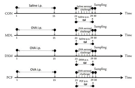
Figure 1 Experimental flow diagram
Drug preparation and administration
The PCF was prepared according to previous studies.8Briefly,water and herbs were first boiled at 6∶1 ratio and then brewed at lower temperature for 1 h.The solution was stabilized for 2 h at 37 ℃before filtered.Next,fresh water was added to the remaining herbs and boiled at 3∶1 ratio and then the brewing and filtration procedures were repeated as above.The filtrate from both brews were combined and concentrated,and the experimental dose was calculated according to the formula in Experimental Animal Methodology:dB=dA × RB/RA × (WA/WB) 1/3 (the dosage is equivalent to 10 times of the clinical dose that commonly applied on 30 kg children).Filtration was repeated after leaving to sit for 24 h,and the filtrate was concentrated to form a watery paste with a final concentration of 5.33 g/mL.
To prepare dexamethasone solution,the dexamethasone acetate tablet (0.75 mg/tablet) was dissolved in distilled water at 0.075 mg/mL.
DETECTION INDICATORS AND METHODS
Respiratory function
After 24 h of the final treatment,mice were anesthetized with an intraperitoneal injection of 0.4% sodium pentobarbital solution at 20 mL/kg and connected to AniRes2005 animal lung function analysis system through tracheal intubation in supine position.The ventilator's breathing frequency was set to 90/min with a breathing ratio at 3∶2.The inspiration resistance(RI),pulmonary dynamic compliance (Cdyn),and maximum ventilation per minute voluntary ventilation(MVV) were measured and compared among different groups.
Specimen collection and processing
After lung function test,bronchial alveolar lavage fluid(BALF) of the left lung was collected and centrifuged at 1200 rpm for 5 min at 4 ℃.The supernatant was stored at-20 ℃until further use.The right lung was collected and divided into two parts:the upper lobe was sliced and fixed in10%formaldehyde for pathological examinations and the lower lobe was froze in liquid nitrogen and stored at-80 ℃until further use.
Lung tissue pathology
The paraffin embedded tissue was stored at 4 ℃overnight and cut into 5-8 μm slices before staining with HE.The morphology of lung was observed under optical microscope.
White blood cells and individual cell counts
Cytospin was prepared with 0.1 mL BALF and fixed as well as stained following a modified Wright method.The total number of white blood cells and each subgroup of cells under a low power microscope was calculated according to the formula.
Detection of α-SMA and TGF-β 1 in BALF of mice in each group by ELISA
The supernatant of BALF solution was collected and used for α-SMA and TGF-β 1 detection following the manufactures'instruction.
Detection of IL-5,IL-13,TNF-αand IFN-γ in lung tissue of mice by ELISA
The right lung(1 g)was collected and homogenized in 2 mL saline at 4 ℃and centrifuged at 2500 r/min for 10 min.The supernatant was collected and used for further cytokine measures following manufactures'protocol.
Protein expression of NF-κBp65,IκBα,p38MAPK,JNK and phosphorylated proteins in lung by Western blot
The lung homogenate was prepared as introduced above.The BCA method was used to load equal amount of protein sample onto SDS-PAGE electrophoresis following standard procedure.Then the protein was transferred to the PVDF membrane,and incubated in 5%BSA solution at room temperature for 1 h before adding primary antibody (1∶1500) Overnight at 4 ℃.The membrane was rinsed before adding HRP-labeled secondary antibody.The membrane was rinsed again and then incubated in the ECL reaction solution at room temperature for 5 min.The film was then exposed to X-ray detection,and measured using Gis analysis software.
Statistical analysis
All statistical analysis was performed with SPSS 25.0 software (MBI Institute,Armonk,NY,USA) and GraphPad Prism 6.0 software (GraphPad Institute,San Diego,CA,USA).Data following normal distribution and uniform variance was compared using analysis of variance and least significant difference multiple comparison.Data conversion was performed first when variance was not uniform before conducting statistical analysis.P<0.05 was considered statistically significant.
RESULTS
General conditions of mice in each group
Our data suggested that there was no significant difference in body weight among different groups at baseline.After sensitization,mice in the MDL,DXM and PCF groups showed significantly lower body weight compared to the CON group (P <0.01).After 7 d of treatment,the body weight of mice in the MDL and DXM groups was significantly lower compared to that of CON and PCF group (P <0.01) and no significant difference was observed between the PCF and CON groups(P>0.05)(Table 1).
Table 1 Bodyweight of mice in the four groups at baseline and after 7 d of treatment(g,±s)

Table 1 Bodyweight of mice in the four groups at baseline and after 7 d of treatment(g,±s)
Notes:group CON:treated with physiological saline at 0.4 mL/20 g for 7 d;group MDL:treated with physiological saline at 0.4 mL/20 g for 7 d;group DXM:treated with dexamethasone at 0.4 mL/20 g for 7 d;group PCF:treated with PCF 0.4 mL/20 g for 7 d.CON:blank control;MDL:model;DXM:dexamethasone;PCF:Pingchuan formula.OVA:oval albumin.Compared with group CON,aP <0.01;compared with group MDL,bP<0.01;compared group DXM,cP<0.01.
After modeling,mice in the MDL,DXM and PCF group showed classic allergic asthma symptoms including shortness of breath,abdominal muscle convulsions,scratching of skin,irritability,eyelid edema,incontinence,damp hair,lacking luster,reduced appetite as well as physical activity.The symptom score was significantly higher compared to the CON group(P<0.05).After 7 d of treatment,the symptoms of mice in the PCF and DXM groups were significantly alleviated compared to the MDL group(P<0.05).Furthermore,our study indicated that the average symptom score of mice in the PCF group was significantly decreased compared to the DXM group(P<0.05)(Table 2).
Respiratory function of mice in each group after treatment
As presented in Table 3,the RI of the DXM and PCF groups was significantly lower compared to the MDL group (P <0.01) after treatment.Furthermore,we discovered that the Cdyn and MVV of the DXM group and the PCF group were significantly higher compared to the MDL group (P <0.01).In addition,the MVVof the PCF group was significantly higher compared to the DXM group(P<0.01).
Table 2 Quantification of the symptoms of mice in the four groups at baseline and after 7 d of treatment(±s)
Table 2 Quantification of the symptoms of mice in the four groups at baseline and after 7 d of treatment(±s)
Notes:group CON:treated with physiological saline at 0.4 mL/20 g for 7 d;group MDL:treated with physiological saline at 0.4 mL/20 g for 7 d;group DXM:treated with dexamethasone at 0.4 mL/20 g for 7 d;group PCF:treated with PCF 0.4 mL/20 g for 7 d.CON:blank control;MDL:model;DXM:dexamethasone;PCF:Pingchuan formula.OVA:oval albumin.Compared with group CON,aP<0.01;compared with group MDL,bP<0.01.
Pathological changes in lung tissue
After 7 d of treatment,the bronchial wall structure of mice in the CON group was intact and the no cellular abnormality was observed.The bronchial mucosa of mice in the MDL group showed obvious inflammatory cell infiltration,thickened tracheal wall,accompanied by obvious congestion as well as edema and showing chrysanthemum-like changes.In addition,the normal structure of bronchial epithelial cells and cilia were disrupted and destroyed.Compared to the MDL group,the overall lung pathology changes in mice from the DXM group and PCF group were ameliorated suggested by the relatively intact bronchial wall structure and limited inflammatory cell infiltration,as presented in Figure 2.
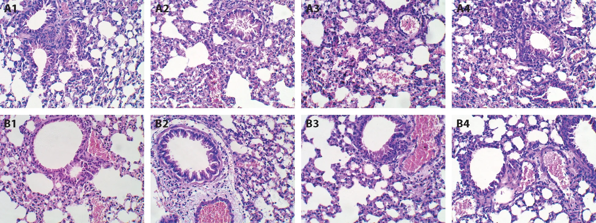
Figure 2 Lung morphology of mice in each group
Cellular changes in BALF
As suggested in Table 4,compared with the CON group,the total number of leukocytes,eosinophils and neutrophil cells in the BALF of mice from other groups were significantly increased (P <0.01).In addition,the total number of BALF white blood cells,eosinophils and neutrophil cells in the DXM group and the PCF group were significantly lower compared to the MDL group (P <0.01).Furthermore,the total number of BALF white blood cells,eosinophils and neutrophil cells in THE PCF group was higher than that of the DXM group(P<0.01).In addition,our data indicated that the number of BALF Lymphocytes in the MDL group was the highest among all four groups(P <0.01) and no significant difference was discovered among other 3 groups(P>0.05).
Levels of α-SMA and TGF-β1 in BALF
Compared with the MDL group,the secretion of α-SMA and TGF-β1 in BALF of mice in the PCF andDXM group was significantly decreased (P <0.01),but still higher compared to that in the CON group(P <0.01).There was no significant difference of α-SMA identified between the DXM and PCF group(P>0.05),while the level of TGF-β1 was significantly lower in the PCF group(P<0.01)(Table 5).
Table 3 Comparison of RI,cdyn and MVV in lung function of mice in each group(±s)
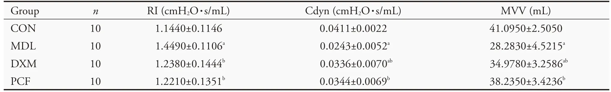
Table 3 Comparison of RI,cdyn and MVV in lung function of mice in each group(±s)
Notes:group CON:treated with physiological saline at 0.4 mL/20 g for 7 d;group MDL:treated with physiological saline at 0.4 mL/20 g for 7 d;group DXM:treated with dexamethasone at 0.4 mL/20 g for 7 d;group PCF:treated with PCF 0.4 mL/20 g for 7 d.CON:blank control;MDL:model;DXM:dexamethasone;PCF:Pingchuan formula;OVA:oval albumin;RI:inspiration resistance;Cdyn:pulmonary dynamic compliance;MVV:maximum ventilation per minute voluntary ventilation.Compared with group CON,aP <0.01;compared with group MDL,bP<0.01.
Table 4 Measurement of white blood cells and individual cells in BALF of each group(±s)
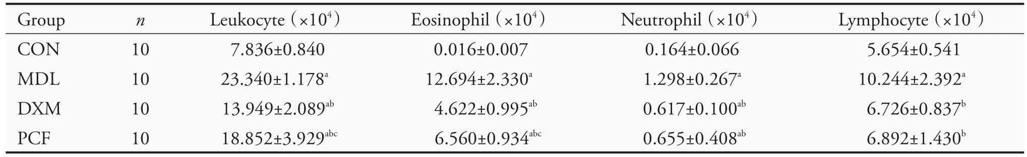
Table 4 Measurement of white blood cells and individual cells in BALF of each group(±s)
Notes:group CON:treated with physiological saline at 0.4 mL/20 g for 7 d;group MDL:treated with physiological saline at 0.4 mL/20 g for 7 d;group DXM:treated with dexamethasone at 0.4 mL/20 g for 7 d;group PCF:treated with PCF 0.4 mL/20 g for 7 d.CON:blank control;MDL:model;DXM:dexamethasone;PCF:Pingchuan formula.Compared with group CON,aP <0.01;compared with group MDL,bP<0.01;compared group DXM,cP<0.01.
Table 5 Expressions of α-SMA and TGF-β1 in serum of each group(pg/mL,±s)

Table 5 Expressions of α-SMA and TGF-β1 in serum of each group(pg/mL,±s)
Notes:group CON:treated with physiological saline at 0.4 mL/20 g for 7 d;group MDL:treated with physiological saline at 0.4 mL/20 g for 7 d;group DXM:treated with dexamethasone at 0.4 mL/20 g for 7 d;group PCF:treated with PCF 0.4 mL/20 g for 7 d.CON:blank control;MDL:model;DXM:dexamethasone;PCF:Pingchuan formula;α-SMA:α-smooth muscle actin;TGF-β1:transforming growth factor-beta 1.Compared with group CON,aP<0.01;compared with group MDL,bP<0.01;compared group DXM,cP<0.01.
Comparison of IL-5,IL-13,TNF-α and IFN-γ levels in lung tissue
Compared with the MDL group,the level of IL-5 and IL-13 in the DXM and PCF group were decreased(P <0.01),and there was no significant difference observed between the two groups (P >0.01).The levels of TNF-α and IFN-γ in the DXM and PCF groups were significantly higher compared to those in the MDL group (P <0.01),which were more prominent in the PCF group(P<0.01)(Table 6).
Expression of NF-κBp65,IκBα,p38MAPK,JNK and their phosphorylated proteins in lung
Compared to the MDL group,the expression of NF-κBp65,IκBα,p38MAPK,JNK and their phosphorylated proteins in the lung tissue of mice from the DXM and PCF groups were significantly reduced.There was no significant difference identified in the expression of p38MAPK and P-P38MAPK between PCF and CON groups(P>0.05)(Tables 7,8,Figures 3,4).
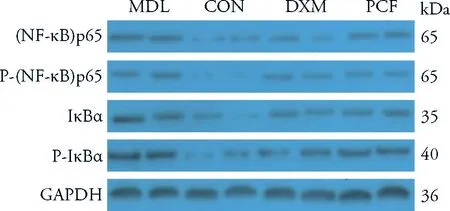
Figure 3 Protein levels of NF-κB signaling pathway
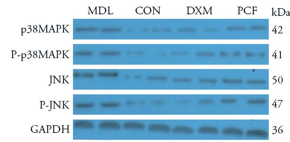
Figure 4 Protein levels of MAPK signaling pathway
Table 6 Expressions of IL-5,IL-13 and TNF-α,TNF-γ in serum(pg/mL,±s)
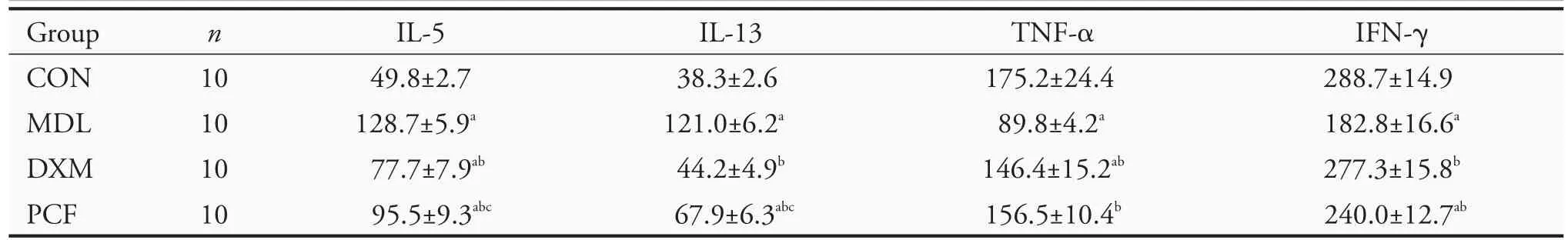
Table 6 Expressions of IL-5,IL-13 and TNF-α,TNF-γ in serum(pg/mL,±s)
Notes:group CON:treated with physiological saline at 0.4 mL/20 g for 7 d;group MDL:treated with physiological saline at 0.4 mL/20 g for 7 d;group DXM:treated with dexamethasone at 0.4 mL/20 g for 7 d;group PCF:treated with PCF 0.4 mL/20 g for 7 d.CON:blank control;MDL:model;DXM:dexamethasone;PCF:Pingchuan formula;IL-5:interleukin-5;IL-13:interleukin-13;TNF-α:tumor necrosis factor-α;IFN-γ:interferon-γ.Compared with group CON,aP<0.01;compared with group MDL,bP<0.01;compared group DXM,cP<0.01.
Table 7 Protein levels of NF-κB signaling pathway(±s)
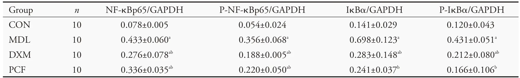
Table 7 Protein levels of NF-κB signaling pathway(±s)
Notes:group CON:treated with physiological saline at 0.4 mL/20 g for 7 d;group MDL:treated with physiological saline at 0.4 mL/20 g for 7 d;group DXM:treated with dexamethasone at 0.4 mL/20 g for 7 d;group PCF:treated with PCF 0.4 mL/20 g for 7 d.CON:blank control;MDL:model;DXM:dexamethasone;PCF:Pingchuan formula;NF-κBp65:nuclear factor-kappa B-p65;IκBα:inhibitor-α of nuclear transcription factor κB.Compared with group CON,aP<0.01;compared with group MDL,bP<0.01.
Table 8 Protein levels of MAPK signaling pathway(±s)

Table 8 Protein levels of MAPK signaling pathway(±s)
Notes:group CON:treated with physiological saline at 0.4 mL/20 g for 7 d;group MDL:treated with physiological saline at 0.4 mL/20 g for 7 d;group DXM:treated with dexamethasone at 0.4 mL/20 g for 7 d;group PCF:treated with PCF 0.4 mL/20 g for 7 d.CON:blank control;MDL:model;DXM:dexamethasone;PCF:Pingchuan formula;p38MAPK:mitogen-activated protein kinase;JNK:c-jun n-terminal kinase.Compared with group CON,aP<0.01;compared with group MDL,bP<0.01.
DISCUSSION
It has been known that NF-κB is a transcription factor that can be regulated by the k-light chain of mouse B lymphocytes.9When activated by virus,cytokine,or oxidant,IκB kinase phosphorylation and ubiquitination of IκB protein occur,hence the release of NF-κB di-mer,and subsequent transfer into the nucleus.NF-κB then binds to corresponding promoter or enhancer regions to regulate the expression of genes such as IL-5,IL-13 and TNF-α,and participates in the process of inflammation and apoptosis.10p38MAPK and JNK belong to the classic signaling pathway of MAPK.11Studies have shown that Staphylococcus aureus can stimulate macrophages to promote JNK phosphorylation to increase the expression of IL-5 and INF-γ12 and p38MAPK can affect macrophages and induce Th0 cells differentiating into Th2 which eventually participate in the pathogenesis of asthma.13Previous study indicated that the measurement of respiratory function in mice can be used as a surrogate to evaluate the airway hyperresponsiveness of asthma.14Hypertrophic smooth muscle is a characteristic change of airway remodeling in asthma which indicated by the increasingα-SMA expression levels.15TGF-β1 is not only involved in airway epithelial injury and goblet cell proliferation,but also involved in airway smooth muscle cell remodeling.16,17
Based on our previous studies,sputum andQias well as Blood stasis all together disrupt the normal functions of lung and upper respiratory tract,leading to a combination of deficiency and excess which resulted inYinandYangimbalance that eventually presented as asthma attack.18As important components in the PCF,Candied Ephedra is the Monarch.Its main component,ephedrine,has the effect of relieving shortness of breath.And it has been known that pseudoephedrine can inhibit the release of allergic media and exert anti-inflammatory and anti-allergic effects.19Bitter almonds and Peach kernels are also important associates in the formula.Bitter almond is effective in restoring the qi and helps eliminating the phlegm.Peach kernel can invigorate the circulation of blood and promote the establishment of new circulation.Perilla seed and Radish seed can warm and dissipate the phlegm and excessive fluid.The 4 herbs introduced above are considered as Minister in the PCF.Lepidium seed can purify the lungs and clear the water passages,which helps to eliminate the phlegm and excessive fluid.Earthworm also has anti-inflammatory,anti-histamine,diastolic bronchial and immunomodulatory effects.20Scutellaria baicalensis not only has anti-inflammatory and immune-regulating effects,but also can help to restrict the dryness of other herbs when used together with the Earthworm,so that excessive phlegm could be expelled without damagingYinand normal bodily fluid,21and they are both the Assistants in the formula.Candied licorice is the Guide in the PCF and it can nourish the spleen,replenishQi,ease coughing,and moderate the properties of all other herbs.The whole formula is designed to promote dissemination of air in lungs,reduce asthma,eliminate phlegm and remove blood stasis.
As introduced in detail in our previous study,the high dosage (5.33 g/mL) of PCF was selected in this experiment to recapitulate the clinical situation.22This study showed that PCF inhibited the increase of leukocytes,eosinophils,neutrophils as well as lymphocytes in BALF and reduced the infiltration of inflammatory exudate and inflammatory cells in lungs and tracheal tissue in the mouse model.Moreover,the formula significantly reduced the expression of Th2 associated cytokines (IL-5 and IL-13) while increased the secretion of Th1 associated cytokines (TNF-α and INF-γ).Furthermore,it significantly reduced the formation of airway remodeling by inhibiting the secretion of a-SMA and TGF-β1 as well as the expression of NF-κBp65,IκBα,p38MAPK and JNK in lung.Our study indicated that PCF improved the airway resistance and restored the lung function of asthmatic mice similarly,if not superior,to dexamethasone.In summary,the mechanism of PCF in the treatment of allergic asthma might be associated with the regulation of Th1/Th2 immune imbalance by inhibiting NF-kB/MAPK signaling pathway.However,more studies are needed to better understand the mechanisms.
猜你喜欢
杂志排行
Journal of Traditional Chinese Medicine的其它文章
- In vivo anti-diarrheal activity of jujube honey on castor oil-induced diarrhea in mice
- Ethanolic extract of Puhuang (Pollen Typhae) modulates lipopolysaccharide-induced inflammatory response through inducible nitric oxide synthase/cyclooxygenase-2 signaling in RAW 264.7 macrophages
- Target prediction and activity verification for the antidepressant action of Huangqin(Radix Scutellariae Baicalensis)
- Can Fig and Olive Ameliorate the toxicity Induced by 2-nitropropane in some organs of mice? role of inflammatory versus anti-inflammatory genes
- Efficacy of Renshen(Radix Ginseng)plus Fuzi(Radix Aconiti Lateralis Preparata)on myocardial infarction by enhancing autophagy in rats
- Anti-hypertensive and endothelia protective effects of Fufang Qima capsule (复方芪麻胶囊) on primary hypertension via adiponectin/adenosine monophosphate activated protein kinase pathway