Isolation and identification of fungi found in contaminated fermented milk and antifungal activity of vanillin
2021-06-05XuPeiMekonenTekliyeMingshengDong
Xu Pei, Mekonen Tekliye, Mingsheng Dong*
College of Food Science and Technology, Nanjing Agricultural University, 1 Weigang Road, Nanjing, Jiangsu 210095, China
ABSTRACT
Fungi contamination is an important factor affecting the quality and commercial value of fermented milk.Vanillin is known as a safe and efficient natural antimicrobial additive with great potential value as a food bio-preservative. In this study, the microbiological quality of fermented milk of different brands retailed in China and the antimicrobial activity of vanillin were investigated. A total of 27 fermented milk samples were purchased and evaluated. Fungal isolates obtained were characterized by DNA sequencing of the internal transcribed spacer (ITS) region. The effects of vanillin on the growth of fungi were studied by plate dilution method, radical colony diameter, and mycelium biomass. The result showed that 14.8% of the samples were contaminated with three different species of fungi with Cladosporium spp. being the most predominant fungal contaminant. The study further showed that vanillin had significant inhibitory activity against the growth of the fungi species. The cell number, radical colony diameter, and the mycelium biomass of the fungal contaminant were significantly reduced by the inhibitory action of the vanillin. The study thus provides reliable data evidence that will help relevant stakeholders in the fermented milk industries to improve their quality control strategies and consider vanillin as a bio-preservative with the potential to increase the shelf-life and safety of fermented milk.
Keywords:
Fermented milk
Fungi
Internal transcribed spacer
Vanillin
Antimicrobial activity
1. Introduction
In recent years, the consumers’ search for natural and healthy fermented milk has increased due to their nutritional and healthpromoting properties [2-4]. At present, a large variety of fermented milk products are produced and consumed in China and across the globe [1]. Several studies have shown that consumption of fermented milk can confer numerous desirable biological functions such as boosting the immune system, modulating intestinal microbiota, and alleviating hypertension [5-7]. However, microbial contamination and spoilage is a major problem affecting the quality and safety of fermented milk products.
Microbial spoilage is a leading problem confronting the food industry because it reduces consumers’ acceptability of food products and consequently leads to significant food waste and economic losses.Fermented milk products have high acidity hence its less vulnerable bacteria spoilage. Therefore, the prominent contaminants and spoilage agents of fermented milk are fungi. In addition to being a significant health risk, contamination of fermented milk by fungal species reduces its chemical and organoleptic characteristics. High numbers of toxigenic molds are often indicative of the presence of mycotoxins.Sources of yeast and mold contamination of dairy products typically appear to be the air and other environmental sources in processing facilities and other environments [8,9]. Moreover, fungal contamination of dairy foods can occur at different stages, from dairy farms to dairy processing units and at consumers’ homes [10,11].
Several studies have shown that antimicrobial agents can be applied to food preservation and antisepsis. Furthermore, the prospect and developing trend of natural healthy food is discussed. Therefore,higher requirements for food preservation have been pointed out. The exploration for chemical preservative substitutes and the development of natural antimicrobial agents has become the focus of many researchers in recent times [12,13]. Vanillin is widely used as a natural food additive. It has been proved that vanillin has a good antimicrobial effect on a variety of microorganisms, such as bacteria, yeast and mould [14]. The effect of vanillin against spoilage microorganisms was concentration dependent. According to the investigations by previous authors reported, radial growth of Aspergillus spp. was inhibited by 20 mmol/L vanillin, while Penicillium spp., Alternaria spp., and Fusarium spp. were inhibited by 12.5 mmol/L to 12.7 mmol/L vanillin, respectively [13]. Besides functioning as an antimicrobial agent, vanilla has also been shown to improve the flavor of fermented dairy products. Consequently, vanillin can be used as a natural food antiseptic and preservation agent in dairy products [15-17].Thus, in this study, the fungal contamination of fermented milk of different brands retailed in China, and the antifungal activity of vanillin was assessed, which can provide theoretical support for the development and application of vanillin in dairy products.
2. Materials and methods
Twenty-seven different commercial brands of fermented milk samples in plastic or paper box containers were purchased from a local supermarket in Nanjing, China and stored at 4 °C until use.The name, address, volume, production, and expiry dates were all indicated on the sample containers. The number of viable cells counts in the product in all samples was not specified on the label. All samples were transported to the laboratory under aseptic and sterile conditions transported to the laboratory. The analysis of all samples was carried out before the expiry date.
2.1 Indicator strain
Actinomucor elegans DCY-1 is a fungus isolated from mildew soybean dregs from Yangxin County, Huangshi City, Hubei Province.It is preserved in the laboratory until use.
2.2 Fungal isolation
The contents of each fermented milk sample container were uniformly mixed in an aseptic condition, then poured into a sterile dish, and plates were incubated without inversion at 25 °C to check for the presence of fungal growth and fungal isolation was carried out by previous method [16]. Fungi were isolated and purified by a three-point inoculation method, and cultured in a tiger red medium supplemented with antibacterial antibiotics. The hyphae or a small number of cells were inoculated on the surface of the plate with an inoculating needle, and the morphology was observed.Isolates obtained from fermented milk samples were characterized by molecular techniques using internal transcribed spacer (ITS)sequencing part of the gene which is a universal barcode for fungal identification that encodes D1/D2 domain of the 26S rRNA gene [18].The sequence (Fig. 1) obtained from each strain was aligned with the BLAST algorithm and submitted to the GenBank database of the National Center for Biotechnology Information (NCBI) website (https://blast.ncbi.nlm.nih.gov/Blast.cgi). Phylogenetic analyses (multiple alignment Mode using Clustalx (phylogenetic reconstruction using the neighbor-joining method) (Fig. 2) were performed with sequences retrieved from the NCBI database using the MEGA6.06 software [19].

Fig. 1 Nucleotide sequences isolated from fermented milk retailed in China: I) Cladosporium spp., II) Penicillium spp., III) Ramularia spp.

Fig. 2 Phylogenetic relationship between isolated fungi spp. from fermented milk-based on D1/D2 26S rDNA sequences analyses. The scale bar means 5% sequence difference.
2.3 Preparation of spore suspension
Fungi was activated on the tiger red medium, when the mycelium grew well and produced a large amount of spores, it could be used to prepare spore suspension. 1 mL of sterile normal saline was added to the plate each time to wash the spores completely. The spore suspension was filtered, centrifuged and diluted to a suitable concentration. The sample was gently blended before counting and 0.1 mL spore suspension was taken, covered with a coverslip, and applied to the counting chamber of a hemocytometer. It was observed and counted at high magnification using a microscope until all 5 sets of 25 corner squares are counted (25 squares of hemocytometer was used in counting) [20,21].

Nindicated number of spores (CFU/mL);N1indicated average spores count for 25 squares;Dindicated dilution multiple.
2.4 Effect of vanillin on growth of fungi
2.4.1 Plate dilution method
The spore suspension of 107CFU was inoculated into 50 mL of PDA medium, mix well and pour it into a plate. After coagulation,0.2 mL vanillin solution of different concentrations was coated on the medium. Plates were incubated at 25 °C for 48 h, then observed.
2.4.2 Determination of radical colony diameter of fungi
A punch was used to remove one piece of activated fungi to ensure that the initial colony diameter was the same. It was inoculated into the center of a PDA medium, and then one filter paper soaked in different concentrations of vanillin solution was pasted on the inner cover of the plate. The filter paper without a vanillin solution was used as the control. The plates were incubated at an inverted position at 25 °C. A vernier caliper was used for the measurement of the diameter variation of the colony. The growth rate at the lag phase of the fungi colony was calculated [22].
The growth rate (mm/D) is the slope of the linear equation obtained by plotting the colony diameter against time, and the lag phase (d) is the days required for the colony diameter to reach 20 mm.
2.4.3 Determination of mycelium biomass of fungi
The spore suspension of 105CFU was inoculated in the PDA medium to ensure the same spore concentration. 0.2 mL vanillin solution of different concentrations was added in each conical flask which was cultured in a shaker at 28 °C, 160 r/min for 72 h. The mycelium was collected by centrifugation and dried for measuring the dry weight, and the inhibition rate of mycelium growth was calculated.
Iindicated inhibition rate (%);M1indicated mycelium biomass of control group (g);M2indicated mycelium biomass of experimental group (g)
2、110KV高压断路器在合与分的过程中很容易出现各种异常现象,而异常现象数据和故障都要经过分析,对故障及时处理,才能确定由于断路器灭弧室的热膨胀出现气缸抱死,导致 110KV断路器合分的过程中发生异常。
3. Results and discussion
3.1 Identification of fungi
Based on Table 1 out of 27 fermented milk samples examined, 4(14.8%) samples were found contaminated with 3 different species of fungi. The OTU-group strains used in the phylogenetic dendrogram were shown in Table 2, and the cultural and morphological characteristics of the isolated fungi species were given in Table 3.Neighbor-Joining phylogenetic tree of sequencing analyses of the 26S rRNA gene D1/D2 domain showed the relationships of isolated fungi spp. from fermented milk, which were depicted in Fig. 2.The result showed that the most prevalent type of contaminant was theCladosporiumspp.with a rate of 7.41% (Table 3). Fungal contaminants encountered in fermented milk may have originated from poor cleaning practices, the use of unhygienic processing techniques and/or packaging, storage, transport and sale [23,24].Moubasher and other authors [25] isolated a variety of filamentous fungi from the collected dairy products, includingAspergillus(17 species + 2 varieties),Penicillium(10 species),Cladosporium(5 species) andMucor(1 species). The most common species wereflavus, circnelloides, chrysogenum, etc., which were similar to the results of this study, indicating that they were the common contaminated fungi in dairy products. In addition, the contaminated fermented milk was all fruit yoghurt, which indicated that the probability of separating contaminating fungi from fruit yoghurt was much higher than that of original yoghurt. It could be inferred that the fungal contamination of fermented milk was related to the asepsis of all kinds of fruit ingredients and the process of putting them in. Therefore, it was suggested that in the production of fermented milk, the sterilized canned fruit could be purchased directly or the fruit ingredients could be pasteurized immediately at first, and the possible fungi contamination during the production and processing should be strictly controlled.

Fig. 3 Effect of vanillin on mycelium growth of (A) Actinomucor elegans DCY-1, (B) Penicillium citrinum CBS 126809, (C) Cladosporium cladosporioides CBS 129108 by plate dilution method.
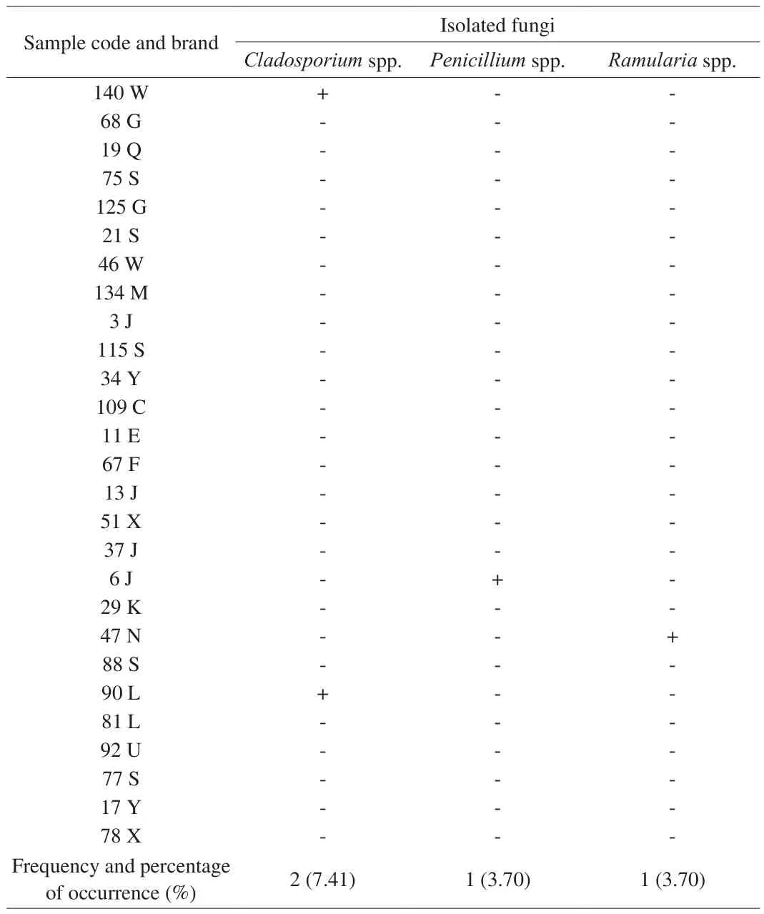
Table 1Frequency and percentage occurrence of isolated fungi spp. in fermented milk samples retailed in China.

Table 2 Out-group strains used in the phylogenetic dendrogram.
3.2 Inhibition of vanillin on the growth of fungi
3.2.1 Determination of vanillin antimicrobial activity by plate dilution method
The inhibition of different concentrations of vanillin on the growth of the fungal isolates and the effect of different concentrations of vanillin on the growth of different fungi were observed and measured directly by the plate dilution method. The result revealed that after 48 h, the inhibition effect of vanillin on fungi growth can be observed obviously. In Fig. 3, different concentrations of vanillin were used 0, 10, 25, 50, 100 mg/mL from left to right respectively.Overall, the growth ofActinomyces elegansandPenicilliumwith vanillin was similar, but compared with the controlCladosporiumgrow less. According to the area of the fungi growth occupied in the plate, the inhibition of fungi growth increased with increased vanillin concentrations. The high concentration of vanillin not only inhibited the growth of fungi, but also made spores unable to germinate. The results demonstrated that the antimicrobial activity of vanillin to different indicator fungi was different, the inhibition effect of vanillin on 3 fungi wereActinomyces elegans,PenicilliumandCladosporiumin ascending order. Therefore, the damage of vanillin to different cells was different, which might be related to the structure and composition of cell wall, the shape and volume of microorganisms [26].
3.2.2 The inhibition of vanillin on growth and morphology of fungi
The addition of vanillin had a damaging impact on the morphology of fungi. Compared to the control, by addition of vanillin, the color ofActinomyces eleganscolony was more yellow,the mycelium grew loosely and abnormal morphology was observed.In the other hand, the fungi in control were more neat, denser and smoother. This is might be due to that vanillin caused irreversible damage to the mycelium morphology.
In the first 24 h which included the spore germination period, the colony diameter ofActinomyces eleganswas increased with 5 and 10 mg/mL vanillin accordingly. The colony diameter of the control group also increased, and there was no significant difference (P< 0.05)with the rest of the colonies. The colony with 50 and 100 mg/mL vanillin began to grow on the 5th and 7th day of culture. Therefore,the addition of vanillin could inhibit the spore germination of fungi,and the higher the concentration of vanillin was, the stronger the inhibition effect was. The inhibition of vanillin on spore germination might be due to the damage of genetic material. Fitzgerald and other authors [16] found that vanillin could indirectly inhibit the activity of enzymes involved in the synthesis and expression of genetic material,then the genetic material was inactive or had incomplete structure,which increased the lag phase of microorganisms, and inhibited the spore germination.
With the increase of culture time, the growth rate of colony diameter with vanillin was lower than that of the control, and the more the amount of vanillin was, the lower the growth rate was as a whole. On the 3rd day of culture, the colony diameter ofActinomyces eleganswith 5 and 25 mg/mL vanillin was 61.12% and 32.27% of the control, respectively. Therefore, the addition of vanillin could significantly change the colony diameter ofActinomyces elegans(P< 0.05). Similar result was obtained regardingCladosporiumgrowth.

Fig. 4 Effect of vanillin on radical colony diameter of Actinomucor elegans DCY-1. Different lowercase letters indicate the comparison of culture time;significance level is P < 0.05.

Fig. 5 Effect of vanillin on radical colony diameter of Cladosporium cladosporioides CBS 129108. Different lowercase letters indicate the comparison of culture time; significance level is P < 0.05.
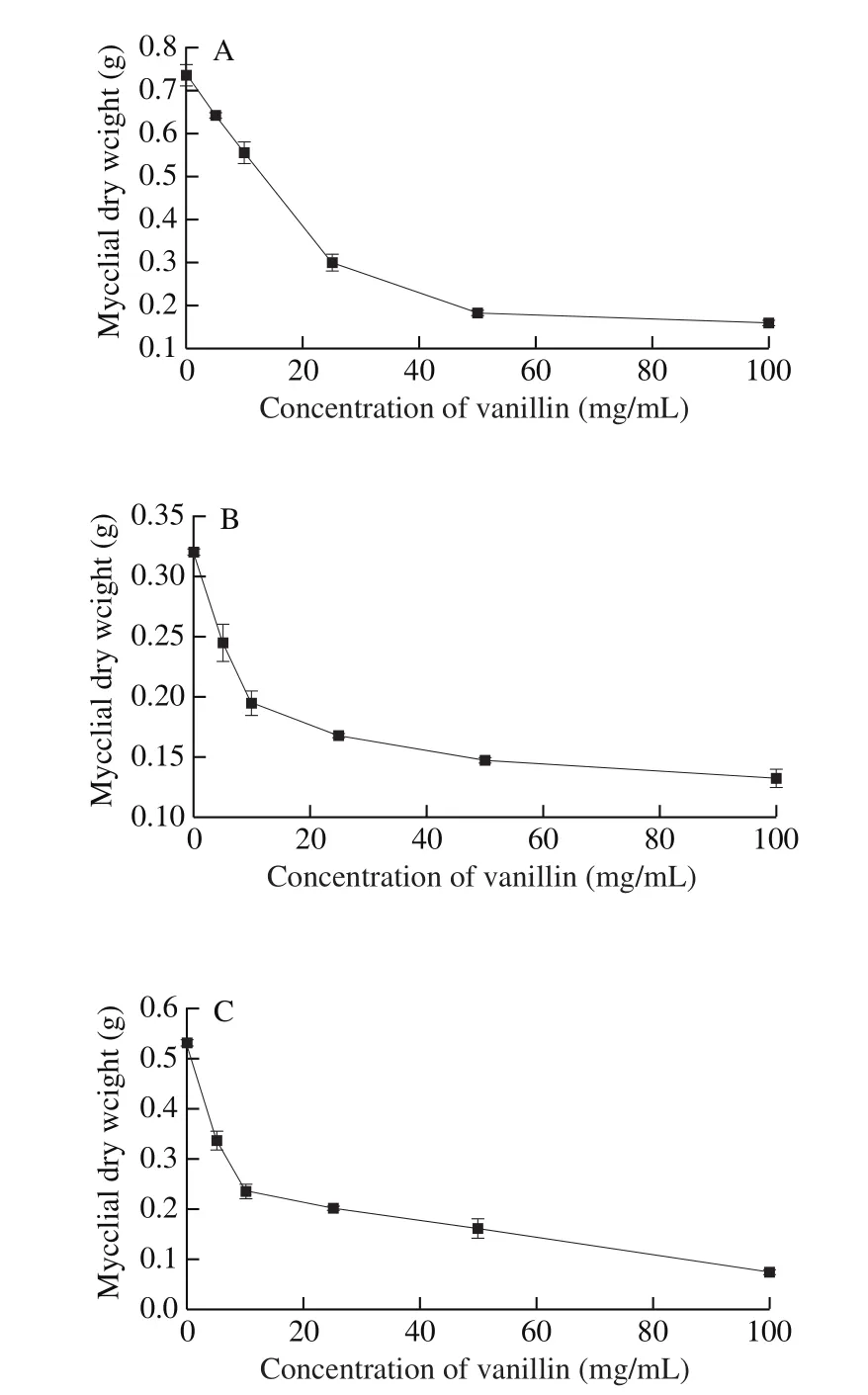
Fig. 6 Effect of vanillin on mycelium biomass of (A) Actinomucor elegans DCY-1, (B) Penicillium citrinum CBS 126809, (C) Cladosporium cladosporioides CBS 129108.
The result further showed that the more the concentration of the vanillin is increased, the slower the growth rate of both fungi was,and the longer the lag phase was which consequently resulted in an increased logarithmic growth phase of the fungi (Table 4 and Table 5).When the concentration of vanillin was 5, 10, 25, 50 and 100 mg/mL,compared with the control, the colony growth rate ofActinomyces elegansdecreased by 21.71%, 30.93%, 45.34%, 75.69% and 88.45%,and the lag phase increased to 1.24, 1.42, 2.38, 5.94 and 8.60 times.The growth rate and lag phase ofCladosporiumspp. also had the same trend. When the concentration of vanillin was 5, 10 and 25 mg/mL,the growth rate of colony decreased by 24.26%, 26.14% and 43.83%,and the lag phase increased to 1.67, 1.69 and 2.13 times. Therefore,the inhibition effect of vanillin on the growth of two kinds of fungi colonies increased significantly (P< 0.05) with the increase of the concentration of vanillin added. However, this method could not be applied to determine the effect of vanillin on fungi with small colony diameter. The vanillin might have inhibited the synthesis and expression of genetic material during the lag phase of the fungal growth, which might be due to the damage of the integrity of the cell membrane. This resulted in the disruption in the homeostasis of ion gradient and pH, indirectly inhibiting the activity of enzymes involved in the synthesis and expression of genetic material [17].

Table 4Effect of vanillin on the growth rate and lag phase of Actinomucor elegans DCY-1.

Table 5Effect of Vanillin on the growth rate and lag phase of Cladosporium cladosporioides CBS 129108.
3.2.3 The inhibition potency of the vanillin on the growth of the fungal mycelium
BesidesActinomyces elegansandCladosporium, the study further revealed that vanilla had an inhibitory effect againstPenicilliumafter 24 h of incubation in a medium containing 50 and 100 mg/mL vanillin. It further showed that after 48 h, the number of mycelial pellets in the fungal medium containing 10 and 25 mg/mL vanillin as well as the control sample increased, while the liquid medium containing 50 mg/mL vanillin was completely turbid. However, the liquid medium containing 100 mg/mL vanillin remained clear with a little turbidity. In general, after 72 h culture, only a few mycelial balls were formed in the liquid of 50 and 100mg/mL vanillin.
Based on the result in Fig. 6 and Table 6, with the increased vanillin addition, the dry weight of 3 fungi decreased. The growth inhibition rates of the 3 fungi werePenicillium,Actinomyces elegansandCladosporiumin ascending order. When the vanillin content was 100 mg/mL, the minimum biomass of mycelium was 0.16, 0.13 and 0.075 g, and the maximum inhibition rates were 58.59%, 78.23% and 85.93%, respectively. When the concentration of vanillin was 5-100 mg/mL, the growth inhibition rate of the 3 kinds of fungi was in the range of 12.9%-85.93%. This further demonstrates that vanillin is a potent inhibitor of fungal growth. With vanillin content from 0 to 100 mg/mL as the abscissa and growth inhibition rate as the ordinate,the logarithmic trend equation was obtained for all 3 kinds of fungi,then with the increase of vanillin concentration, the rate of increase in the growth inhibition rate increased first then decreased and tended finally to zero. Therefore, it clearly shows that vanillin had a significant inhibition effect on the fungal biomass and mycelium growth as well as the fungal morphology.
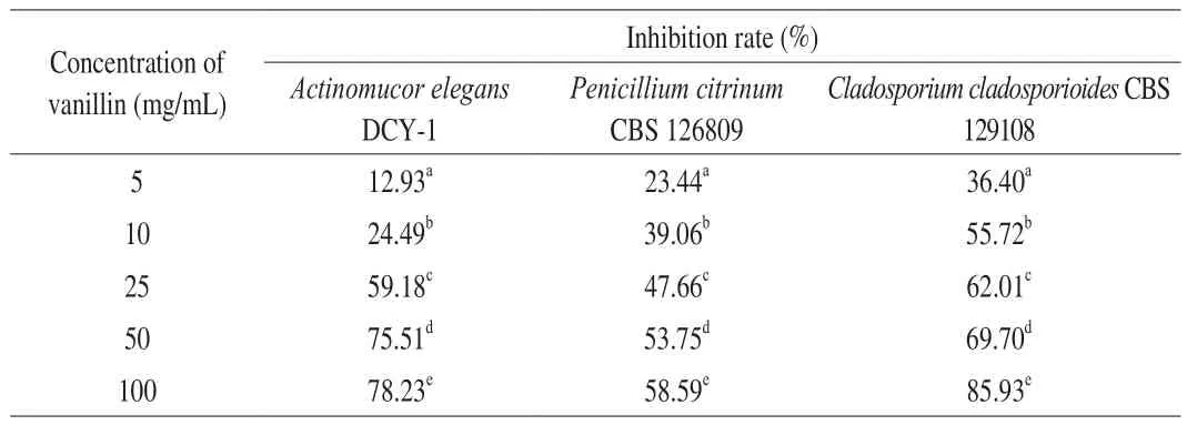
Table 6Effect of vanillin on the growth inhibition rate.
4. Conclusion
The result of this study revealed that about 14.8% of the samples were found contaminated with 3 different species of fungi. The most predominant type of contaminant wasCladosporiumspp.In this study,Actinomyces elegans, which was commonly used in food fermentation, was selected as the indicator fungi. The results showed that vanillin had inhibition effect on fungi, mainly reflected on prolonging effectively the germination time of spores, decreasing the growth rate, increasing the lag phase and decreasing the biomass of mycelium. The anti-fungal effect of vanillin varied on different strains, and the effect increased as its concentration increased. When the vanillin content was 100 mg/mL, the colony growth rate of Actinomyces elegans decreased by 88.45%, the lag phase increased to 8.60 times, the inhibition rate of mycelium growth was 85.93%,and the morphology of mycelium was damaged. The study showed that vanillin could be a potent bio-preservative for fermented dairy products. However, more study is needed to further explore the antifungal mechanisms vanillin and promotes the use of vanillin in the preservation of fermented dairy products.
Therefore, this study provides useful data evidence that will help relevant stakeholders in the fermented milk industry, to better monitor the quality and control microbial contaminants in fermented milk.
Conflict of interest
The authors declare that there are no conflicts of interest.
Acknowledgments
This research work was supported by the Jiangsu Agricultural Industry Technology System (No JATS-2018-296).
猜你喜欢
杂志排行

食品科学与人类健康(英文)的其它文章
- Bioactive compounds and probiotics-a ray of hope in COVID-19 management
- Approaches to evaluate nutrition of minerals in food
- The role of glutamine in supporting gut health and neuropsychiatric factors
- Anti-hyperglycemic effects of dihydromyricetin in streptozotocin-induced diabetic rats
- Aroma profile of two commercial truffle species from Yunnan and Sichuan, China:inter- and intraspecific variability and shared key compounds
- Stability of phenolic compounds and drying characteristics of apple peel as affected by three drying treatments