电针对早期膝骨关节炎兔模型关节软骨病理及黏弹性力学性能的影响
2021-04-29史晓伟乌云额尔顿王东峰王彤王春久郭长青史术峰
史晓伟 乌云额尔顿 王东峰 王彤 王春久 郭长青 史术峰
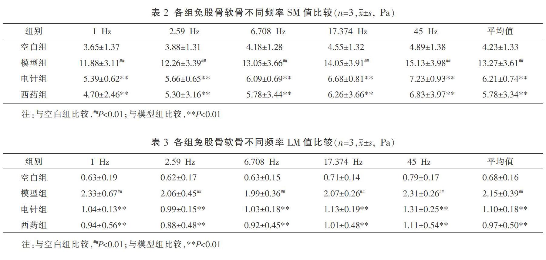
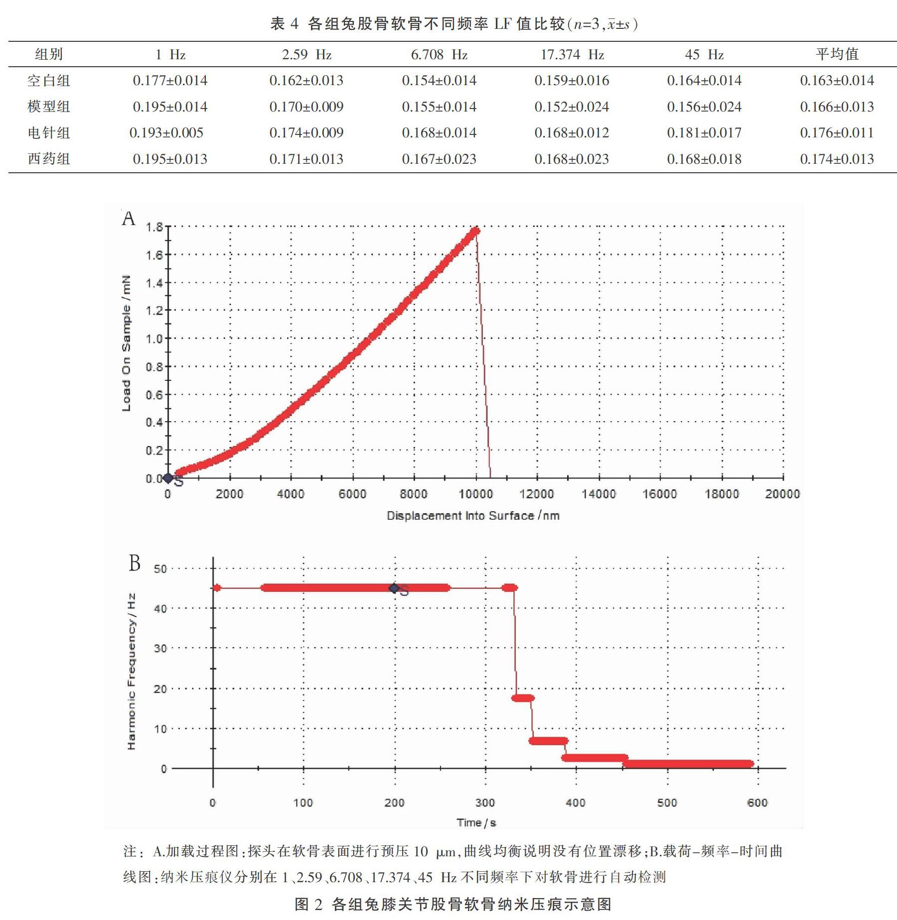
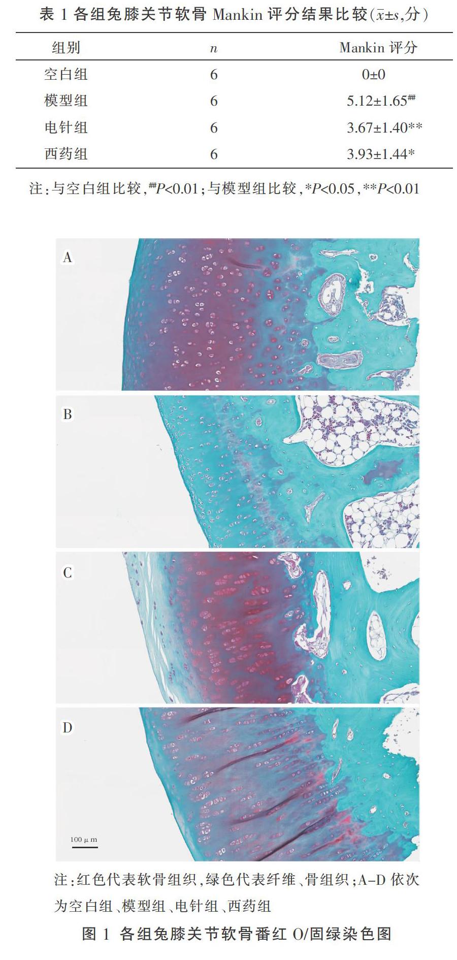
〔摘要〕 目的 探索電针治疗对早期膝骨关节炎(knee osteoarthritis, KOA)兔模型关节软骨病理及黏弹性力学性能的影响。方法 33只新西兰兔按体质量随机分为空白组(6只)及KOA造模组(27只),KOA造模组应用改良Videman法(过伸位制动)制动4周建立早期KOA兔模型,拆除固定装置后根据Lequesne评分再随机分为模型组、电针组和西药组(每组9只),均干预4周。干预结束后,兔股骨内侧髁软骨分别进行番红0/固绿染色及黏弹性力学性能(纳米压痕仪)检测。结果 (1)与空白组相比,模型组Mankin评分显著升高(P<0.01);干预治疗后与模型组相比,电针组Mankin评分显著降低(P<0.01),西药组Mankin评分明显降低(P<0.05)。(2)与空白组相比,模型组股骨软骨储存模量(storage modulus, SM)平均值显著升高(P<0.01);干预治疗后与模型组相比,电针组、西药组股骨软骨SM平均值表达显著降低(P<0.01)。(3)与空白组相比,模型组软骨损失模量(loss modulus, LM)平均值显著升高(P<0.01);干预治疗后与模型组相比,电针组、西药组软骨平均LM值表达显著降低(P<0.01)。(4)与空白组相比,模型组损失因子(loss factor, LF)表达升高;干预治疗后与模型组相比,电针组和西药组LF值表达升高,但差异均无统计学意义(P>0.05)。结论 电针干预早期KOA兔模型,具有减轻关节软骨病理损伤、改善关节软骨SM和LM的作用。
〔关键词〕 膝骨关节炎;关节软骨;电针;番红O/固绿染色;纳米压痕
〔中图分类号〕R245.9 〔文献标志码〕A 〔文章编号〕doi:10.3969/j.issn.1674-070X.2021.01.013
〔Abstract〕 Objective To explore the effects of electroacupuncture on articular cartilage pathology and viscoelastic mechanical properties in a rabbit model of early knee osteoarthritis (KOA). Methods 33 New Zealand rabbits were randomly divided into the blank group (6 rabbits) and the KOA model group (27 rabbits) according to the body weight. In the KOA model group, the modified Videman method (hyperextension braking) was applied for 4 weeks to establish the early KOA rabbit model. After removing the fixed device, the KOA rabbits were randomly divided into the model group, the electroacupuncture group and the drug group (9 rabbits in each group). All groups were intervened for 4 weeks. After the intervention, the cartilage of medial femoral condyle was stained with safranin O/fast green and its viscoelastic properties were detected (nanoindentation instrument). Results (1) Compared with the blank group, the Mankin score of the model group was significantly increased (P<0.01); after intervention, compared with the model group, the Mankin score in the electroacupuncture group (P<0.01) and drug group (P<0.05) were significantly lower. (2) Compared with the blank group, the average storage modulus (SM) of femoral cartilage in the model group was significantly increased (P<0.01); after intervention, compared with the model group, the average SM of femoral cartilage in the electroacupuncture group and drug group were significantly lower (P<0.01). (3) Compared with the blank group, the expression of average loss modulus (LM) of cartilage was significantly increased (P<0.01); after intervention, compared with the model group, the expression of average LM of femoral cartilage in the electroacupuncture group and drug group were significantly lower (P<0.01). (4) Compared with the blank group, the expression of loss factor (LF) of the model group was increased; after intervention, compared with the model group, the expression of LF in the electroacupuncture group and drug group were increased. but the difference was not statistically significant (P>0.05). Conclusion Electroacupuncture could reduce the pathological damage of articular cartilage and improve the SM and LM of articular cartilage in a rabbit model of early KOA.
〔Keywords〕 knee osteoarthritis; articular cartilage; electroacupuncture; saffron O/fast green staining; nanoindentation
膝骨关节炎(knee osteoarthritis, KOA)是一种临床常见的慢性关节退行性疾病,主要临床表现为膝关节疼痛及功能活动障碍等,其典型病理改变为膝关节软骨退变以及病情进行性加重[1]。导致KOA的高危因素众多,比如年龄、性别、职业等,其中膝关节承受异常的生物力是致病的重要原因[2]。在膝关节的稳定中,肌肉发挥动态保护作用,肌肉功能异常如肌萎缩、肌无力等会加速KOA进展[3]。既往研究证实,KOA患者多存在膝关节周围肌肉萎缩或肌力降低的临床表现,如股四头肌、腘绳肌功能减退[4]。因此,通过改善膝周肌肉功能、调整膝关节异常的生物力学环境,可能是缓解KOA关节软骨退变的良好策略。中医诊疗专家共识(2015年版)推荐针灸治疗KOA[5],但其对膝关节软骨是否具有保护作用有待研究。……