Aphrodisiac potential of Polyalthia bullata(Tongkat Ali)in fowl
2021-04-01JayaVejayanYasminAmiraCheYahyaSrikumarChakravarthiHalijahIbrahimAidaYun
Jaya Vejayan,Yasmin Amira Che Yahya,Srikumar Chakravarthi,Halijah Ibrahim,Aida Yun
1Faculty of Industrial Sciences & Technology,Universiti Malaysia Pahang,Lebuhraya Tun Razak,26300,Gambang,Kuantan,Pahang Darul Makmur,Malaysia
2Faculty of Medicine,Biomedical Sciences and Nursing,MAHSA University,Jalan SP2,Bandar Saujana Putra,42610 Jenjarom Selangor,Malaysia
3Institute of Biological Sciences,University of Malaya,50603 Kuala Lumpur,Malaysia
4Ubat Tradisional Orang Asli,Dataran Sungai Perak Sebelah Utara,North-South Expressway,Kampung Menora,33000 Kuala Kangsar,Perak Darul Ridzuan,Malaysia
ABSTRACT
KEYWORDS:Male hormone; Testosterone; Steroids; Leydig cells; Domestic chicken; Cock; Medicinal plant; Polyalthia bullata
1.Introduction
The use of aphrodisiac plants dates back centuries and includes plants either in their original form or made into consumable beverages,food or supplements for improving one's libido and sexual performance[1].Not surprisingly,men are more inclined to resort to the use of aphrodisiacs.Many plants have been used for this purpose,with some being more popular than others in certain countries.Among them are Microstylis wallichi(Jeevak)and Withania somnifera(Ashwagandha)in India[2],Butea superba in Thailand[3],Cynomorium coccineum(Desert Thumb),Prunus amygdalus(almond)and Zingiber officinale(ginger)in Arab nations[4],and Epimedium koreanum(Epimedium leaf),Dioscorea polystachya(Chinese potato),Ophiocordyceps sinensis(catepillar fungus),Panax ginseng,Ginkgo biloba and Ganoderma lingzhi in China and other pan-Asian countries[5].In Malaysia,many plants have also been claimed to be aphrodisiacs,with some popularly used traditionally by the Malays and the indigenous people residing in the forest.Only a few of the aphrodisiac plants endemic to Malaysia have gone beyond the boundaries of its borders and been popularized internationally,including Eurycoma(E.)longifolia(Tongkat Ali)and Smilax myosotiflora(Ubi Jaga)for men and Labisia pumila(Kacip Fatimah)for women.Not known to many users of E.longifolia is that the term “Tongkat Ali” is synonymous with at least three plants in Malaysia.Perhaps the synonym originated through cultural practices over the years in defining any plant with roots with a peculiar shape and claims of being an aphrodisiac.The three most widely used plants include E.longifolia,Polyalthia(P.)bullata and Stema(S.)tuberosa[6-8].
Of the three plants,P.bullata is the main focus of this study due to a lack of scientific investigations on its aphrodisiac claims.The whole plant is rarely seen in the modern landscapes of Malaysia but rather is confined to wild growth in forests located at moderate altitudes below 1 200 m[9].The distinctive differences between the more popular Tongkat Ali,i.e.,E.longifolia,and the lesser known P.bullata roots,where the colour is markedly visible as white or fairer for the former(hence simply termed as Tongkat Ali Putih in Malay),while P.bullata grows with black roots(Tongkat Ali Hitam)[10].The root of P.bullata has been sold worldwide mostly in its original form of dry cut chips to users by e-commerce with claims of increasing sexual performances once its boiled preparations have been consumed regularly[7,11].At times,this plant has been combined with other plants claimed to be aphrodisiacs,including the two other Tongkat Ali plants,i.e.,E.longifolia and S.tuberosa,to become products of premixed coffee,capsules or tablets and other beverages or preparations[12,13].Under such conditions,it has not been possible to ascertain whether the action of the products is due to this plant or merely due to the more established libido and testosterone boosting plant of E.longifolia.The latter has been well studied both clinically and in laboratory environments[13-18].A study in 64 human subjects with confirmed moderate stress levels that were given E.longifolia extract for 4 weeks showed an increased testosterone level(+37%)and reduced cortisol level(-16%)compared to placebo[19].
P.bullata may not be a mere pseudosexual enhancer,since it has been well-known that this plant has been sold to repeat buyers and users worldwide for many years.As previous reports have almost no data on its aphrodisiac potential,this current in vitro and in vivo investigation was undertaken.This work used domesticated fowls similar to the study previously done for the other two Tongkat Ali plants i.e.E.longifolia and S.tuberosa[10].
2.Materials and methods
This work was conducted for a duration of 10 months(April 2019 through January 2020).
2.1.The in vitro Leydig cell evaluations
2.1.1.Extraction of crude plant samples
The highest quality roots of P.bullata were acquired by the indigenous Temiar group from the Perak forest,Malaysia,near Kampung Orang Asli Bukit Cermin(specimen deposited in University of Malaya with voucher number of HI1445).The group of 8 to 11 families belonging to a larger Senoic cluster have for generations harvested the thick tropical jungle of Malaysia for medicinal plants for their own use and as their livelihood in selling to herbal manufacturers involved in encapsulating them into registered products.
The raw Tongkat Ali(P.bullata and E.longifolia)was obtained in the form of roots of various sizes and lengths.The roots were cut by using an electric saw into smaller pieces,followed by a slicing step that transformed the plant samples into chips.The chips were ovendried at a temperature of 50 ℃ with convection.Tongkat Ali chips were turned into powder with a blender.The choice of the positive control for the in vitro studies was based on many reports on E.longifolia being a testosterone booster.Fifty grams of each powdered plant material was mixed with 500 mL of water and refluxed for 6 h.Upon completion,the particulates were filtered,and the filtrate was freeze-dried and kept until further use.
2.1.2.Growth of Leydig cells
TM3 Leydig cells(mouse origin),purchased from the US-based ATCC,were cultured at 37 ℃ in a 5% COincubator with complete medium consisting of Dulbecco's modified Eagle's medium/nutrient mixture F-12 medium(Nacalai Tesque,JP),5% horse serum(Thermo Scientific,UK),2.5% fetal bovine serum(Thermo Scientific,UK)and 1% penicillin-streptomycin(Nacalai Tesque,JP).The cells were allowed to reach approximately 80% confluence before passaging.Passaging of the cells was carried out according to Erasmus with slight modifications[20].Briefly,the culture media was discarded,and the cells were washed with 4 mL Dulbecco-phosphate buffered saline(Nacalai Tesque,JP).Next,1 mL of 10 ×diluted 0.25%(w/v)trypsin with 0.53 mM ethylene diamine tetraacetic acid(Nacalai Tesque,JP)was added and incubated at 37 ℃ with 5% COfor 5 min until the cells detached.A volume of 2 mL complete medium was added,and the cells were centrifuged at 730 ×g for 5 min at 37 ℃.Thereafter,the cell sediment(after discarding the supernatant)was resuspended in 1 mL complete medium,and 0.25 mL of the suspension was mixed with 4.75 mL of complete medium.
2.1.3.Evaluation of testosterone production
The cells(4×10cells/well)were seeded in a 96-well plate,treated with 50 µg/mL of P.bullata(test)and E.longifolia(a positive control)and incubated at 37 ℃ with 5% COfor 24 h.After 72 h,the production of testosterone was assessed by using a testosterone enzyme linked immunosorbent assay kit according to the manufacturer's instructions(Elabscience,US).Briefly,the absorbances obtained at 450 nm were converted into the corresponding testosterone concentration with a plotted testosterone standard curve.The percentage of testosterone secretion was calculated with the following formula:

2.2.In vivo evaluations
2.2.1.Fowl(Gallus gallus)
The animals selected for this experiment were domesticated red junglefowl(Gallus gallus),a type of rooster commonly found in Malaysia provided by Sapphire Enterprise(specialized in supplying experimental animals).The period of acclimatization was approximately 10 days following delivery.The control and test groups consisted of four fowls each.The fowls were 25-27 weeks of age,i.e.,prior to reaching their fertile maturity(usually 28-38 weeks of age).The weights of the fowl ranged from 1.3-2.0 kg.The experiment conducted in controlled environment as indicated within the Animal Institutional Care and Use Committee approval of UMPIACUC_2018_01.The fowls were housed in cages at Experimental Animal Laboratory of Universiti Malaysia Pahang and fed ad libitum of chicken feed and drinking water.
2.2.2.Capsule preparation
The dosing was performed by using capsules containing the active ingredient of finely powdered P.bullata roots.A commercially available capsule maker used number “3” sized capsules to generate dosages of approximately 10 mg per capsule(adjusted to the average weight of fowl,based on capsules containing 250 mg of active ingredient per Bio-Root Tok Ali capsule that were given to adults weighing 70-90 kg).The type “3” capsule was able to contain up to 200 mg,and hence any empty space was filled with corn bran(blended chicken feed).
2.2.3.Dosing
The test and control fowl groups were made to swallow one capsule twice a day for a total of 50 days,a duration randomly selected considering no discernible increase of testosterone level at 30 days dosing was achieved(results not included).The fowls in the test group were given capsules containing 10 mg P.bullata,while the controls were given capsules with only blended chicken feed.The dose selected was similar to that used in the investigations of other Tongkat Ali plants in fowls[10].The delivery of the capsule was performed by inserting it on the tongue of the forced opened beak,followed by induction of swallowing with a supply of water squirted gently and shutting the beak tightly.Consequently,the fowl was inspected by opening the mouth for successful dosing.
2.2.4.Evaluating the mating behaviours
Upon completing the dosing,the fowls,one at a time,were released to mate with a healthy hen and observed carefully for indices known as mating behaviours(wing flapping,body shakes,crowing and beak pecking)for fowl[21].The frequencies of the mating behaviours were compared with the control fowls.The duration of observation was limited for an hour,and for accuracy in determining the frequencies,video recordings were performed for subsequent viewing.The mating experiment conducted in a closed area(dimension:3 m ×10 m)fitted with a glass window within the Experimental Animal Laboratory of Universiti Malaysia Pahang.
2.2.5.Blood sampling
To determine changes in biochemical parameters of vital organs after 50 days of dosing,approximately 4 mL blood was carefully taken into a vacutainer by positioning the fowl to expose the wing,next identified the brachial veins(located at the ventral humoral region)followed by careful drawing of the blood using a 25 G needle connected to a 5 mL syringe.The vacutainer containing the blood was sent immediately to a diagnostic lab to measure the various biochemical parameters.
2.2.6.Microanatomy of the organs
To determine the histological changes in the organs,both the liver and testis were removed and preserved in 10 mL of 10% formalin.All tissue preparations and evaluations were performed as previously described[22].Briefly,the tissue was cut into small blocks and stained in a xylene-ethanol mixture before being waxed in paraffin.A microtome was used to make a thin slice of approximately 4-5 microns,which was placed onto a microscope slide before being stained with haematoxylin and eosin(H & E).Consequently,a trained pathologist observed the slides under a light microscope(Nikon TS100 equipped with Eclipse software)at magnification of 400×and graded the tissues for changes in structure and function in comparison to the controls.Consequently,photos were taken using a camera linked to the microscope.
2.2.7.Statistical analysis
All data requiring statistical analysis were appropriately analysed by using IBM SPSS Statistics version 21.Analysis of variance was applied to determine the statistical significance of the data by calculating the variance in the data collected.A P-value less than 0.05 was considered as significant.To test the means of the population,the least significance formulae was used.
2.2.8.Ethics approval
This study was approved by the Institutional Animal Care and Use Committee of the Universiti Malaysia Pahang(ethics approval number:UMPIACUC_2018_01).
3.Results
3.1.In vitro evaluations of testosterone production
Initially,TM3 Leydig cells were used to examine the effects of P.bullata on testosterone production.As shown in Figure 1,E.longifolia(denoted as the positive control),which was the most popularly studied Tongkat Ali plant,had higher percentage of increase in testosterone secretion,at(0.80±0.02)nmol/L(350%),followed by P.bullata at(0.48±0.04)nmol/L(170%)compared to the untreated control cells with only(0.18±0.02)nmol/L(0%).The outcome showed almost twice as more testosterone produced by TM3 Leydig cells by E.longifolia compared to P.bullata in the in vitro test conducted.The results were significantly different compared to the testosterone produced by the untreated cells(P<0.05).
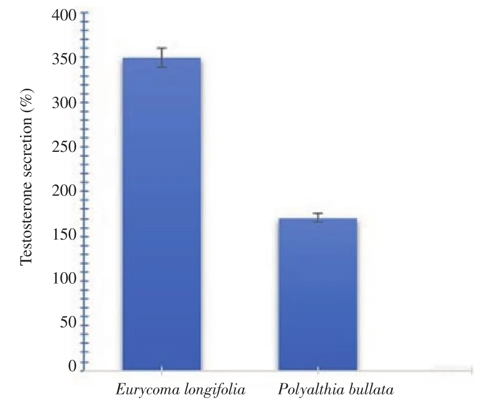
Figure 1.Percentage of testosterone production by TM3 Leydig cells after treatment with 50 µg/mL Polyalthia bullata and Eurycoma longifolia for 72 h compared to the untreated control cells.The increases in the Polyalthia bullata and Eurycoma longifoliais groups are significantly different compared to the untreated control cells(P<0.05).
3.2.Frequency of mating behaviours in the fowl
A total of four mating behaviours were observed,and their frequencies were shown in Table 1.The mating behaviours included wing flaps(flapping with the head held high and chest outwards),body shakes(wing dropped down and held inside,followed by dancelike motion circling around the hen),crows(distinctive chicken noise to attract attention of hen)and beak pecking(using the beak to peck the back of the hen's neck).Based on five observations from video recordings of fowl mating with the hens,the total frequencies for all the behaviours were 23 counts for fowl dosed with P.bullata and only approximately 15 counts for the control fowl.

Table 1.Mating frequency observed in fowls treated with Polyalthia bullata.
3.3.Blood diagnostics
The levels of the selected blood diagnostics were given in Table 2.To date,no known normal range is available for any of the biochemical parameters for chickens,unlike for humans.The testosterone measured for the control fowl averaged(4.05±0.84)nmol/L,which more than doubled to(9.72±1.1)nmol/L for P.bullata-treated fowl.There was a significant difference in testosterone between the two groups(P=0.02).Apart from the markedly lower detection of aspartate aminotransferase(AST),alanine aminotransferase(ALT)and bilirubin,all other parameters were within the levels of the controls and hence considered as insignificant and safe.Interestingly,the decrease in AST and ALT(biomarkers generalized for liver failure)and in bilirubin [approximately three times below the average of the controls,(50.74±2.97)µmol/L]suggested that this was not the result of acute toxicity but rather due to protective capabilities generally observed in some medicinal plants.

Table 2.Biochemical parameters of the Polyalthia bullata-dosed and the control groups.
3.4.Histological profiles of liver and testis
The results gave the overall impressions of the P.bullata-treated fowl having livers with focal areas of congestion in the central veins and scattered inflammation within the hepatocytes,sinusoids and portal triads.However,only insignificant to relatively mild congestion was seen in all livers(Table 3).
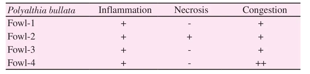
Table 3.Histopathological evaluations of the livers of the fowls treated with Polyalthia bullata.
A photomicroscopic section of the control liver microanatomy,shown in Figure 2a,showed no discernible evidence of congestion,a central vein that appeared normal and a portal triad geometrically well-organized throughout.The inspection of the sinusoidal capillary showed that it was similar to the natural structure found in liver cells.Figure 2b showed fowl-4 was graded as the most extreme among the four P.bullata-treated fowls,which had focal regions of congestion in the central vein and scattered periportal inflammation,collectively indicating that the dose given was able to lead to an initial change in the liver; however,the drug evaluated was not strong enough to cause severe necrosis or impairment.

Figure 2.Histology of normal liver(A)and fowl-4(B)liver tissues stained with H & E under a magnification of 400×.The circled area highlights comparison between a selected region of the portal triad for the control(A)and fowl-4(B)liver tissues.The outcome indicates no visible necrosis,mild inflammation and tolerable congestions within the tissues of the most extremely graded of the 4 fowls tested i.e.belonging to fowl-4.
The normal testicular tissue(Figure 3a)examined showed abundant seminiferous tubules with dimensions estimated to be approximately 150 microns but not exceeding 200 microns,while the spermatogonia,primary and secondary spermatocytes,spermatids and spermatozoa were structurally uniform and normal.Any Leydig cells visible were without inflammation or fibrosis.The testicular microanatomy of the fowl dosed with P.bullata was also examined,with that of fowl-4 was again chosen as the representative(Figure 3b).Some degree of increased seminiferous tubule size was evident,estimated to be up to 250-300 microns,which was apparently slightly larger than the control shown.The tubules were concluded to be apparently increased cellularly as well as in numbers.The same results were observed for the spermatogonia,primary and secondary spermatocytes,spermatids and spermatozoa,all of which indicated a progressive development in comparison to the controls.Throughout the inspection,no signs of inflammation or fibrosis were observed.Furthermore,the spermatogonia were intensely dense in the core of the lumen with long tails and a close grouping.All of these results indicated that P.bullata was capable of encouraging the positive development of seminiferous tubules and germ cell division followed by maturation better than the control.

Figure 3.Histology of normal testis(A)and a selected dosed fowl,fowl-4(B)tissues stained in H & E under a magnification of 400×.The circled area highlights the increased activity within the seminiferous tubules.Overall,there is a progressive development of seminiferous tubules,spermatogonia,primary and secondary spermatocytes,spermatids and spermatozoa of fowl-4 compared to those belonging to the control.
4.Discussion
P.bullata is capable of releasing testosterone in TM3 Leydigtreated cells,although to levels not as high as the more popularly studied Tongkat Ali plant of E.longifolia.No similar study on this cell line has been reported for P.bullata.Mohamed et al reported that E.longifolia possesses a greater ability to promote serum testosterone increase in rats than P.bullata[23].
Additionally,P.bullata provided a significant libido improvement based on sexual mating behaviours observed in the treated fowl.The fowl also showed an increased testosterone production by approximately 138%.The microanatomy of specific testicular tissue regions further supported the capability of P.bullata to stimulate the male sexual reproductive organ of the fowl.There are no available results assessing the ability of P.bullata to improve sexual function in chickens.However,it is known that the inclusion of testosterone alone by direct injection into fowl causes an increase in sexual behaviours such as crows and rooster dances upon facing hens[24].
The current work on P.bullata indicates that it has the potential to elevate testosterone and mating behaviours.In fowl,these effects were achieved only after a total of 50 days of dosing with a capsule containing 10 mg of active ingredient and required dosing twice a day.Such an activity can be considered as late-onset comparison to drugs such as Viagra or Cialis,which are effective within hours after consumption.Herbal aphrodisiacs in fact have a late-onset activity,as shown previously by similar work in fowl given capsules containing 12 mg of E.longifolia and S.tuberosa,both capable of increasing testosterone levels after 30 days when dosed individually[10].The indigenous people of Kampung Orang Asli of Bukit Cermin revealed that some of their customers purchasing and using P.bullata for the very first time experienced none of the aphrodisiac effects of this plant in the first month of consumption.Due to this explanation given by the indigenous people as well as the initial trial of dosing the fowl twice a day with a 10 mg capsule for 30 days provided negative results(data not included),hence prompting another trial of up to 50 days.Importantly,the results of this study provided evidence of the testosterone-boosting capabilities of P.bullata in fowl,similarly shown in only one other study,where this plant was administered to rats[23].In contrast,E.longifolia,which shares the same name of Tongkat Ali,has been proven in numerous studies[25].Some examples include the study of male rats and wild pigs fed E.longifolia that showed an increase in sexual desires and other important sexual parameters,establishing the usefulness of this plant in its aphrodisiac capabilities[26,27].
Personal communications with the families of indigenous people also highlighted that their returning customers had never complained about any adverse effects after consuming P.bullata.In this study of administering P.bullata for a duration of 50 days,none of the biochemical markers assessed were alarmingly elevated,and even in the worst graded fowl,the histology examination by the pathologist did not report severe findings,suggesting that overall,for shortduration or acute investigations,the plant is considerably safe.A study detected high levels of heavy metals in 25% of a total of 100 herbal products with P.bullata as the active ingredient[11].This sort of metal ion accumulation is due to the root leaching water containing industrial contaminated soil[28].The samples of P.bullata obtained by the indigenous people within virgin forests that were administered to the fowl are unlikely to be polluted by heavy metals;however,herbal use has been demonstrated to be toxic towards vital organs such as the liver,kidney or heart.Hence,P.bullata still requires chronic toxicity studies before concluding that it is safe.Popular Indian medicinal plants such as Eclipta alba and Phyllanthus niruri have apparently been shown to have adverse effects on the liver at high doses[29,30].
ALT and AST,if elevated to more than 5 times the highest values of their normal ranges,are considered to indicate the onset of likely liver failure according to the Association for the Study of Liver Diseases[31].To date,unlike humans,there is no range for these biomarkers in chickens; hence,they can only be compared with the control fowl.In this study,the biochemical markers of the liver,namely,ALT and AST,were not elevated; instead,they were lowered,as was bilirubin.In chickens,abnormally elevated bilirubin levels,accompanied by obstructive jaundice,are indicators of active liver disease[32].However,not only were the levels of ALT,AST and bilirubin lower than those of the controls,but histology examinations were also supportive of normal structural architectures and functions of the hepatocytes.Plants have already been reported to be able to lower bilirubin that was initially elevated dangerously high due to carbon tetrachloride exposure in liver cells[16,33,34].Although none of these liver protective studies have ever been reported for P.bullata,it is likely that it may possess such potential.Importantly,P.bullata was found to be safe in comparison to E.longifolia,which was demonstrated in a similar study to induce an elevation in ALT that was noticeable after 30 days of dosing with 12 mg E.longifolia per capsule twice daily in fowl[10].
This study limited by the ability to use a larger number of animals due to 3Rs(Replacement,Reduction and Refinement)requirement imposed by Institutional Animal Care and Use Committee.The researchers understood and accepted the requirement as it is for the welfare of the animal in study.This study of evaluating for 50 days was only an acute investigation on the efficacy and safety of P.bullata.A longer duration of at least 6 months can potentially provide the chronic outcomes.
In conclusion,P.bullata showed an increase in efficacy indicators in both in vitro(Leydig cells)and in vivo investigations performed in fowl(elevated frequencies of selected sexual mating indicators,boosted testosterone concentrations in blood,and evidence of a positive active testicular microanatomy).The onset of aphrodisiac activities nevertheless required up to 50 days of dosing in the fowl.
Conflict of interest statement
The authors declare that there is no conflict of interest.
Funding
This work was funded by grants awarded by Malaysian Technological University Network(MTUN)with grant number of UIC191201 as well as grants awarded by Universiti Malaysia Pahang with grant numbers of PDU203209(product development grant)and PGRS1903203(postgraduate grant).
Authors' contributions
The main author,Assoc.Prof Dr Jaya Vejayan,was the project leader,involved in conceptualizing,designing and executing the project,as well as in writing the manuscript from an initial rough draft stage to its current status and in editing and submitting the manuscript.The second author,Yasmin Amira Che Yahya,contributed to performing the bench work to obtain all raw data and ensure statistical validity.She also contributed to collating the results into the initial rough draft of the manuscript for the purpose of refining by other authors.Professor Dr Srikumar Chakravarthy,with expertise as a pathologist,contributed to preparing the histology tissues and examining and grading them.Next,Professor Dr Halijah Ibrahim was involved in the initial conceptualization of the research into the aphrodisiac plants,editing the manuscript draft and providing her expertise in data analysis.Last,Aida Yun,as an indigenous person,was of the utmost assistance in acquiring precise plants from the wild for the numerous samples required for the completion of the study.
杂志排行
Asian Pacific Journal of Reproduction的其它文章
- Association between estradiol levels and clinical outcomes of IVF cycles with single blastocyst embryo transfer
- Antifertility effects of 60-day oral gavage of ethanol extract of Spondias mombin leaves in guinea pigs:A biochemical,reproductive and histological study
- Combined effects of Gymnema sylvestre and Pergularia daemia on letrozole-induced polycystic ovarian syndrome in rats
- Semen characteristics of the three genetic types of boars reared in Benin
- Evaluation of pig oocyte in vitro maturation and fertilization using three gonadotropin-based hormonal compounds