肺结核空洞与肺癌空洞的多层螺旋CT鉴别诊断分析
2020-11-10孔令周
孔令周
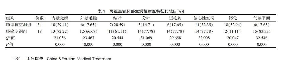

[摘要] 目的 分析多層螺旋CT对肺结核空洞与肺癌空洞的鉴别诊断价值。方法 方便选取2017年8月—2019年7月在该院就诊的肺部空洞性病变患者52例,根据其疾病类型的不同将该组患者分成肺结核空洞组(34例)与肺癌空洞组(18例),对其均行多层螺旋CT鉴别诊断,观察并比较两组患者肺部空洞性病变特征以及病灶邻近组织的结构特征。结果 肺结核空洞组患者肺部空洞内壁光滑:10例(29.41%)、外壁毛糙:6例(17.65%)、结叶:7例(20.59%)、分叶:5例(14.71%)、短毛刺:6例(17.65%)、偏心性空洞:11例(32.35%)、钙化:18例(52.94%)、气液平面:6例(17.65%)等比例均低于肺癌空洞组:13例(72.22%)、12例(66.67%)、11例(61.11%)、14例(77.78%)、14例(77.78%)、14例(77.78%)、2例(11.11%)、15例(83.33%),差异有统计学意义(χ2=21.036、23.467、20.544、31.069、29.658、22.008、20.047、32.546,P<0.05),但是肺结核空洞组患者病灶钙化比例高于肺癌空洞组,差异有统计学意义(P<0.05);在病灶邻近组织的结构特征方面,肺结核空洞组周围组织炎症、纵膈淋巴结肿大、纵膈淋巴结钙化、周围卫星灶、胸膜凹陷等特征占比均高于肺癌空洞组,差异有统计学意义(P<0.05),但是肺结核空洞组胸膜粘连所占比例与肺癌空洞组,差异无统计学意义(P>0.05)。结论 对肺结核空洞与肺癌空洞患者肺部空洞病变特征及病灶邻近组织的结特征进行进行区分鉴别时,采用多层螺旋CT进行,诊断准确率较高,有较高的推广价值。
[关键词] 肺结核空洞;肺癌空洞;淋巴结钙化;多层螺旋CT;结构特征
[中图分类号] R521;R816.4 [文献标识码] A [文章编号] 1674-0742(2020)08(c)-0183-03
[Abstract] Objective To analyze the value of multi-slice spiral CT in the differential diagnosis of pulmonary tuberculosis cavity and lung cancer cavity. Methods Fifty-two patients with cavitary lung disease who were treated in the hospital from August 2017 to July 2019 were selected, and the patients in this group were divided into tuberculosis cavity group (34 cases) and lung cancer cavity group (18 cases) according to their disease types, the differential diagnosis of multi-slice spiral CT was performed to observe and compare the characteristics of lung cavity lesions and the structural characteristics of adjacent tissues of the two groups of patients. Results In the pulmonary tuberculosis cavity group, the inner wall of the pulmonary cavity was smooth:10(29.41%), the outer wall was rough: 6(17.65%), the lobes:7(20.59%), the lobes: 5(14.71%), and short burrs: 6cases(17.65%), eccentric cavity: 11cases(32.35%), calcification: 18cases (52.94%), gas-liquid level: 6cases(17.65%), etc, lower than those of the lung cancer cavity group: 13cases(72.22%), 12cases(66.67%), 11cases(61.11%), 14cases (77.78%), 14cases(77.78%), 14cases(77.78%), 2cases(11.11%), 15cases(83.33%), the difference was statistically significant (χ2=21.036,23.467,20.544,31.069,29.658,22.008,20.047,32.546,P<0.05), but the proportion of calcification in the pulmonary tuberculosis cavity group was higher than that of the lung cancer cavity group, and the difference was statistically significant(P<0.05); in terms of the structural characteristics of the adjacent tissues of the lesion, the proportions of the surrounding tissue inflammation, mediastinal lymph node enlargement, mediastinal lymph node calcification, peripheral satellite lesions, and pleural depression in the pulmonary tuberculosis cavity group were higher than those in the lung cancer cavity group, the difference was statistically significant (P<0.05), but the proportion of pleural adhesions in the pulmonary tuberculosis cavity group was not statistically significantly different from that of the lung cancer cavity group(P>0.05). Conclusion When distinguishing the characteristics of pulmonary cavity lesions and the nodal characteristics of the adjacent tissues of patients with pulmonary tuberculosis cavity and lung cancer cavity, the use of multi-slice spiral CT has a high diagnostic accuracy and has a high promotion value.
[Key words] Pulmonary tuberculosis cavity; Lung cancer cavity; Lymph node calcification; Multi-slice spiral CT; Structural features
肺部空洞性病变疾病在临床上比较常见,其是由于患者肺部组织病变出现了明显的液化坏死,其从支气管排出的过程中吸入了气体,由空气代替了坏死组织后形成了空洞[1]。其引发机制比较复杂,常见的有结核、肿瘤等。目前,临床上比较常见的两种肺部空洞性病变包括肺癌空洞和肺结核空洞,由于病变特征的不同,其治疗方式也大为不同。因此,早期对患者的空洞特征进行鉴别诊断,能够为临床治疗提供准确的指导意见。随着我国临床诊断技术的不断改进,多层螺旋CT被广泛的应用在了肺部空洞性病变疾病的诊断中[2]。……
