基于Calpain-ERK信号通路研究Calpeptin对雌激素诱导人乳腺上皮细胞MCF-10A转化及干性标志物表达的影响
2020-07-17张艳王旭东金爱何艳詹云惠沈敬堃董宇华宛蕾
张艳 王旭东 金爱 何艳 詹云惠 沈敬堃 董宇华 宛蕾
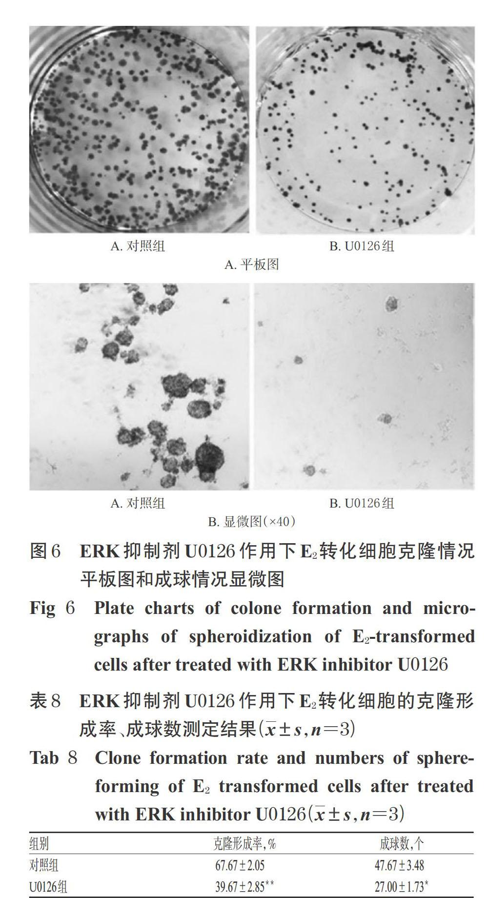
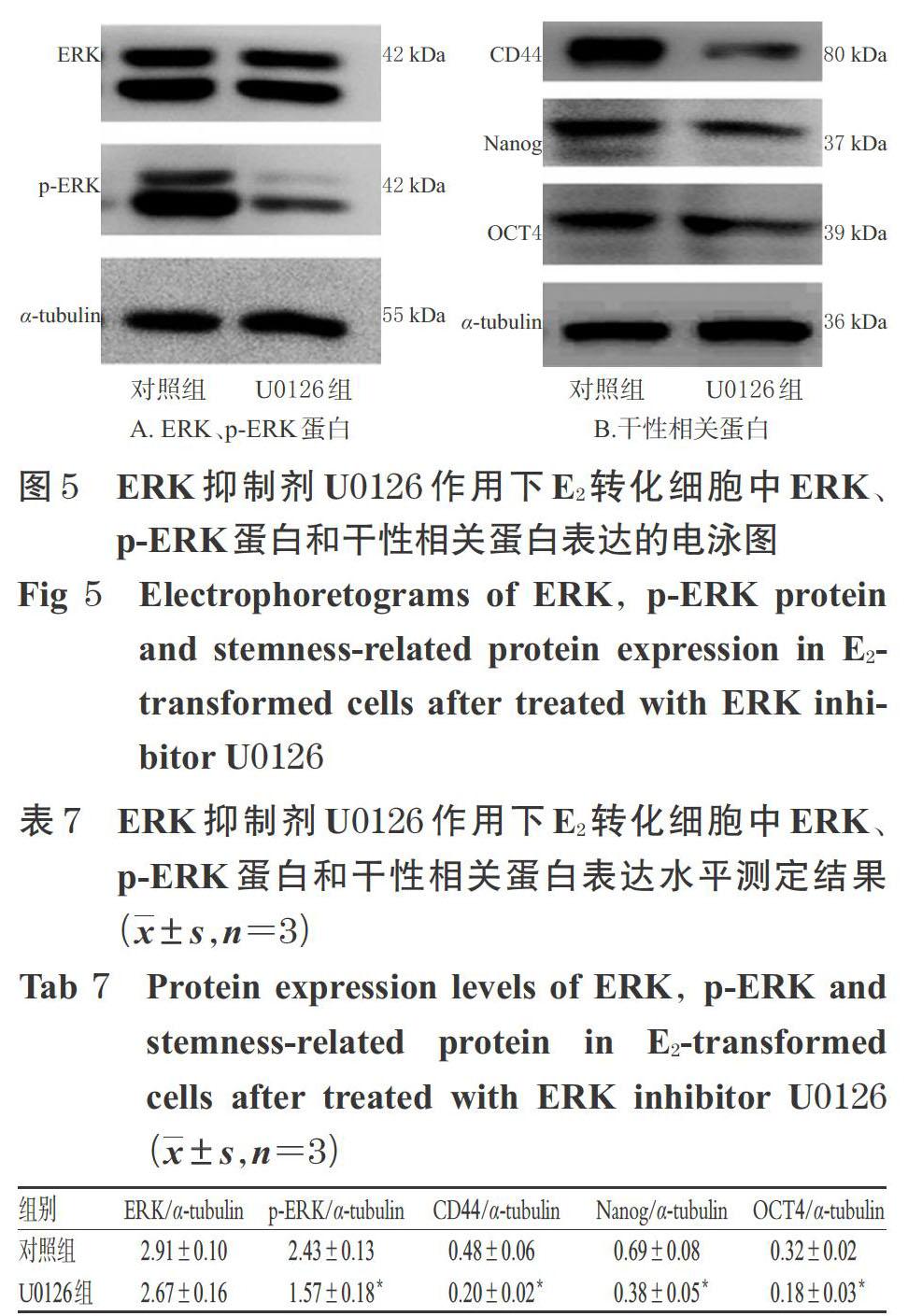
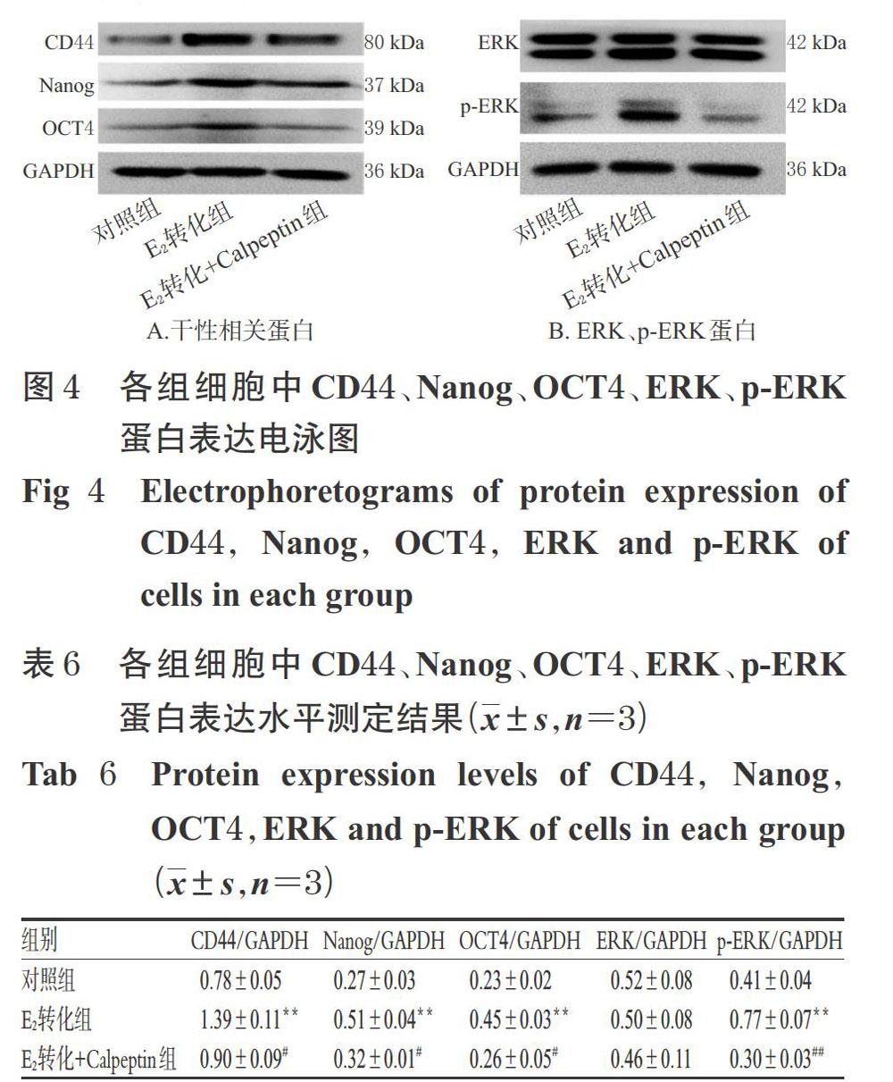
摘 要 目的:研究钙激活中性蛋白酶(Calpain)抑制剂Calpeptin对雌二醇(E2)诱导人乳腺上皮细胞MCF-10A转化及干性标志物表达的影响,并探讨其作用机制。方法:以人乳腺上皮细胞MCF-10A为研究对象,采用E2诱导制备转化细胞模型。将细胞分为对照组[0.1%二甲基亚砜(DMSO)]、E2转化组(50 nmol/L)、E2转化+Calpeptin组(50 nmol/L E2+1 μmol/L Calpeptin),以相应含药培养基连续培养15代。然后采用MTT法检测细胞的增殖率(24、48 h),采用平板克隆试验检测细胞的克隆形成率,采用悬浮成球试验检测细胞的成球数;采用实时荧光定量-聚合酶链式反应法检测细胞中干性标志物(CD44、Nanog、OCT4)和细胞外信号调节激酶(ERK)mRNA表达水平,并采用Western blotting法检测细胞中CD44、Nanog、OCT4、ERK和磷酸化ERK(p-ERK)蛋白表达水平。另取E2转化细胞分为对照组(0.1%DMSO)和U0126(ERK抑制剂)组(10 μmol/L),按上述方法测定细胞的克隆形成率、成球数以及细胞中CD44、Nanog、OCT4、p-ERK蛋白表达水平,以验证ERK表达抑制与转化细胞生物学行为及干性标志物表达之间的关系。结果:与对照组比较,E2转化组细胞的增殖率(24、48 h)、克隆形成率均显著升高(P<0.01),细胞成球数均显著增加(P<0.01),细胞中CD44、Nanog、OCT4、p-ERK mRNA表达水平及CD44、Nanog、OCT4、p-ERK蛋白表达水平均显著升高(P<0.01)。与E2转化组比较,E2转化+Calpeptin组细胞的增殖率(24、48 h)、克隆形成率均显著降低(P<0.01),细胞成球数显著减少(P<0.05),细胞中CD44、Nanog、OCT4、ERK mRNA表达水平及CD44、Nanog、OCT4、p-ERK蛋白表达水平均显著降低(P<0.05或P<0.01)。加入ERK抑制剂U0126后,E2转化细胞的克隆形成率、成球数以及细胞中p-ERK、CD44、Nanog、OCT4蛋白表达水平均显著增加或升高(P<0.05或P<0.01)。结论:Calpeptin可抑制E2诱导的人乳腺上皮细胞MCF-10A转化及干性标志物表达,其機制可能与抑制Calpain-ERK信号通路的激活有关。
关键词 Calpeptin;雌二醇;人乳腺上皮细胞MCF-10A;细胞转化;干性标志物;细胞外信号调节激酶;机制
ABSTRACT OBJECTIVE: To study the effects of Calpeptin inhibitor Calpeptin on the transformation and stemness markers expression induced by estradiol (E2), and to investigate its mechanism. METHODS: Taking human mammary epithelial cells MCF-10A as research object, transformed cells were induced by E2 treatment. Cells were divided into control group(0.1%DMSO), E2-transformed group (50 nmol/L), E2-transformed+Calpeptin group (50 nmol/L E2+1 μmol/L Calpeptin), then continuously treated with corresponding drug-containing culture medium for 15 generations. Then, MTT assay was used to determine the proliferation rate of cells (24,48 h); plate colony test was used to detect the Clone formation rate of cells; the number of sphere-forming cells was measured by suspension spheroidization test;mRNA expressions of stemness marker (CD44, Nanog, OCT4) and extracellular sigal-regulated kinase (ERK) were detected by RT-qPCR,and protein expressions of CD44, Nanog, OCT4 , ERK and p-ERK were detected by Western blotting assay. Another E2-transformed cells were divided into control group (0.1%DMSO) and U0126 (ERK inhibitor) group (10 μmol/L). Clone formation rate, the number of sphere-forming, protein expressions of CD44, Nanog, OCT4,ERK and p-ERK were determined with above methods, and to validate the relationship of ERK inhibition with transformed cell behavior and the expression of stemness markers. RESULTS:Compared with control group, proliferation rate and clone formation rate of E2 transformed group were increased significantly (P<0.01), and the number of sphere-forming was increased significantly (P<0.01); mRNA expression levels of CD44, Nanog, OCT4,ERK and protein expression levels of CD44, Nanog, OCT4 and p-ERK in cells were increased significantly (P<0.01). Compared with E2-transformed group, proliferation rate (24, 48 h) and clone formation rate of E2-transformed+Calpeptin group were decreased significantly (P<0.01), and the number of sphere-forming was decreased significantly (P<0.05); mRNA expression levels of CD44, Nanog, OCT4 , ERK and protein expression levels of CD44, Nanog, OCT4, p-ERK in cells were decreased significantly (P<0.05 or P<0.01). After treated with ERK inhibitor U0126, clone formation rate of E2-transformed cells, the number of sphere-forming, protein expression levels of CD44, Nanog, OCT4 and p-ERK were increased significantly (P<0.05 or P<0.01). CONCLUSIONS: Calpeptin can inhibit the transformation and the expression of stemness markers of human mammary epithelial cells MCF-10A, and the mechanism of it may be associated with inhibiting the activation of Calpain-ERK signaling pathway.
KEYWORDS Calpeptin; Estradiol; Human mammary epithelial cells MCF-10A; Cell transformation; Stemness marker; Extracellular sigal-regulated kinase; Mechanism
雌二醇(E2)是诱导乳腺癌发生的主要风险因素[1],其可通过激活雌激素受体(ER)或代谢产生有毒代谢产物,从而诱导乳腺上皮细胞转化以及癌变发生[2-3]。相关研究显示,乳腺肿瘤干细胞(BCSCs)在乳腺癌的发生、维持、转移、治疗抵抗和復发中起着重要作用,而E2与BCSCs的产生密切相关[4]。人乳腺上皮细胞MCF-10A是一种非致瘤性乳腺上皮细胞,正常情况下其呈不规则多边形贴壁生长,且增殖缓慢;但经E2长期诱导后,MCF-10A细胞将失去上皮细胞相关特性,胞体变大,呈长梭形贴壁生长,增殖能力增强,且具有一定的间质特征[5]。……