分根术在前牙区断根微创拔除中的临床研究
2020-07-14陈冶
陈冶
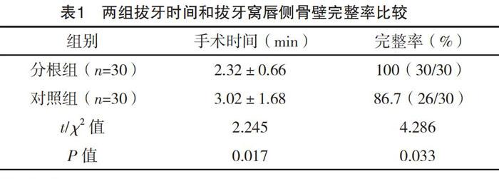
【摘要】 目的:评估分根术在前牙区断根微创拔除中的临床应用效果。方法:收集2017年10月-2019年8月来武汉科技大学附属汉阳医院口腔科门诊就诊的上前牙断根患者60例,依就诊顺序单数纳入分根组,双数纳入对照组,各30例。分根组先分根,再结合微创拔牙刀拔除;对照组不分根,采用常规微创拔牙法拔除断根。比较两组手术时间及拔牙窝唇侧骨壁完整率。结果:分根组手术时间平均为(2.32±0.66)min,短于对照组的(3.02±1.68)min,差异有统计学意义(P<0.05);分根组拔牙窝唇侧骨壁无破损,完整率100%,优于对照组的86.7%,差异有统计学意义(P<0.05)。结论:分根术应用于前牙区断根微创拔除可缩短拔牙时间,更好地保持拔牙窝唇侧骨壁完整,有利于后期的即刻种植修复,值得推广。
【关键词】 分根术 前牙区 断根 微创拔牙
doi:10.14033/j.cnki.cfmr.2020.15.017 文献标识码 B 文章编号 1674-6805(2020)15-00-02
Clinical Research of Minimally Invasive Extraction of Anterior Tooth Broken Root by Root Separation/CHEN Ye. //Chinese and Foreign Medical Research, 2020, 18(15): -42
[Abstract] Objective: To investigate the clinical results of minimally invasive extraction of anterior tooth broken root after root separation. Method: A total of 60 cases of anterior tooth broken root were collected in the Department of Dentistry in Affiliated Hanyang Hospital of Wuhan University of Science and Technology from October 2017 to August 2019, then they were divided into root seperation group and control group. In the root seperation group, anterior tooth broken roots were seperated by high speed turbine before using mimimally invasive extraction tool, while in the control group, broken roots were extracted only using minimally invasive extraction tool. The surgical duration, postoperative damage rate of the lip side plate in the two groups were analyzed. Result: The surgical duration was shorter in the root seperation group of (2.32±0.66) min, compared with the control group of (3.02±1.68) min (P<0.05). The postoperative damage rate of the lip side plate was lower in the root seperation group of 100% than in the control group of 86.7% (P<0.05). Conclusion: The surgical duration is shorter, and the postoperative damage rate of the lip side plate is lower in minimally invasive extraction of anterior tooth broken root after root separation. Smaller trauma is conductive to the immediate implant afterwards.
[Key words] Root separation Anterior tooth Broken root Mimimally invasive extraction
First-authors address: The Affiliated Hanyang Hospital of Wuhan University of Science and Technology, Wuhan 430050, China
斷根是由于牙折断或广泛的龋坏所致。口腔门诊常见上前牙外伤导致的牙折,形成较多深度达牙槽嵴以下的断根。由于难以钳夹拔除,其中部分断根为死髓牙,脆性较大,与周围牙槽骨粘连等情况,临床拔除难度较大,又因前牙区唇侧骨壁较薄,拔除时常常导致唇侧骨壁破坏较多[1-2]。随着即刻种植修复技术的广泛开展,尤其在上前牙美学区,对微创拔牙提出了严格要求,且拔牙窝唇侧骨壁的完整对后续的种植修复意义重大[3-4]。笔者将分根术应用于上前牙断根的微创拔除,并与传统断根微创拔除法进行比较。现总结报告如下。
1 资料与方法
1.1 一般资料
收集2017年10月-2019年8月来武汉科技大学附属汉阳医院口腔科门诊就诊的上前牙断根患者60例,其中男42例,女18例,年龄18~52岁。纳入标准:断根顶距牙槽嵴顶≥2 mm,仅有一颗前牙区断根,经临床评估无松动、无急性炎症、无瘘管、影像学检查无牙槽骨骨折及根尖暗影、无拔牙禁忌证。……