基于卷积神经网络的左心室超声图像特征点定位
2019-08-01周玉金王晓东张力戈朱锴姚宇
周玉金 王晓东 张力戈 朱锴 姚宇
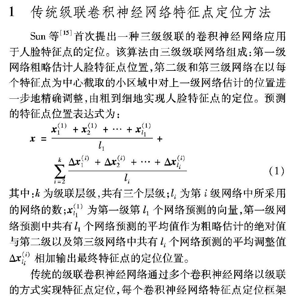
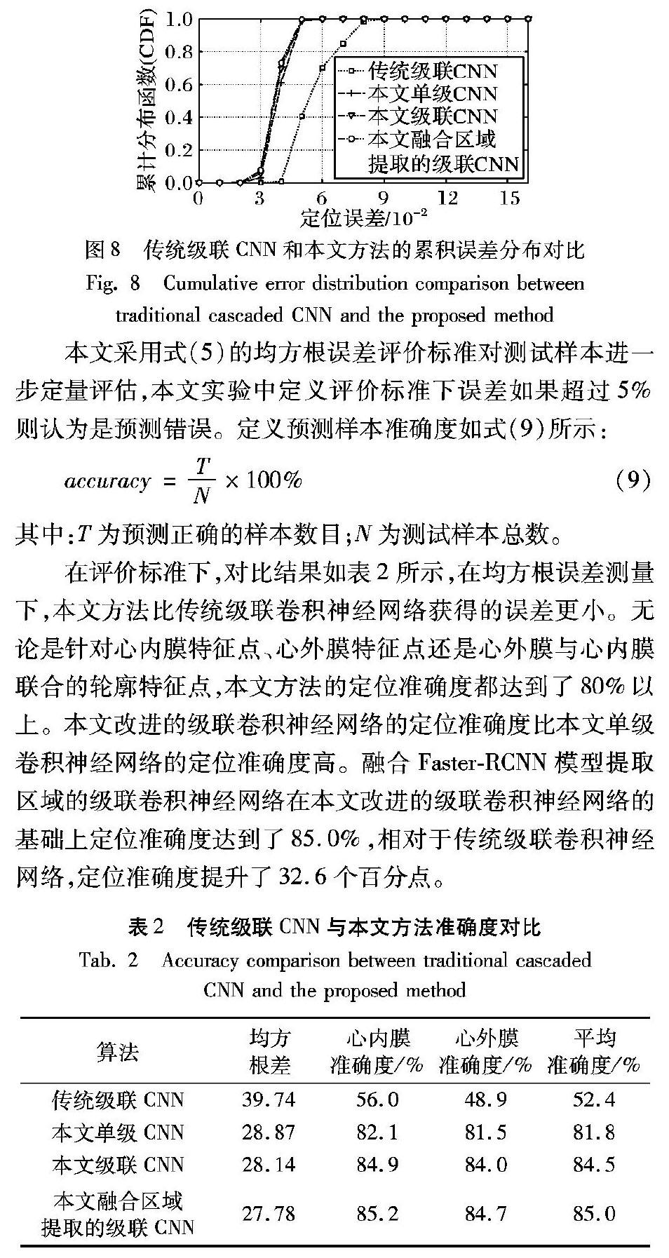
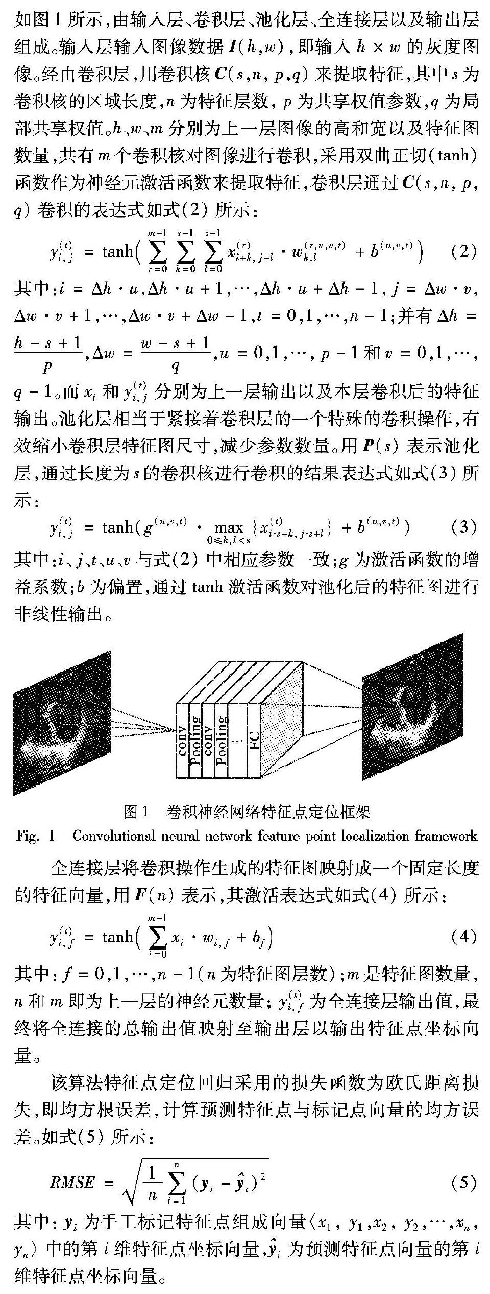
摘 要:针对传统级联卷积神经网络(CNN)在左心室超声图像中定位准确度较低的问题,提出一种融合更快速区域卷积神经网络(Faster-RCNN)模型提取区域的级联卷积神经网络,实现对超声图像中左心室心内膜和心外膜轮廓特征点的定位。首先,采用两级级联的方式改进传统级联卷积神经网络的网络结构,第一级网络利用一个改进的卷积网络粗略定位左心室心内膜和心外膜联合的特征点,第二级网络使用四个改进的卷积网络分别对心内膜特征点和心外膜特征点进行位置微调,之后定位输出左心室心内膜和心外膜联合的轮廓特征点位置;然后,将改进的级联卷积神经网络与目标区域提取融合,即利用Faster-RCNN模型提取包含左心室的目标区域并将目标区域送入改进的级联卷积神经网络;最后,由粗到细对左心室轮廓特征点进行定位。实验结果表明,与传统级联卷积神经网络相比,所提方法在左心室超声图像上的定位效果更好,更逼近真实值,在均方根误差的评价标准下,特征点定位准确度提升了32.6个百分点。
关键词:超声心动图;左心室;特征点定位;卷积神经网络;级联卷积神经网络
中图分类号: TP391.41
文献标志码:A
文章编号:1001-9081(2019)04-1201-07
Abstract: In order to solve the problem that the traditional cascaded Convolutional Neural Network (CNN) has low accuracy of feature point localization in left ventricular ultrasound image, an improved cascaded CNN with region extracted by Faster Region-based CNN (Faster-RCNN) model was proposed to locate the left ventricular endocardial and epicardial feature points in ultrasound images. Firstly, the traditional cascaded CNN was improved by a structure of two-stage cascaded. In the first stage, an improved convolutional network was used to roughly locate the endocardial and epicardial joint feature points. In the second stage, four improved convolutional networks were used to fine-tune the endocardial feature points and the epicardial feature points separately. After that, the positions of joint contour feature points were output. Secondly, the improved cascaded CNN was merged with target region extraction, which means that the target region containing the left ventricle was extracted by the Faster-RCNN model and then was sent into the improved cascaded CNN. Finally, the left ventricular contour feature points were located from coarse to fine. Experimental results show that compared with the traditional cascaded CNN, the proposed method is much more accurate in left ventricle feature point localization, and its prediction points are closer to the actual values. Under the root mean square error evaluation standard, the accuracy of feature point localization is improved by 32.6 percentage points.
Key words: echocardiography; left ventricle; feature points location; Convolutional Neural Network (CNN); cascaded convolutional neural network
0 引言
超聲心动图因其便携、成本低廉成为医学中心脏腔室测量可视化最常用的一种医疗成像方式。超声专家们往往关心超声图像上关键部位的生理参数信息,通常对左心室超声成像进行分析并对收缩末期和舒张末期阶段心室轮廓边界进行检测,用它来评价心功能的好坏进而预判诊断心脏疾病[1]。……