人参总皂苷抑制肺纤维化大鼠血清氧化水平的研究
2019-05-30陈盼盼
杨 露,陈盼盼,罗 敏,2,高 杨,邓 江
(1.遵义医科大学 基础药理教育部重点实验室暨特色民族药教育部国际合作联合实验室,贵州 遵义 563099;2.遵义市第一人民医院,贵州 遵义 563000)
Pulmonary fibrosis is a chronic,progressive and a variety of diffuse interstitial lung diseases,with high morbidity and mortality rate[1-3].Most of Patients died of respiratory failure eventually and there is no effective treatment[4].Therefore,the disease is harmful to human health.Inflammation and immune response are one of the main pathogenesis of lung tissue injury and fibrosis.A large number of studies have shown that oxygen free radicals play an important role in inflammation and immune-mediated tissue damage[5].
Total ginsenosides(TG)is the main active component of ginseng,which is one of the most precious traditional chinese medicinal herbs,and has been shown to exert beneficial pharmacology effects on the respiratory system[6-8].TG has been reported to inhibit acute lung injury[9-10].TG is effective in inhibiting oxidative and anti-inflammatory injury.At the same time,TG can regulate the immune function[11-13].However,it is unclear whether TG can inhibit pulmonary fibrosis.In this study,we observed the protective effect and mechanism of TG on pulmonary fibrosis induced by monocrotaline(MCT)and preliminarily determined whether it was related to antioxidant damage.
1 Materials and Methods
1.1 Animals and reagents Male Sprague Dawley rats(n=75,weight 180 to 220 g,SPF,Certificate Number:SCXK 2017-0005)were purchased by Experimental Animal Center of the Third Military Medical University(Chongqing,China).Rats were fed rodent feed and distilled water in the laboratory animal barrier system facilities of the Key Laboratory for Basic Pharmacology of Ministry of Education(Zunyi Medical University).TG(purity:90%)was obtained from Beijing Institute of Natural Medicine(Beijing,China).MCT was product of Sigma Corporation of Germany.The research protocol was approved by the ethics committee of Zunyi Medical University.
1.2 Pulmonary fibrosis model and experimental protocol The rat model of pulmonary fibrosis was produced with the method reported by Molteni[14].Briefly,the rats were given single subcutaneous injection with either MCT 50 mg/kg or equal saline as sham control after they were acclimatized for a week.Then,all rats were randomly divided into 5 groups(n=15 per group):sham operation control group(Sham),model control group(Model)and TG 40,80 and 160 mg/kg group.The rats of model and sham groups were given distilled water,and the other rats were administered TG 40,80,and 160 mg/kg/day.The dosage of TG and the course of treatment were determined by our preliminary experiment.All animals were given intragastric administration following the operation for 21 days,and were weighed every 5 days to enable dose adjustment during this period.
1.3 Pulmonary indexes analysis Sixty-nine surviving rats were anesthetized chloral hydrate(350 mg/kg,intraperitoneal injection)after the last administration.The lung samples were separated.The weights of lung were recorded to calculate pulmonary indexes.The pulmonary indexes are equal to the weight of the lung divided by the body weight[15].
1.4 Histologic analysis To observe the histopathology of lung samples,the rat lung specimens were fixed with 4% formaldehyde,embedded in paraffin.Sections(5μm thick)were dyed with hematoxylin-eosin(HE),Masson and TUNEL for morphological analysis by LEICA image analysis system(LEICA,Germany).HE staining is carried out at room temperature and can assess the degree of alveolitis in lung tissue.Masson staining can assess the degree of pulmonary fibrosis and TUNEL staining can assess the degree of cell apoptosis in lung tissue.Masson and TUNEL dyeing are carried out at 37°C.
1.5 Detection of SOD,MPO,NOS,T-AOC and ·OH in rats serum Blood was collected from abdominal aorta of the rats under anesthesia and the serum was prepared by centrifugation at 3 000 rpm for 15 min.The activity of superoxide dismutase(SOD),myeloperoxidase(MPO)and nitric oxide synthase(NOS),the level of total antioxidant ability(T-AOC)and hydroxyl radical(·OH)in rats serum were measured with Multiskan Sky Microplate Spectrophotometer(Thermo Fisher Scientific Inc.,USA)according to manufacturer's instruction of the test kits(Nanjing Jiancheng Bioengineering Institute,China)at room temperature.
1.6 Statistical analysis All data were analyzed by SPSS 18.0 statistics software(SPSS Inc.,Chicago,Illinois,USA)and presented as mean±SEM.The normal distributed data firstly were analyzed statistically via one-way analysis of variance(ANOVA),and the statistical significance of difference between two groups was determined using LSD method if equal variance or Dunnett's T3 method if missing variance.Statistical significance was accepted atP<0.05.
2 Results
2.1 TG administration reduced pulmonary indexes of pulmonary fibrosis induced by MCT in rats At 21 days after MCT injection,the rats showed shortness of breath,decreased activity and unsmooth hair and weight loss(P<0.05).Meanwhile,pulmonary indexes were increased significantly(P<0.05).The conditions of the rats were improved and the pulmonary indexes were reduced by TG(P<0.05).The effects of TG on pulmonary indexes are shown in Fig 1.
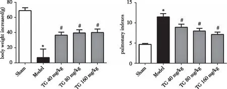
Male rats were injection of MCT(50 mg/kg).TG 40,80,160 mg/kg/day intragastric administration for 21 d.The weight decreased in the model group(Model).Total ginsenoside(TG) treatment could improve significantly the weight.The pulmonary indexes are defined as the weight of the lung divided by the weight.(Sham,Model,and TG-40:total ginsenoside 40 mg/kg/day,TG-80:total ginsenoside 80mg/kg/day,TG-160:total ginsenoside 160 mg/kg/day).Data are mean±SEM of 11-15 rats(significant at *:P<0.05 against Sham control and #:P<0.05 against Model control). Fig 1 Effect of total ginsenoside on the pulmonary indexes of pulmonary fibrosis induced by monocrotaline in rats
2.2 TG treatmenta meliorates the histomorphologic changes of pulmonary fibrosis induced by MCT in rats As shown in Fig 2,compared with the sham group,there was obvious pulmonary congestion and consolidation in the model group.TG can significantly improve the general morphological changes of lung tissue.The pulmonary histomorphology is shown in Fig 3.It was shown that the rats get a significant degree of pulmonary fibrosis after MCT injection,which was characterized by structural disorders,alveolar septum widening,collapse fusion,inflammatory cell infiltration and fibroblast proliferation.Masson staining showed a lot of blue collagen fibers were deposited(Fig 4).TUNEL staining showed a lot of brown apoptotic cells in the tissue of pulmonary fibrosis induced by MCT(Fig 5).Compared with model group,TG administration significantly improved the lung pathological injury induced by MCT.
2.3 TG administration reduced oxidative damage of pulmonary fibrosis induced by MCT in rats As shown in Fig 6,compared with sham group,the activity of SOD was decreased but MPO and NOS were increased significantly(P<0.05).Moreover,the level of T-AOC was decreased but ·OH were increased significantly(P<0.05).TG can effectively improve the changes of these indexes caused by MCT(P<0.05).

The surface of the lung tissue of the rats was smooth and ruddy in the sham group,and obvious pulmonary congestion and consolidation appeared in model group.TG could obviously improve the general morphological changes of lung tissue in model rats.Fig 2 Effect of total ginsenoside on the general morphological changes of pulmonary fibrosis induced by monocrotaline in rats
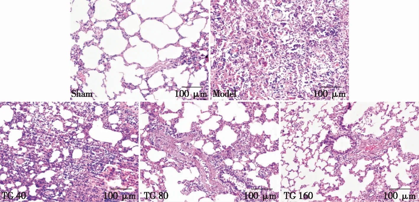
The sections demonstrated normal morphology in the sham group(Sham) and structural disorder and alveolar septum widening in the model group(Model).Total ginsenoside treatment significantly improved these histopathological lesions(TG-40:total ginsenoside 40 mg/kg/day,TG-80:total ginsenoside 80 mg/kg/day,TG-160:total ginsenoside 160 mg/kg/day).(Original magnification,Bar=100 μm,Magnification 20×).Fig 3 Representative pulmonary tissue sections stained with H.E.

The lung structure was hardly any collagen accumulation in the sham group(Sham),and the numerous collagen fibers were observed in the model group(Model).The degree of collagen accumulation was significantly alleviated after treatment with total ginsenoside.(TG-40:total ginsenoside 40 mg/kg/day,TG-80:total ginsenoside 80 mg/kg/day,TG-160:total ginsenoside 160 mg/kg/day).(Original magnification,Bar=100 μm,200×).Fig 4 Representative pulmonary tissue sections stained with masson

The lung structure was few apoptotic in the sham group(Sham),and apoptotic were significantly in the model group(Model).The degree of apoptotic was significantly alleviated after treatment with total ginsenoside.(TG-40:total ginsenoside 40 mg/kg/day,TG-80:total ginsenoside 80mg/kg/day,TG-160:total ginsenoside 160 mg/kg/d).(Original magnification,Bar=50 μm,400×).Fig 5 Representative pulmonary tissue sections stained with TUNEL

Male rats were injection of MCT(50 mg/kg).TG 40,80,160 mg/kg/day intragastric administration for 21d.TG can effectively improve the changes of these indexes caused by monocrotaline(Sham,Model,and TG-40:total ginsenoside 40 mg/kg/day,TG-80:total ginsenoside 80 mg/kg/day,TG-160:total ginsenoside 160 mg/kg/day).Data are mean±SEM of 8 rats(significant at *:P<0.05 against Sham group and #:P<0.05 against Model group). Fig 6 Effect of total ginsenoside on oxidative damage-related indexes of rats pulmonary fibrosis induced by monocrotaline
3 Discussion
It has been reported pulmonary fibrosis is a serious threat to human health.Most patients died of pulmonary hypertension,pulmonary heart disease and right heart failure[16-19].Previous studies have shown that MCT causes pulmonary fibrosis as well as pulmonary hypertension,which is similar to many patients with pulmonary hypertension complicated with pulmonary fibrosis[14,20].In this study,pulmonary fibrosis in rats was induced by MCT of subcutaneous injection and the effect of TG was observed on pulmonary interstitial fibrosis.
MCT was transformed into MCT pyrrole by cytochrome P450 monooxidase in the liver of rats.MCT pyrrole is a bioactive substance,which it reached the lung through blood circulation,and caused delayed and progressive damage to pulmonary vascular endothelial cells.Inflammatory cells,such as monocytes/macrophages and lymphocytes infiltrate around the involved vessels and release a large number of cytokines[21].These cytokines can promote NOS synthesis and the release of nitric oxide(NO)and participate in oxidative damage[22].At 21 days after MCT injection,some pulmonary vascular endothelial cells even necrosis and the whole pulmonary artery became fibrosis[23].Our study found that TG reduced the pulmonary indexes and improved the change of histologyinrats by subcutaneous injection MCT.The results indicated that TG could inhibit the occurrence and development of pulmonary fibrosis.
Many studies have demonstrated that oxidative stress plays an important role in the formation of pulmonary fibrosis,and it can induce apoptosis of epithelial cells,which may be an important factor to development of pulmonary fibrosis in the early occurrence[24].In physiological state,system is in dynamic equilibrium.When the body is affected by the damaging factors,the oxidation-antioxidant system is out of balance.A large number of free radicals and degradation products will be produced,and it eventually leads to the damage of plasma embrane,promoting the proliferation of fibroblasts and collagen synthesis[25-27].Some studies have shown that SOD is the main antioxidant in lung tissue,which distributes on the cell surface and extracellular matrix,and plays an important role in protecting alveolar cells[28].As the most abundant antioxidants in cells,T-AOC plays a key role in the process of scavenging oxide,which can directly reflect the activity of antioxidant enzymes[29].As a key enzyme,NOS can catalyze L-arginine to produce ammonia acid and nitric oxide.NO and its metabolites not only directly cause lung injury,but also mediate the proliferation and apoptosis of alveolar epithelial cells,macrophages and fibroblasts[30].The main function of MPO is to eliminate microorganisms in phagocytes and produce hypochlorite by hydrogen peroxide and chloride ions and form free radicals with oxidation ability.It has been reported that the activity of MPO increased is responsible for lung fibers.The ·OH is known to be the most oxidizing and toxic oxygen free radical,and it is also one of the important factors leading to oxidative injury of lung tissue[29].In this study,TG(80,160 mg/kg)could improve pulmonary fibrosis.The activity of MPO,NOS and the level of·OH were reduced,meanwhile,the activity of SOD and the level of T-AOC in serum of pulmonary fibrosis rats were increased.These results suggest that TG could inhibit the experimental pulmonary fibrosis induced by MCT by regulating the antioxidant capacity.
In summary,TG could inhibit pulmonary fibrosis induced by MCT.The mechanism may be related to the increased antioxidant capacity of organisms.
