动态增强MRI与SPECT肾动态显像测定GFR评估肾移植术肾功能的临床价值比较
2019-03-18顾虹刘丹李云鹤
顾虹 刘丹 李云鹤

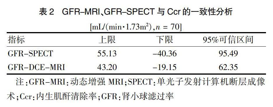
[摘要] 目的 比較动态增强MRI(DCE-MRI)与单光子发射计算机断层成像术(SPECT)肾动态显像测定肾小球滤过率(GFR)评估肾移植术肾功能的临床价值。 方法 选取2015年4月~2018年4月于内蒙古包钢医院接受肾移植术的患者70例为研究对象,分别采用DCE-MRI及SPECT测定其移植肾GFR(分别为GFR-MRI、GFR-SPECT),并与内生肌酐清除率(Ccr)进行比较分析。评估DCE-MRI检测中造影剂的安全性。 结果 检查测得患者GFR-MRI与Ccr值的偏倚高于GFR-SPECT,30%及50%精确度高于GFR-SPECT,差异有统计学意义(P < 0.05);Pearson相关性分析结果显示,GFR-MRI、GFR-SPECT值均与Ccr呈正相关(P < 0.05),GFR-MRI与Ccr相关系数高于GFR-SPECT与Ccr相关系数,差异有统计学意义(P < 0.05)。通过Bland-A1tman分析,GFR-SPECT的95%可信区间为95.49 mL/(min·1.73m2),GFR-MRI的95%可信区间为62.35 mL/(min·1.73m2),GFR-MRI的可信区间更窄。70例患者中仅2例出现轻微皮疹,且在未经任何治疗的情况下自行好转。 结论 与SPECT比较,DCE-MRI测得的GFR与Ccr偏倚较大,但与Ccr比较具有更高的精确度、相关性及一致性,可信区间更窄,DCE-MRI能更准确通过测定GFR评估肾移植术肾功能,且具有较高的安全性。
[关键词] 肾移植;动态增强MRI;单光子发射计算机断层成像术;肾动态成像
[中图分类号] R692 [文献标识码] A [文章编号] 1673-7210(2019)01(c)-0167-04
[Abstract] Objective To compare the clinical value of dynamic contrast-enhanced MRI (DCE-MRI) and single photon emission computed tomography (SPECT) renal dynamic imaging of glomerular filtration rate (GFR) measurement in the evaluation of renal function in renal transplantation. Methods A total of 70 recipients who underwent renal transplantation in Baogang Hospital in Inner Mongolia from April 2015 to April 2018 were selected research as objeets. The transplanted renal GFR was measured by DCE-MRI and SPECT (GFR-MRI, GFR-SPECT respectively), and was compared with endogenous creatinine clearance rate (Ccr). The safety of contrast agents was evaluated in DCE-MRI detection. Results The GFR-MRI and Ccr values were more biased than GFR-SPECT, and the accuracy of 30% and 50% were higher than GFR-SPECT, and the difference was statistically significant (P < 0.05). Pearson correlation analysis showed that GFR-MRI and GFR-SPECT values were positively correlated with Ccr (P < 0.05), and GFR-MRI and Ccr correlation coefficients were higher than GFR-MRI and Ccr correlation coefficients, and the difference was statistically significant (P < 0.05). By Bland-A1tman analysis, the 95% confidential interval of GFR-SPECT was 95.49 mL/(min·1.73m2), and the 95% confidential interval of GFR-MRI was 62.35 mL/(min·1.73m2), and the confidential interval of GFR-MRI was narrower. Only 2 cases developed mild rash among 70 patients, and improved on their own without any treatment. Conclusion Compared with SPECT, the bias of GFR measured by DCE-MRI and Ccr is larger, but it has higher accuracy, correlation and consistency with Ccr, and has narrower confidential interval. DCE-MRI can more accurately evaluate renal function in renal transplantation through measuring GFR, and it has high safety.
[Key words] Renal transplantation; Dynamic contrast-enhanced MRI; Single photon emission computed tomography; Renal dynamic imaging
目前,肾移植已经成为终末期肾病最有效的治疗措施,而肾移植术后肾功能监测及移植肾解剖结构的及时评估是有效延长移植肾存活时间的关键[1]。肾小球滤过率(GFR)是反映肾脏滤过功能最直接指标,也是评价肾脏功能最为敏感的指标之一[2]。但是GFR不能直接测定,只能通过某些标志物的清除率来间接测得。菊粉清除率是公认的测定GFR的金标准,但其操作复杂、费时,不适用于临床推广[3]。单光子发射计算机断层成像术(SPECT)肾动态显像方法是目前评价肾移植供体GFR的常规检查方法,简易方便、安全无创,可直观反映GFR及尿路排泄情况,但是SPECT无法提供肾脏的血管、尿路解剖结构[4]。核磁共振(MRI)常被用于了解肾脏结构,其中动态增强MRI(DCE-MRI)已逐步被应用于评估肾脏GFR[5]。然而,DCE-MRI在评估肾移植术后移植肾GFR中的准确性及安全性的研究尚不多见。本研究选取70例肾功能良……