胸部多排螺旋CT扫描诊断职业性尘肺病的应用及其临床意义分析
2019-01-06胡碧华曹子文陈丽琨周建中伍健芝
胡碧华 曹子文 陈丽琨 周建中 伍健芝
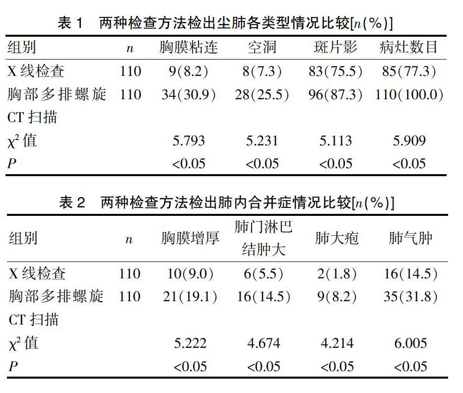
[摘要] 目的 探讨胸部多排螺旋CT扫描诊断职业性尘肺病的应用及其临床意义。 方法 选取2017年8月~2018年8月我院收治的职业性尘肺病患者110例,所有患者均进行X线检查与胸部多排螺旋CT扫描。比较两种检查方法尘肺各类型情况、肺内合并症情况。 结果 胸部多排螺旋CT扫描检查出的胸膜粘连、空洞、斑片影、病灶数目多于X线检查(P<0.05);胸部多排螺旋CT扫描检查出的胸膜增厚、肺门淋巴结肿大、肺大疱、肺气肿多于X线检查(P<0.05)。 结论 职业性尘肺病诊断中,胸部多排螺旋CT扫描的诊断结果更准确,诊断率更高。
[关键词] 职业性;尘肺病;胸部多排螺旋CT扫描;X线检查
[中图分类号] R445.3 [文献标识码] B [文章编号] 1673-9701(2019)32-0104-03
Application and clinical significance of chest multi-slice spiral CT scan in diagnosis of occupational pneumoconiosis
HU Bihua CAO Ziwen CHEN Likun ZHOU Jianzhong WU Jianzhi
Department of Radiology, Occupational Health Surveillance Institute, Guangdong Occupational Disease Prevention and Treatment Institute, Guangzhou 510300, China
[Abstract] Objective To investigate the application and clinical significance of chest multi-slice spiral CT scan in the diagnosis of occupational pneumoconiosis. Methods A total of 110 patients with occupational pneumoconiosis admitted to our hospital from August 2017 to August 2018 were enrolled. X-ray examination and chest multi-slice spiral CT scan were performed among all the patients. Types of pneumoconiosis and intrapulmonary complications of the two examination methods were compared. Results The number of cases of pleural adhesions, cavities, patchy shadows, and lesions detected by multi-slice spiral CT scan was larger than that detected by X-ray examination (P<0.05). The number of cases of pleural thickening, hilar lymph node enlargement, pulmonary bulla and pulmonary emphysema detected by multi-slice spiral CT scan was larger than that detected by X-ray examination (P<0.05). Conclusion In the diagnosis of occupational pneumoconiosis, chest multi-slice spiral CT scan has more accurate diagnosis results and higher diagnosis rate.
[Key words] Occupational; Pneumoconiosis; Chest multi-slice spiral CT scan; X-ray examination
塵肺在我国是常见的职业疾病,多发于长期进行粉尘工作的人群,主要分为石棉肺、煤工尘肺、矽肺三种,发病原因为细小粉尘长时间堆积在肺部组织中刺激组织发生病变[1]。近几年,多层螺旋CT(Multisliecs helieal CT,MSCT)技术发展迅速,该方法扫描所需时间较短,通过多种重建、薄层等可以获得更加清晰、高质量的图像,有利于尽早诊断出尘肺,为患者提供治疗[2]。我院在职业性尘肺病诊断中,使用胸部多排螺旋CT扫描进行诊断,诊断结果准确,现报道如下。
1 资料与方法
1.1一般资料
选取2017年8月~2018年8月我院收治的职业性尘肺病患者110例,所有患者均进行X线检查与胸部多排螺旋CT扫描。纳入标准:(1)符合职业性尘肺病相关诊断标准;(2)有明确的粉尘接触史;(3)通过肺功能检查确诊为职业性尘肺病;(4)知晓同意此次研究。排除标准:(1)心肝肾功能衰竭者;(2)存在肺部急性感染疾病者;(3)肺癌患者;(4)近期接受过相关治疗者。110例患者中,男63例,女47例,年龄42~64岁,平均(47.4±2.3)岁;粉尘接触时间7~21年,平均(10.5±2.7)年;煤工尘肺20例,矽肺75例,其他15例。本研究经我院伦理委员会审核批准。