频域光学相干断层扫描观察早期小切口白内障摘除联合人工晶体植入术后黄斑区的形态变化
2017-12-11贾绍友丁明红赵金亮
贾绍友 丁明红 赵金亮
【摘要】 目的 應用频域光学相干断层扫描(SD-OCT)观察小切口白内障摘除联合人工晶体植入术后早期黄斑中心区的形态变化, 探讨该术式对黄斑区的安全性。方法 选取160例(160眼)接受小切口白内障摘除联合人工晶体植入术患者作为病例组, 另选取29例健康志愿者作为正常对照组。对两组受检者的双眼采用SD-OCT行黄斑中心凹视网膜厚度及黄斑区容积的检测并进行比较。结果 正常对照组右眼黄斑中心凹视网膜厚度为(224.40±14.81)μm, 黄斑区容积为(8.62±0.40)mm3;左眼黄斑中心凹视网膜厚度为(221.05±13.25)μm, 黄斑区容积为(8.43±0.46)mm3, 正常对照组右、左眼黄斑中心凹视网膜厚度及黄斑区容积比较差异无统计学意义(P>0.05)。病例组术后3 d及1个月右眼和左眼黄斑中心凹视网膜厚度、黄斑区容积比较差异无统计学意义(P>0.05)。病例组术后3 d及1个月右眼黄斑中心凹视网膜厚度、黄斑区容积与正常对照组比较差异无统计学意义(P>0.05), 病例组术后3 d及1个月左眼黄斑中心凹视网膜厚度、黄斑区容积与正常对照组比较差异无统计学意义(P>0.05)。结论 小切口白内障摘除联合人工晶体植入术对黄斑中心区的形态影响较小, 安全性较高。SD-OCT是观察小切口白内障摘除术后早期黄斑中心区形态变化的有效方法。
【关键词】 黄斑区容积;黄斑中心凹视网膜厚度;白内障手术;频域光学相干断层扫描
DOI:10.14163/j.cnki.11-5547/r.2017.34.003
Morphological changes of macula in early small incision cataract extraction combined with intraocular lens implantation by spectral-domain optical coherence tomography JIA Shao-you, DING Ming-hong, ZHAO Jin-liang. Affiliated Hospital of Qiingdao University, Qingdao 266003, China
【Abstract】 Objective To observe the morphological changes of macula in early small incision cataract extraction combined with intraocular lens implantation by spectral-domain optical coherence tomography (SD-OCT) and discuss the safety of this operation for macular region. Methods There were 160 cases (160 eyes) receiving small incision cataract extraction combined with intraocular lens implantation as case group, and
29 healthy volunteers as normal control group. The macular foveal thickness and macular volume were measured by SD-OCT in the eyes of two groups were compared. Results Normal control group had macular foveal retinal thickness in the right eye as (224.40±14.81) μm, macular volume in the right eye as (8.62±0.40) mm3, and macular foveal retinal thickness in the left eye as (221.05±13.25) μm, macular volume in the left eye as (8.43±0.46) mm3. There was no statistically significant difference in macular foveal retinal thickness and macular volume between right eyes and left eyes (P>0.05). The case group had no statistically significant difference in macular foveal retinal thickness and macular volume between right eyes and left eyes in postoperative 3 d and 1 month (P>0.05). The case group had no statistically significant difference in macular foveal retinal thickness and macular volume in the right eyes in postoperative 3 d and 1 month, comparing with the normal control group (P>0.05). The case group had no statistically significant difference in macular foveal retinal thickness and macular volume in the left eyes in postoperative 3 d and 1 month, comparing with the normal control group (P>0.05). Conclusion Combination of small incision cataract extraction and intraocular lens implantation has little influence on the shape of morphology of the central region of macula, and has higher safety. SD-OCT is an effective method to observe the morphological changes of central macular in early stage after small incision cataract extraction.endprint
【Key words】 Macular volume; Macular foveal retinal thickness; Cataract surgery; Spectral-domain optical coherence tomography
随着白内障手术的进步和手术设备、材料的不断完善, 手术并发症明显减少, 手术效果显著提高, 多数患者迅速恢复近乎正常的视力。为观察小切口白内障摘除术后早期黄斑中心区光学相干断层扫描(OCT)的形态变化及小切口白内障摘除术对黄斑区的安全性, 本文对小切口白内障摘除联合人工晶体植入术患者进行黄斑中心区频域光学相干断层扫描(spectral-domain optical coherence tomography, SD-OCT)检查, 现分析报告如下。
1 资料与方法
1. 1 一般资料 选取2016年1~6月在本院接受小切口白内障摘除联合人工晶体植入手术、术中无并发症并资料完整的160例(160眼)患者作为病例组, 其中男83例, 女77例, 年龄最大92岁, 最小55岁, 平均年龄(71.43±7.51)岁;术眼:右眼者82例, 左眼者78例。所有患者既往无眼部疾病, 无眼部手术史, 无糖尿病、高血压、高血脂等全身疾病史。另选取29例健康志愿者作为正常对照组, 其中男16例, 女13例,
年龄50~80岁, 平均年龄(67.54±5.70)岁;裸眼视力均≥0.8;
所有志愿者均无眼部疾病。两组年龄、性别等一般资料比较差异无统计学意义(P>0.05), 具有可比性。
1. 2 方法
1. 2. 1 小切口白内障摘除联合人工晶体植入手术方法 常规消毒铺巾, 2%利多卡因+布比卡因3 ml球后注射, 置上直肌缝线, 做以上穹窿为基底结膜瓣, 及角膜缘后1 mm做巩膜隧道切口至透明角膜内1 mm, 连续环形撕囊, 水法分离, 扩大切口至5.5 mm, 挽出晶体核, 吸皮质至清, 植入人工晶状体, 切口自行关闭, 术后给予庆大霉素 8万单位和地塞米松5 mg冲洗结膜囊, 包扎术眼。
1. 2. 2 SD-OCT检测方法 使用德国Heidelberg公司Spectralis OCT仪, 对两组受检者的双眼行扫描检查。受检者检查时不散瞳。扫描时指示被检者注视镜头内的固视点, 对被检眼以长度为8.8 mm的扫描线段对后极部黄斑中心凹0°和90°方
位进行扫描, 每张OCT像均由100张图片叠加而成, 测量中心凹视网膜厚度值。每只眼分别获得2个高质量的0°和
90°方位黄斑中心凹扫描图像。读取所有眼0°和90°方位黄斑中心凹视网膜厚度, 取两者平均值作为最终数据。数据由OCT自带测量软件测出。行黄斑区黄斑中心凹6.0 mm2區容积扫描, 扫描面积8.8 mm×8.8 mm, 扫描模式512×496。每张OCT像叠加16张图片。中心凹6.0 mm2区容积数据由OCT自带测量软件测出。
1. 3 观察指标 观察两组右眼和左眼的黄斑中心凹视网膜厚度、黄斑区容积, 并进行比较。
1. 4 统计学方法 采用SPSS22.0统计学软件进行数据统计分析。计量资料以均数±标准差( x-±s)表示, 采用t检验;计数资料以率(%)表示, 采用χ2检验。P<0.05表示差异具有统计学意义。
2 结果
正常对照组右眼黄斑中心凹视网膜厚度为(224.40±
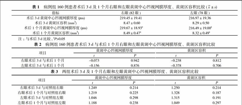
14.81)μm, 黄斑区容积为(8.62±0.40)mm3;左眼黄斑中心凹视网膜厚度为(221.05±13.25)μm, 黄斑区容积为(8.43±0.46)mm3, 正常对照组右眼和左眼黄斑中心凹视网膜厚度及黄斑区容积比较差异无统计学意义(P>0.05)。病例组术后3 d及1个月右眼和左眼黄斑中心凹视网膜厚度、黄斑区容积比较差异无统计学意义(P>0.05)。见表1, 表2。病例组术后3 d及1个月右眼黄斑中心凹视网膜厚度、黄斑区容积与正常对照组比较差异无统计学意义(t=1.249、1.219, 1.250、1.328, P>0.05), 病例组术后3 d及1个月左眼黄斑中心凹视网膜厚度、黄斑区容积与正常对照组比较差异无统计学意义(t=1.046、1.188, 1.315、1.049, P>0.05)。见表1, 表3。
3 讨论
SD-OCT技术是一种具有高分辨率、非接触性和无创伤性的组织断层成像方法, 不仅可以从三维角度观察黄斑区组织的细微结构变化, 并且可定量测量黄斑中心凹视网膜的厚度, 定性描述黄斑区视网膜各层组织结构变化, 它有利于对黄斑区病变病情进行客观评价[1]。
黄斑水肿是白内障摘除术后影响视力恢复的常见原因[2]。患者视功能的异常与黄斑中心凹视网膜厚度的改变有关[3]。白内障摘除术中血-视网膜屏障的破坏与术后视网膜黄斑区的异常有密切相关, 其发病机制尚无定论, 目前主要认为白内障摘除术对于正常患者的血-视网膜屏障功能均有一定的破坏作用, 而造成视网膜厚度增加[4-8]。另外小切口白内障摘除术中血-房水屏障破坏, 其炎性物质如前列腺素、p物质血清素、内毒素、缓激肽、白血病素等在白内障手术的刺激下释放入房水, 引起黄斑部毛细血管的通透性增加以及视网膜色素上皮细胞排水功能的减退, 从而破坏了血-视网膜屏障, 可导致黄斑部视网膜厚度增加[9-12]。作者基于探究上述的观点, 对160例(160眼)单纯老年性白内障患者小切口摘除联合人工晶体植入术术后3 d及1个月的黄斑中心凹视网膜厚度进行了测量, 发现术眼术后平均黄斑中心凹视网膜厚度及黄斑区容积值较正常同龄人群组比较, 差异无统计学意义(P>0.05)。因此, 无任何并发症的小切口白内障摘除联合人工晶体植入术术后短期内对视网膜黄斑区无明显影响。endprint
综上所述, 黄斑中心凹视网膜厚度的测量可以反映视网膜局部断层情况, 黄斑区容积测量可以反映黄斑区域的形态特征。SD-OCT能够直观、清晰显示小切口白内障摘除联合人工晶体植入术术后早期黄斑中心凹视网膜厚度及黄斑区容积的形态变化, 提示小切口白内障摘除术对黄斑区的影响较小。小切口白内障摘除术仍是基层医院恢复白内障患者视功能安全、有效的手术方式。
参考文献
[1] Biro Z, Balla Z, Kovacs B. Change of foveal and perifoveal thickness measured by OCT after phacoemulsification and IOL implantation. Eye, 2008, 22(1):8.
[2] Takamura Y, Kubo E, Akagi Y. Analysis of the effect of intravitreal bevacizumab injection on diabetic macular edema after cataract surgery. Ophthalmology, 2009, 116(6):1151-1157.
[3] Leung CK, Cheung CY, Weinreb RN, et al. Comparison of macular thickness measurements between time domain and spectral domain optical coherence tomography. Invest Ophthalmol Vis Sci, 2008, 49(11):4893-4897.
[4] Ashwin PT, Shah S, Wolfsohn JS. Advances in cataract surgery. Clin Exp Otom , 2009, 92(4):333-342.
[5] Ghosh S, Roy I, Biswas PN, et al. Prospective randomized comparative study of macular thickness following phacoemulsification and manual small incision cataract surgery. Acta Ophthalmol, 2010, 88(4):102-106.
[6] 谢娟, 王瑞妹, 张素华, 等. 老年性白内障术后黄斑病变的光相干断层扫描观察. 国际眼科杂志, 2005, 5(2):268-269.
[7] 李鹏, 安洁, 李昆, 等. 小切口白内障囊外摘除术后黄斑区光学相干断层扫描观察. 西北国防医学杂志, 2016(4):243-246.
[8] 陈志强, 谢平, 刘新亚, 等. 频域光学相干断层扫描对自闭式小切口白内障摘除术后黄斑厚度的测量研究. 实用老年医学, 2016(8):641-643.
[9] 周正申, 孙静芬. 白内障超声乳化术后黄斑区光学相干断层扫描观察. 眼科新进展, 2013, 33(2):184-186.
[10] 庞燕华, 赵桂玲, 朱敏怡, 等. 应用频域光学相干断层扫描比较超聲乳化白内障吸除术及小切口白内障摘除术术后黄斑厚度. 齐齐哈尔医学院学报, 2013, 34(21):3175-3177.
[11] 孔凡宏, 王艳玲, 吴胜卫, 等. 白内障术后黄斑区光学相干断层扫描动态观察. 国际眼科杂志, 2014, 14(6):1023-1025.
[12] 尹江波, 刘宝海, 陈军, 等. 超声乳化白内障摘除及小切口白内障囊外摘除术后黄斑囊样水肿的临床研究. 山东医学高等专科学校学报, 2016, 38(5):395-398.
[收稿日期:2017-08-14]endprint