青少年与成人正畸患者腭部植入部位骨皮质密度的CBCT研究
2017-09-25孟庆芳吾尔古力·阿不都热依木侯军衡热那古丽·伊那木
孟庆芳 吾尔古力·阿不都热依木 侯军衡 热那古丽·伊那木
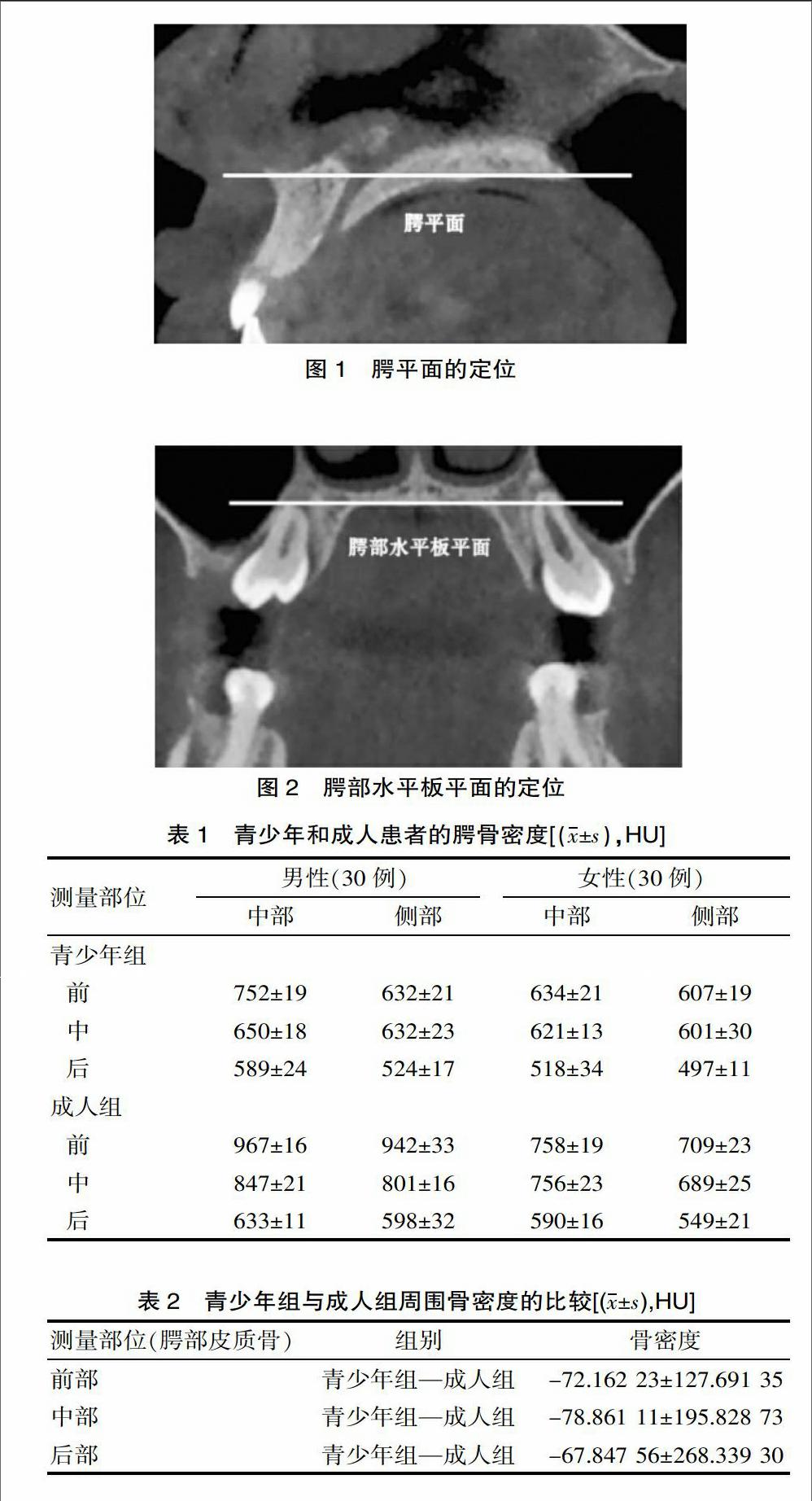
[摘要] 目的 应用锥形束CT评价并测量青少年与成年患者腭部骨皮质密度,为临床微种植体的应用提供参考。方法方便选择乌鲁木齐市口腔医院正畸科2016年3—6月在此期间进行治疗的青少年(60例)和成人患者(60例)进行CBCT扫描。将所有患者的腭部进行骨皮质密度测量,将得出的结果进行统计学分析。 结果 青少年组(613±27)HU小于成年人组的骨皮质密度(733±5)HU,(P<0.01);同组患者男性骨密度大于女性骨密度;所有患者腭骨两侧面的骨密度(733±21)HU都要比中间区域骨密度小,即青年组前侧(632±21)、(607±19)HU,后侧(524±17)、(497±11)HU比中间区域的(632±23)、(601±30)HU小;成年组前侧(942±33)、(709±23)HU,后侧(598±32)、(549±21)HU比中间区域的(801±16)、(689±25)HU小(P<0.01)。 结论 利用CBCT结合图像分析技术对牙槽骨腭部骨皮质密度进行测量,具有无创性、可行性及可重复性的优点。
[关键词] 正畸治疗;腭骨;骨密度; 锥形束CT;微种植体
[中图分类号] R783 [文献标识码] A [文章编号] 1674-0742(2017)07(a)-0194-03
CBCT Research on Bone Cortical Density in Palatal Implantation Site of Teenagers and Adult Orthodontics Patients
MENG Qing-fang, Wueguly·Abudrmy, HOU Jun-heng, Rennagolia·Yinnamoo
Orthodontic Department, Urumqi Stomatological Hospital, Urumqi, Xinjiang, 830002 China
[Abstract] Objective To evaluate the bone cortical density in palatal implantation site of teenagers and adult orthodontics patients and provide reference for the application of clinical micro-implants. Methods 60 cases of teenagers and 60 cases of adult patients treated in our hospital from which conveniently selected March to June 2016 adopted the CBCT scanning, and the pars palatalis of all patients were given the bone cortical density measurement, and the results were statistically analyzed. Results The bone cortical density in the teenager group was less than that in the adult group[(613±27)HU vs (733±5)HU](P<0.01), and the bone cortical density in males was bigger than the female bone cortical density, and the lateral bone density was smaller than that in the middle region, and the anterior and posterior bone densities in the teenagers group were smaller than those in the middle regions in the teenagers group[(632±21),(607±19)HU,(524±17),(497±11)HU vs (632±23),(601±30)HU], and the anterior and posterior bone densities in the teenagers group were smaller than those in the middle regions in the adult group [(942±33),(709±23)HU, (598±32),(549±21)HU vs (801±16),(689±25)HU](P<0.01). Conclusion The measurement of CBCT and imaging analysis technology in bone cortical density alveolar bone pars palatalis has the advantages of no wound, feasibility and repeatability.
[Key words] Orthodontic treatment; Palate bone; Bone density; Cone beam CT; Micro-implant
口腔正畸治療中治疗成功关键在于支抗控制,传统方法加强支抗对患者的依从性要求较高,制作装置需较长时间;正畸微种植钉的出现及应用,能在三维方向上起到所谓强支抗的效果[1]。CBCT可以清晰显示出颌骨硬性组织的细微结构,具有常规二维影像所不具备的高精度[2],该研究方便选取选择乌鲁木齐市口腔医院正畸科2016年3—6月间进收治的120例作为研究对象,现报道如下。endprint
1 资料与方法
1.1 一般资料
方便选择在乌鲁木齐市口腔医院正畸科在此期间进行治疗的青少年(60例)和成人患者(60例)进行CBCT扫描。青少年组:男性和女性各30例,平均年龄(12.9±2.6)岁,成人组:男性和女性各30例,平均年龄(22.8±6.5)岁。
1.2 实验方法
对研究对象进行CBCT扫描,嘱患者头矢状面与地面垂直,且听眶线与听鼻线的夹角平分线也需保持与地面垂直,然后调整矢状定位扫描线使其与正中矢状面重合;再水平扫描定位标志点使其对准听口线的中点,咬合要保持在正中合位,扫描全程保持静止;……
