Self-assembling peptide nanofibrous hydrogel as a promising strategy in nerve repair after traumatic injury in the nervous system
2016-12-02NaZhang,LiuminHe,WutianWu
PERSPECTIVE
Self-assembling peptide nanofibrous hydrogel as a promising strategy in nerve repair after traumatic injury in the nervous system
Following injury in central nervous system (CNS), there are pathological changes in the injured region, which include neuronal death, axonal damage and demyelination, inflammatory response and activation of glial cells. The proliferation of a large number of astrocytes results in the formation of glial scar, with which nerve regeneration after injury is inhibited. Other factors such as inhibitory molecules and poor blood supply around the injured area further intensify the difficulty of nerve repair. Peripheral nervous system has the ability to regenerate at some extent, however, the capacity is limited. Direct end-to-end surgical reconnection becomes a common strategy for the treatment of nerve transection in peripheral nervous system (PNS) when the gap is small (< 1 cm). In addition, nerve autografting is another common used therapy when the gap is large (> 1 cm). However, the shortage of donor grafts, the loss of function at the donor sites as well as the requirement of complex surgical procedure restrict the application of autografts (Cao et al., 2009). Another therapeutic measure of PNS injury is allografts and xenografts, however, these treatments are hampered by immunological rejections and chances of disease transfer (Evans et al., 1994). Based on these difficulties in CNS regeneration and shortcomings in PNS repair, biomaterial has shown its potential in tissue repair in recent years.
What we focused on at the present is a kind of biomaterial named self-assembling peptide (SAP). SAP is made of nanofiber, which is the self-organization of the molecules and components into structure without intervention (Whitesides and Grzybowski, 2002). Molecules are linked with weak non-covalent bonds, in particular with hydrogen bonds, electrostatic interactions, hydrophobic interactions, van der Waals interactions, as well as water-mediated hydrogen bonds. Although these bonds are relatively isolated, when combined together, they dominate all structures of biological shapes including fibrils, fibril network, gels and membranes. These biological shapes could influence other molecules, which is rather important in living system (Wang et al., 2012). Traditional SAP, RADA16-I (Ac-(RADA) 4-CONH2) which belongs to the family of ionic self-complementary peptides, could self-assemble into nanofibers at ~10 nm in diameter and eventually form a 3D hydrogel. It was reported that RADA16-I could support neuronal cell attachment, promote differentiation and enhance neurite outgrowth and the formation of synapse (Holmes et al., 2000). The most interesting finding related to RADA16-I was its function of hemostat. RADA16-I could achieve hemostasis immediately when applied directly to a wound in the brain (Ellis-Behnke et al., 2006). The decomposition products of RADA16-I were amino acids, which served as building blocks for tissues and could be used to repair the site of injury. Since the pH value of 1% RADA16-I was about 3.5 and the extracellular matrix has a positive charge, fibers could absorb wound edges by electrostatic interactions to seal the wound. Further, RADA16-I was highly hydrated and exhibited a water content of about 99%. It is assembled prior to filling the gap irregularly and then assembled to form a nanofiber scaffold molecule (Nune et al., 2013). However, RADA16-I has been suffering from its main drawback caused by low pH (pH 3.5), which damages cells and host tissues after direct exposure. Researchers have modified its pH into neutral by changing the medium frequently in cell culture process, which reduces damages to cultured cells and enhances the survival rate after grafted into injured spinal cord (Guo et al., 2007). Also, considerable effects have been devoted by us to the modification of the pH of RADA16-I. In our strategy, we prepare nanofiber hydrogels from two designer SAPs at neutral pH. RADA16-I was appended with functional motifs containing cell adhesion peptide RGD and neurite outgrowth peptide IKVAV. The two SAPs were specially designed to have opposite net charges at neutral pH, the combination of which created a nanofiber hydrogel (-IKVAV/RGD). The two functional SAPs were named as RADA16-RGD (Ac-(RADA) 4-DGDRGDS) and RADA16-IKVAV (Ac-(RADA) 4-RIKVAV). These two designer SAPs were opposite charged at physiological pH in aqueous solution, and when combined together they could form a three-dimensional (3D) nanofiber hydrogel. The two bioactive motifs were thereby incorporated in three-dimensional hydrogel scaffold, yielding a synergistic promotion effect on nerve cell response. Constant neutral pH was maintained during the process of self-assembly so that bioactive molecules and cells could be fully embedded in 3D environment. In addition, the designer SAP hydrogel could be transferred directly to living tissues.
Both in vitro and in vivo studies were done in order to test SAP hydrogel. What we cultured were neural stem cells (NSCs) and neural progenitor cells (NPCs), which were obtained from the E14.5 embryos of GFP-transgenic rats. Results indicated that spheres did not change their shapes when cultured inside the RADA16-I hydrogel while could grow out when cultured inside the RADA16-IKVAV/RGD hydrogel. To further evaluate cell viability, single NPC or NSC were embedded within these two SAPs and Live/Dead Cell Viability Assay Kit was used, finding showed that more dead cells were labeled in RADA16-I group than that in RADA16-IKVAV/RGD group. Positive immunofluorescent staining with β-tubulin for neurons showed that NPCs/ NSCs could differentiate inside RADA16-IKVAV/RGD. This was in sharp contrast to conventional 2D cell culture, in which case a variety of growth factors were needed to maintain cell viability. In vivo studies were done in both CNS and PNS injury models, including spinal cord injury, intracerebral hemorrhage and sciatic nerve defect. In spinal cord injury models, SAP solutions were injected into the lesion site immediately after completely transection with 2 mm of spinal cord removed and nerve fibers were found to regenerate into RADA16-IKVAV/RGD graft two month after injury. However, the regenerated nerve fibers grew along the wall surface of cavities formed in RADA16-I hydrogel, which was not parallel to the spinal cord thus the regenerated fibers could not grow longer. A similar phenomenon was detected in sciatic nerve defect model, axons were also found to grow around the whole transplanted RADA16-I rather than crossing the SAP (Sun et al., 2016). Intracerebral hemorrhage model was induced by intracranial injection of collagenase IV to break blood vessel wall. After collagenase injection, local hemopoietic focus forms and functional deficits have been detected in mice such as circling around on one side and forelimbs paralyzed. At 3.5 hours after collagenase injection, SAP hydrogel solutions were delivered to corpus striatum by local injection after hematoma aspiration. Histomorphology analysis was done using Nissl staining, results showed that collagenase injection induced intracerebral hemorrhage and considerable loss of neurons. However, the injection of SAP filled the voids and the boundary between SAP and the host fits tightly. Hematoma volume was measured three days after injury and local delivery of RADA16-IKVAV/RGD did not reduce the amount of hematoma, hematoma was significantly reduced by RADA16-I, which has been verified as a hemostatic hydrogel. In addition, differentmarkers including IBA-1, CD11b and NeuN have been used to detect inflammatory response and cell survival. Apoptosis was detected by TUNEL staining. Findings showed that local delivery of RADA16-IKVAV/RGD reduced acute brain injury by lowering the number of apoptotic cells, reducing inflammatory response as well as promoting cell survival. Then behavioral tests were done in weekly interval using three different systems including catwalk test, Rota rod test and Grip strength test. Results showed that four weeks later mice treated with RADA16-IKVAV/RGD showed better functional recovery than that in RADA16-I group. Two month after surgery, nerve fibers inside RADA16-IKVAV/RGD were detected using its specific marker, antibody against neurofliment-200. Results showed that two months later RADA16-IKVAV/ RGD could attach well with the host and new nerve fibers have grown into the new SAP hydrogel. RADA16-I, also could attach with the host very well, however, no fibers were detected inside the hydrogel. Transmission electron microscope analysis was done to further confirm the results. Findings showed that at the boundary between RADA16-IKVAV/RGD and the host, a few nerve fibers were found, however, no fibers were detected in RADA16-I group and there were a lot of glial cells around the injured site (Zhang et al., 2016). These results indicated that RADA16-IKVAV/RGD could serve as a bridge for fibers to grow through.
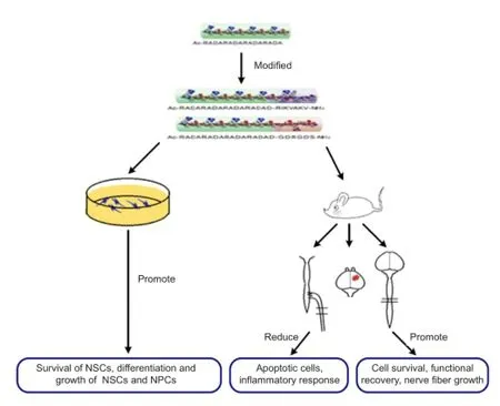
Figure 1 The function of assembling peptide nanofibrous hydrogel in both in vitro and in vivo studies.
In conclusion, these two oppositely charged SAPs carrying IKVAV and RGD epitopes could undergo self-assembly process into a stable nanofiber hydrogel at neutral pH. The mixed hydrogel RADA16-IKVAV/RGD could promote the survival and differentiation of neural stem cells and neural precursor cells. In addition, with its good biocompatibility, RADA16-IKVAV/RGD could lower the number of apoptotic cells, reduce inflammatory response and promote functional recovery. The most important finding was that RADA16-IKVAV/RGD could serve as a bridge for fibers to grow through (Figure 1).
SAP nanofibrous hydrogels provide a true biomimetic platform with their nanofibrous structure, which serve as a powerful tool in the regeneration process of central nervous system. Although SAP hydrogels have many advantages, its limitations that the mechanism of cell proliferation and differentiation inside the SAP is not clear, the design of the structure and properties needs to be improved to accommodate the different tissue types could not be ignored. Despite its shortcomings, SAP hydrogel still plays an important role in tissue repair and nerve regeneration. Due to the huge space in the design of SAP nanofibrous hydrogel, we believe SAP will become a kind of ideal biomaterial in the treatment of various diseases.
This work was supported by National Basic Research Program of China (973 Program, 2014CB542205), Hong Kong RGC grant, Hong Kong Health and Medical Research Fund, foundation for Distinguished Young Talents in Higher Education of Guangdong (Yq2013023) and the Leading Talents of Guangdong Province (87014002).
Na Zhang, Liumin He, Wutian Wu*
Guangdong-Hongkong-Macau Institute of CNS Regeneration, Jinan University, Guangzhou, Guangdong Province, China (Zhang N, Wu W) School of Biomedical Science, LKS Faculty of Medicine, the University of Hong Kong, Pokfulam, Hong Kong Special Administrative Region, China (Wu W)
State Key Laboratory of Brain and Cognitive Sciences, Te University of Hong Kong, Pokfulam, Hong Kong Special Administrative Region, China (Wu W)
Key Laboratory of Biomaterials of Guangdong Higher Education Institutes, Department of Biomedical Engineering, College of Life Science and Technology, Jinan University, Guangzhou, Guangdong Province, China (He L)
*Correspondence to: Wutian Wu, M.D., Ph.D., wtwu@hku.hk.
Accepted: 2016-04-12
Cao H, Liu T, Chew SY (2009) The application of nanofibrous scaffolds in neural tissue engineering. Adv Drug Deliv Rev 61:1055-1064.
Ellis-Behnke RG, Liang YX, Tay DK, Kau PW, Schneider GE, Zhang S, Wu W, So KF (2006) Nano hemostat solution: immediate hemostasis at the nanoscale. Nanomedicine 2:207-215.
Evans PJ, Midha R, Mackinnon SE (1994) The peripheral nerve allograft: a comprehensive review of regeneration and neuroimmunology. Prog Neurobiol 43:187-233.
Guo J, Su H, Zeng Y, Liang YX, Wong WM, Ellis-Behnke RG, So KF, Wu W (2007) Reknitting the injured spinal cord by self-assembling peptide nanofiber scaffold. Nanomedicine 3:311-321.
Holmes TC, de Lacalle S, Su X, Liu GS, Rich A, Zhang SG (2000) Extensive neurite outgrowth and active synapse formation on self-assembling peptide scaffolds. Proc Natl Acad Sci U S A 97:6728-6733.
Nune M, Kumaraswamy P, Krishnan UM, Sethuraman S (2013) Self-assembling peptide nanofibrous scaffolds for tissue engineering: novel approaches and strategies for effective functional regeneration. Curr Protein Pept Sci 14:70-84.
Sun Y, Li W, Wu X, Zhang N, Zhang Y, Ouyang S, Song X, Fang X, Seeram R, Xue W, He L, Wu W (2016) Functional self-assembling peptide nanofiber hydrogels designed for nerve degeneration. Curr Protein Pept Sci 8:2348-2359.
Wang T, Zhong XZ, Wang ST, Lv F, Zhao XJ (2012) Molecular mechanisms of RADA16-1 peptide on fast stop bleeding in rat models. Int J Mol Sci 13:15279-15290.
Whitesides GM, Grzybowski B (2002) Self-assembly at all scales. Science 295:2418-2421.
Zhang N, Luo Y, He L, Zhou L, Wu W (2016) A self-assembly peptide nanofibrous scaffold reduces inflammatory response and promotes functional recovery in a mouse model of intracerebral hemorrhage. Nanomedicine 12:1205-1217.
10.4103/1673-5374.182687 http∶//www.nrronline.org/
How to cite this article: Zhang N, He L, Wu W (2016) Self-assembling peptide nanofibrous hydrogel as a promising strategy in nerve repair after traumatic injury in the nervous system. Neural Regen Res 11(5):717-718.
杂志排行

中国神经再生研究(英文版)的其它文章
- Possible application of apolipoprotein E-containing lipoproteins and polyunsaturated fatty acids in neural regeneration
- Recovery of injured fornical crura following neurosurgical operation of a brain tumor: a case report
- Antibody-based neuronal and axonal delivery vectors for targeted ligand delivery
- Coordination of the axonal cytoskeleton during the emergence of axon collateral branches
- Alzheimer's disease: the silver tsunami of the 21stcentury
- Clinical trial perspective for adult and juvenile Huntington's disease using genetically-engineered mesenchymal stem cells