Coronary Artery Chronic Total Occlusion
2016-05-25CalvinChoiMDMSNayanAgarwalMDKiParkMDMSandDavidAndersonMDMSDivisionofCardiovascularMedicineUniversityofFlorida600SWArcherRoadGainesvillefl32600277USA
Calvin Choi, MD, MS, Nayan Agarwal, MD, Ki Park, MD, MS and R. David Anderson, MD, MSDivision of Cardiovascular Medicine, University of Florida, 600 SW Archer Road, Gainesville, fl3260-0277, USA
Abbreviations
CABG – coronary artery bypass grafting
CAD – coronary artery disease
CTO – coronary artery chronic total occlusions
LAD – left anterior descending artery
LM – left main coronary artery
LVEF – left ventricular ejection fraction
MI – myocardial infarction
PCI – percutaneous coronary intervention
RCA – right coronary artery
TIMI – Thrombolysis in Myocardial Infarction
Introduction
Coronary artery chronic total occlusions (CTO)are defined as occluded coronary artery segments with thrombolysis in myocardial infarction (TIMI)0 flow for at least three months [1, 2]. Stone et al. in a consensus document published in 2005 proposed that CTO be classified into “true” or “functional”based on the degree of lumen narrowing and anterograde blood flow [3]. A “true” CTO is defined as complete interruption of coronary flow (TIMI 0 flow), whereas occlusions with minimal contrast penetration through the lesion without distal-vessel opacification (TIMI 1 flow) are classed as a “functional” CTO. However, in the literature, the distinction between “true” and “functional” CTO is rarely declared. Moreover, the period of time for which a CTO has been present is often difficult to ascertain.Therefore, the age of the occlusion is often determined based on careful assessment of a patient’s medical history and cardiac symptoms in the previous 3 months [3].
Pathophysiology
Percutaneous revascularization of CTO is often complicated by the inability to cross or dilate the lesion. An understanding of the histopathology of these lesions helps provide insight into the development of new revascularization strategies.
CTO most often arise from thrombotic occlusion of vessels and subsequent thrombus organization and tissue remodeling [4]. This may occur after failed revascularization or subsequent reocclusion of the vessel in patients who have had a myocardial infarction [5, 6]. However, a majority of patients with CTO (60%) do not have a history of myocardial infarction (MI) and an alternative mechanism must be involved [7]. For example,there could be recruitment of collateral vessels to counterbalance ischemia due to an obstructed vessel, which might gradually progress to complete occlusion. Insufficient blood flow via collateral circulation results in ischemia and angina.However, due to the insidious nature of the disease progress, symptoms may be mild and/or atypical in some patients [8].
Pathological Considerations
The typical atherosclerotic plaque of CTO consists of intracellular and extracellular lipids, smooth muscle cells, extracellular matrix, and calcium [9].Extracellular matrix is rich in collagens [10, 11]with predominance of types I and III (and minor amounts of IV, V, and VI) [12]. The concentration of collagen-rich fibrous tissue is particularly dense at the proximal and distal ends of the lesion, contributing to a column-like lesion with calcified, resistant fibrous tissue surrounding a softer core of organized thrombus and lipids.
Srivatsa and colleagues demonstrated through an autopsy study that angiographic CTO frequently corresponded to less than 99% stenosis by histology criteria [13]. They demonstrated that key histopathological attributes of CTO are calcification,inflammation, and neovascularization. Thrombotic occlusion progresses over time from a “soft” to a“‘hard” lesion composition. Soft plaque consists of cholesterol-laden cells and foam cells with loose fibrous tissue and neovascular channels which is more common in “new” occlusions (less than 1 year old). “Soft” plaque is more likely to allow wire passage either directly through tissue planes or via neovascular channels into the distal lumen. Conversely,“hard” plaques are characterized by dense fibrous tissue and often contain large fibrocalcific regions without neovascular channels. These occlusions are thus more likely to deflect guidewires into the subintimal space, resulting in dissection planes.“Hard” plaques are more prevalent with “old” CTO(greater than 1 year old). Of note, however, areas of calcification frequently occur even in CTO less than three months of age, although the extent and severity of calcification tend to increase with occlusion duration.
综上可见,孟子独标“仁者无敌”的武德思想,以“明明德于天下”的内圣取代“平天下”的外王实践,对于暴力与霸道进行彻底的否定,绝端的反对战争和刑杀,导致其政治理想失去了现实的抓手而缺乏可操作性。齐宣王所言“吾惛,不能进于是矣”,便是治政者对于道德理想不知如何转化落实为实操层面的慨叹。《史记》称孟子“迂远而阔于事情”[9](P2343),就连十分推重孟子的朱熹也不得不指出:“孟子所论,自世俗观之,则可谓无谋矣。”[4](P226)孟子武德观念在当时的碰壁,在于其“以所如者不合”[9](P2343),无法应对战国之世的现实危局。
Another hallmark of CTO is extensive neovascularization, which occurs throughout the extent of the vessel wall and increases with the age of occlusion.In CTO less than 1 year old, new capillary formation is greatest in the adventitia. In CTO greater than 1 year old, the number and size of capillaries in the intima have increased to a similar or greater extent than those present in the adventitia. Lymphocytes and monocytes/macrophages may play an active role in both angiogenesis and atherosclerotic lesion progression by producing a variety of mitogenic and angiogenic factors [14]. A rich neovasculature network often traverses the CTO vessel wall, arising from the adventitial vasa vasorum across the media and into the intima which suggests that the adventitial vessels initiate the formation of intimal neovascular channels [15]. The channels in this neovasculature often are perpendicular to the long axis of the artery and may be a contributing factor in guidewire diversion into the extravascular space.Post-mortem studies support this mechanism. The channels in the neovasculature mostly lead into the adventitia, small side branches, or vasa vasorum.However, they may also extend longitudinally from the proximal to the distal lumen. Such channels may serve as a route for guidewires and hence may have a therapeutic value.
?
Epidemiology
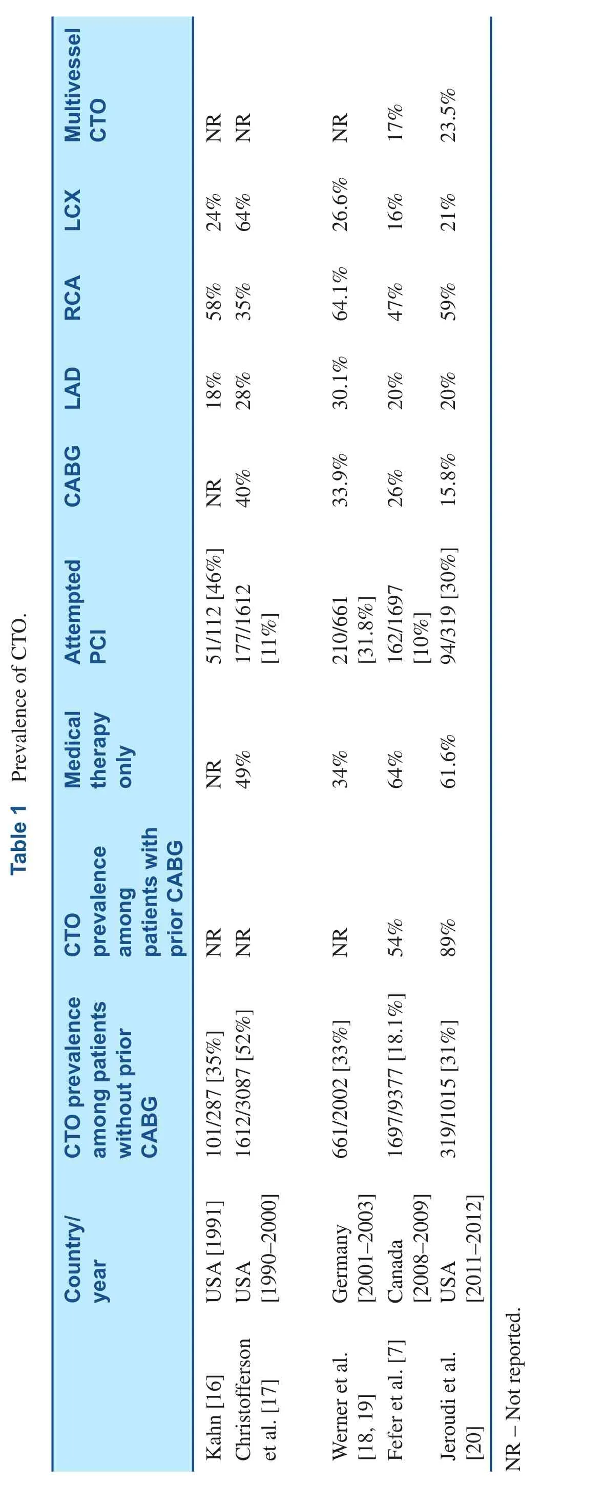
The reported prevalence of CTO among patients undergoing a cardiac catheterization varies widely as illustrated in Table 1 [7, 16–19]. In the largest study reported to date (prospective three center Canadian registry) [7], the prevalence of CTO was 18%among patients with coronary artery disease (CAD,defined as greater than 50% stenosis in at least one vessel) without prior coronary artery bypass grafting (CABG) and 54% among patients with prior CABG. Using the same definition of CAD, Kahn[16] identified a CTO in 35% of 287 patients with CAD at a single institution in one year. In a German multicenter prospective registry spanning 64 hospitals, Werner et al. [18] reported a CTO prevalence of 33% in 2002 patients presenting with stable angina and first angiographic diagnosis of CAD. In a study investigating the frequency of CTO in a veteran population, Christofferson et al. [17] reported a CTO prevalence of 52% in patients with CAD (defined as greater than 70% stenosis in at least one vessel) and an overall prevalence of 24.5% in 8004 non-CABG patients undergoing diagnostic angiography over a ten year period. In another study in a veteran population by Jeroudi et al. [20], the prevalence of CTO was 31% among non-CABG patients presenting with CAD, and 89% in patients with prior CABG.It has also been reported that patients with a CTO have more cardiac risk factors than those with CAD without CTO on coronary angiography, including a higher prevalence of diabetes mellitus (34% vs. 26%),hypertension (75% vs. 68%), hyperlipidemia (82%vs. 78%), heart failure (12% vs. 9%), and peripheral artery disease (8% vs. 4%) [7]. Furthermore, patients with CTO are more likely to have a previous history of myocardial infarction and undergo CABG than non-CTO patients [7, 16–18, 20]. Moreover, patients with CTO are an undertreated cohort with reports indicating that in general only about 10–20% of patients with CTO are treated with PCI [21].

Table 2 Benefits of CTO PCI.
Treatment Goals
Successful CTO revascularization is associated with improved clinical outcomes, and several retrospective studies have shown various benefi-cial effects (Table 2). Angina relief, improvement in exercise tolerance, reduction in ventricular arrhythmia burden, improvement in left ventricular function; reduced need for CABG surgery,enhanced tolerance for future coronary events,reduced future coronary events and survival benefit have all been associated with successful CTO revascularization procedures [22–26, 31].
To date, there is a paucity of prospective, randomized clinical trial data to support the potential benefits of CTO revascularization. The reported benefits of CTO percutaneous coronary intervention(PCI) are mostly derived from registry data where a comparison is made between successful and failed PCI [21].
Due to accumulating interest in CTO PCI, a record number of studies are being pursued to shed a light on the precise benefits and limitations of CTO PCI. Recently, Henrique et al. presented randomized prospective clinical trial data on nonculprit vessel CTO PCI in STEMI patients at the Transcatheter Cardiovascular Therapeutics (TCT)2015 meeting. This study revealed that routine CTO PCI did not improve left ventricular ejection fraction (LVEF) when compared to the control group. A subgroup analysis, however, revealed that for the left anterior descending artery CTO cohort, PCI resulted in improved LVEF [32].
Evidence from additional prospective, randomized trials is needed to confirm the clinical benefits and limitations of CTO revascularization.
Treatment Indications and Decision Making
The European and North American guidelines have adopted CTO revascularization with a class 2a recommendation (level of evidence B) [33, 34].Symptom relief is considered the main indication for the procedure. However, a number of other potential benefits of CTO revascularization have been identified and are reasonable indications for CTO revascularization [35].
Historically low procedural success rates, technical challenges associated with CTO PCI, lack of operator experience, apparent inefficient resource utilization, potential increase in complication rates,uncertain clinical benefits, and the presence of well-developed collaterals have dissuaded interventional cardiologists from pursuing CTO PCI.Although the presence of collateral circulation has been associated with improved survival [36], the collateral flow may be insufficient to preserve ventricular function and meet the metabolic demand in CTO patients which results in ventricular dysfunction, ischemia and angina [19]. With advances in novel and innovative techniques and equipment, the procedural success rate for CTO PCI is reaching 90% and overall complication rates have decreased in the hands of experienced CTO operators [21, 37].
The decision to pursue CTO PCI depends on several factors. First, patient selection is critical.Ischemic burden, myocardial viability, severity of symptoms, and patient preferences need to be taken into consideration. Second, suitable anatomy should be identified. Based on lesion and target vessel characteristics – presence of calcification,stents, tortuosity, bifurcation, branch vessels, size,collateral vessels, location and shape of proximal and distal cap, length of the occlusion – the odds of successful revascularization may be estimated. For example, the J-CTO score which is based on lesion characteristics including the shape of proximal cap,calcification, angulation and length, and repeat attempt can help predict the procedural success rate[38]. Third, operator experience is crucial. CTO PCI requires in-depth knowledge, a unique skill set and experience; (e.g., collateral wiring, dissection and re-entry technique, wire externalization,contemporary algorithm) and complication management. In addition, patients need to be informed of the idiosyncrasies associated with CTO PCI. In general, procedures tend to be longer and radiation and contrast exposure tend to be higher when compared to non-CTO PCI, and multiple staged attempts may be required. For an appropriate candidate with suitable anatomy, CTO PCI should be discussed as a treatment option.
Treatment Options
Based on the severity of symptoms, concomitant CAD, ischemic burden, and patient risk profile and preferences, treatment options may be tailored to each patient. However, irrespective of the presence of CTO, patients with CAD should be counseled about risk profile modification and receive aggressive medical therapy for CAD. Medical management is a reasonable option for patients with no symptoms or mild symptoms. For patients with large ischemic burden or refractory symptoms despite optimal medical therapy, revascularization should be considered. CABG should be considered for patients with concomitant multi-vessel or left main CAD or in need of cardiac surgery for other reasons; valve surgery, etc. CTO PCI may be considered in patients with isolated CTO or those who are poor candidates for CABG. A number of publications dedicated to CTO PCI technique and strategy have significantly simplified and standardized the procedures for CTO operators [39–43]. Since procedural success in CTO PCI is highly dependent on operator experience, a referral to a CTO PCicenter should be considered when local treatment options do not favor CTO PCI.
Salient Features of CTO PCI
Several salient features unique to CTO PCI are worth highlighting. In general, CTO PCI is a time consuming, labor intensive, and a resource heavy endeavor. As such, patient comfort must be taken into consideration. Conscious sedation with anesthesiology support or even general anesthesia may be required to allow patient comfort during a long procedure. Furthermore, for patients with heart failure, volume status should be carefully monitored during the procedure to avoid heart failure exacerbation due to a large volume of contrast used during the procedure; (e.g., left ventricular end diastolic pressure measurement at the beginning of the procedure may be beneficial). Operator fatigue needs to be considered as well. The duration of the procedure needs to be monitored such that beyond a set time, unless the procedure is near completion,the procedure should be terminated and reattempted at another time. A number of CTO PCicenters practice a team approach to avoid operator fatigue;i.e., two operators working as a team. Additionally,repeat attempts may be required for CTO PCI. Due to the radiation dose, the contrast volume and the duration of the procedure, a procedure may not be completed on the initial attempt. However, with the initial attempt a lot can be gained. For example, CTO lesion characteristics can be better appreciated and understood such that device and equipment selection may be streamlined during future attempts.Plaque modification during the initial attempt may increase the likelihood of success with a subsequent CTO PCI.
For practitioners interested in developing a CTO PCI program, a discussion regarding resource utilization is imperative. Based on 2015 Medicare physician reimbursement rate, a single vessel CTO PCI is reimbursed at the same rate as a single vessel non-CTO PCI for acute myocardial infarction[44]. A CTO PCI is an elective procedure whereas a non-CTO PCI for acute myocardial infarction is an emergent or urgent procedure. As such, overall resource utilization may be higher for a non-CTO PCI for acute myocardial infarction due to the additional cost associated with hospitalization.However, for the index procedure, resource utilization may be higher for CTO PCI. According to Grantham et al., for CTO PCI, both the duration of the procedure and fluoroscopy are about twice as long when compared to non-CTO PCI, and balloon and stent utilization for CTO PCI is significantly higher than non-CTO PCI [45].
Traditionally, concerns over potential CTO PCI associated complication risks have deterred cardiologists from pursuing CTO PCI as a treatment option for patients with CTO. Due to recent advances in technique and dedicated CTO PCI equipment, a number of CTO PCI associated complication risks may be comparable to non-CTO PCI. In-hospital death (1.3% vs. 0.8%), Q-wave MI (0.5% vs.0.6%), and urgent CABG (0.7% vs. 1.1%) have been reported as similar between CTO PCI and non-CTO PCI [45].
However, other risks have been reported to be higher in CTO PCicompared to non-CTO PCI.Because of greater contrast volume used during CTO PCI, the risk for contrast-induced nephropathy (CIN) is higher with CTO PCI than non-CTO PCI. This is influenced by pre-existing chronic kidney disease and the contrast volume used during the procedure. Renal protective strategies such as pre-hydration and contrast volume minimization during the procedure may mitigate the risk for CIN [45]. Particularly for patients with pre-existing chronic kidney disease, a discussion regarding the risk for CIN is essential. Radiation injury is another dose-dependent risk associated with CTO PCI. Fluoroscopy time and dose are directly proportional to the radiation injury. As in any PCI, a radiation dose minimization strategy should be utilized. For example, collimation use, image intensifier angle changes to avoid prolonged direct exposure to the same area and lower frame rate for fluoroscopy (7.5 frames per second rather than 15 frames per second) during the procedure can mitigate radiation injury to the patient.For the operator, lead shielding, keeping a minimal distance between the patient and the image intensifier, and maintaining a maximal distance from the radiation source can minimize the radiation exposure [45]. For radiation dose greater than 5 Gy, a follow up evaluation is required to check for radiation dermatitis [45]. Coronary artery dissection and perforation are also reported to be more common in CTO PCI than in non-CTO PCI. Coronary artery dissection is significantly higher in CTO PCI than in non-CTO PCI (17.8%vs. 13.3%) [45]. Interestingly, the contemporary CTO PCI algorithm includes intentional dissection and re-entry technique. Coronary artery perforation is significantly higher in CTO PCI than in non-CTO PCI (0.9% vs. 0.2%) [45]. A strategy to minimize the risk for coronary artery perforation should be practiced. For example, appropriate wire selection, use of reversible pharmacotherapy, and prompt recognition and treatment when perforation is identified are key features [45]. Of note, the incidence of CTO PCI associated risks are somewhat variable depending on the data type (observational vs. prospective), operator experience (expert vs. all-comer) and data time frame (contemporary vs. historical). For example, “Outcomes, patient health status and effi-ciency in CTO hybrid procedures (OPEN CTO)”[ClinicalTrials.gov Identifier: NCT02026466]is an on-going prospective study, and preliminary results presented at TCT 2015 meeting indicate that clinically significant perforation and in-hospital myocardial infarction and death for CTO PCI are 4.9%, 2.4% and 0.9%, respectively.A number of on-going prospective studies will provide a greater insight into the incidence of risks associated with CTO PCI.
CTO PCI Technique
A detailed discussion regarding procedural techniques and strategies is beyond the scope of this manuscript. A review of general concepts is presented below. It is crucial to clearly define a CTO segment for a successful PCI. In order to clearly define CTO segment, a bilateral simultaneous coronary angiogram is required. That is, two guides are simultaneously engaged in the right coronary(RCA) and left main coronary arteries (LM), and contrast is injected into both guides (Figure 1). This technique allows clear visualization of the CTO segment and its collaterals, and this is often ambiguous on a single vessel contrast injection. Bilateral contrast injection also allows the operator to set up the coronary guides such that both anterograde and retrograde CTO PCI techniques can be utilized.
CTO PCican be approached anterograde and retrograde. With the anterograde approach, a CTO segment is crossed with a coronary wire traversing anterograde from the proximal to the distal segment.Thereafter, PCI is performed in a traditional fashion. With the retrograde approach, a CTO segment is crossed with a coronary wire traversing retrograde from the distal to the proximal segment. The wire reaches the distal segment of CTO through collateral vessels which originate from a contralateral donor vessel. For example, Figure 2 shows RCA CTO which is crossed with a wire that traverses the septal perforator from the left descending artery(LAD). Thereafter, the retrograde wire is advanced into the RCA guide and then externalized (Figure 3).That is, the retrograde wire is advanced through the entire length of the RCA guide and externalized outside the RCA guide. Over the externalized segment of the retrograde wire, PCican be performed through the RCA guide in an anterograde fashion(Figures 4 and 5).
Further categorization can be made into true lumen entry and dissection and re-entry. With the true lumen entry, a coronary wire traverses the CTO segment from true lumen on one side of the CTO segment to true lumen on the other side of the CTO segment (Figure 6). With the dissection and re-entry technique, a coronary wire traverses the CTO segment through the sub-intimal space.That is, the wire in true lumen on one side of the CTO segment enters the sub-intimal space adjacent to the CTO segment then re-enters the true lumen on the other side of the CTO segment (Figure 7).

Figure 1 Bilateral Contrast Injection Shows Left Anterior Descending Artery (LAD), Septal Perforator (SP) and Right Coronary Artery Chronic Total Occlusion (RCA CTO).

Figure 2 Retrograde Wire from LAD is AdvancedThrough SP into the Distal Segment of RCA Then Toward the RCA Guide.Blue arrows indicate the direction of the retrograde wire movement.
The decision to pursue either anterograde or retrograde approaches or true lumen entry or dissection and re-entry depends on the anatomical features of CTO, the presence of calcification, stents, tortuosity,bifurcation, branch vessels, size, collateral vessels,location and the shape of the proximal and distal cap,as well as the length of the occlusion. CTO operators need working knowledge of CTO PCI algorithms and techniques, and familiarity with different wire characteristics and dedicated equipment.

Figure 3 Retrograde Wire is Advanced Through the Entire Length of the RCA Guide and Externalized Outside the RCA Guide.

Figure 4 Over the Externalized Retrograde Wire (blue arrows), a Balloon is Advanced Anterograde Through the RCA Guide (Red Arrow).

Figure 5 Finished RCA CTO PCI via Retrograde Approach.

Figure 6 True Lumen Entry.Coronary wire traverses the CTO segment from true lumen on one side of the CTO segment to true lumen on the other side of the CTO segment. Red area indicates true lumen of the vessel. Black area indicates CTO segment of the vessel. Yellow area indicates vessel wall. Blue arrow indicates coronary wire direction.

Figure 7 Dissection and Re-Entry.Coronary wire traverses the CTO segment from true lumen on one side of the CTO segment to true lumen on the other side of the CTO segment through sub-intimal space. Red area indicates true lumen of the vessel. Black area indicates CTO segment of the vessel. Yellow area indicates vessel wall. Blue arrow indicates coronary wire direction.
The learning curve for contemporary CTO PCI algorithm and techniques is steep. However, a CTO PCI program may be a worthy pursuit for a dedicated team. Interested readers are encouraged to study dedicated manuscripts for CTO PCI technique [35–39], and participate in conferences dedicated to CTO PCI and educational programs such as www.CTOfundamentals.org.
Future Direction
Patients with CTO are an underserved cohort in need of a full complement of treatment options.Despite significant strides gained in CTO PCI,the procedure is often viewed by general cardiology community as a “high risk” procedure and often not discussed or offered to patients. In order to change this misconception and overcome this barrier, more educational effort is required. In particular, more prospective, randomized clinical trial data are needed to elucidate the benefits and limitations of CTO PCI. Results from prospective studies such as “A Randomized Multicentre Trial to Evaluate the Utilization of Revascularization or Optimal Medical Therapy for the Treatment of Chronic Total Coronary Occlusions (EuroCTO)”[ClinicalTrials.gov Identifier: NCT01760083],“Drug-Eluting Stent Implantation Versus Optimal Medical Treatment in Patients With Chronic Total Occlusion (DECISION-CTO)” [ClinicalTrials.gov Identifier: NCT01078051], and “Outcomes,patient health status and efficiency in CTO hybrid procedures (OPEN CTO)” [ClinicalTrials.gov Identifier: NCT02026466] will be enlightening.More educational effort is needed to improve the awareness in the general cardiology community regarding CTO such that CTO PCI is considered as a standard treatment option for appropriate candidates. Additional efforts are needed to increase the number of trained CTO operators so that patient care is not limited by the accessibility of an experienced operator. Continued development of dedicated equipment is needed to improve the success rate for CTO PCI. Given time and resource heavy nature of the procedure, policy and administrative support is needed so that CTO PCI is a financially viable endeavor for current and future CTO operators.
Conclusion
In the past decade, there has been an exponential growth in novel and innovative techniques, and development of dedicated equipment for CTO PCI which has resulted in a significant improvement in procedural success. Furthermore, due to growing interest in CTO PCI, studies dedicated to understanding the benefits and limitations of CTO PCI are being pursued in record numbers. Collaboration among CTO operators and industry in an effort to improve procedural success and patient care has been a fruitful endeavor. Dedicated CTO conferences and educational effort will increase the number of CTO operators which in turn will improve patient care and accessibility.
Conflict of Interest
The authors declare no conflict of interest.
Disclosures
Dr. Anderson is a consultant for BioSense Webster,a Johnson & Johnson company.
REFERENCES
1. Sianos G, Werner GS, Galassi AR,Papafaklis MI, Escaned J, Hildick-Smith D, et al. Recanalisation of chronic total coronary occlusions:2012 consensus document from the EuroCTO club. EuroIntervention 2012;8:139–45.
2. Carlino M, MagricJ, Uretsky BF,Brilakis ES, Walsh S, Spratt JC,et al. Treatment of the chronic total occlusion: a call to action for the interventional community. Catheter Cardiovasc Interv 2015;85:771–8.
3. Stone GW, Kandzari DE, Mehran R,Colombo A, Schwarz RS, Bailey S,et al.Percutaneous recanalization of chronically occluded coronary arteries: a consensus document: part I. Circulation 2005;112:2364–72.
4. Katsuragawa M, Fujiwara H,Miyamae M, Sasayama S.Histologic studies in percutaneous transluminal coronary angioplasty for chronic total occlusion:comparison of tapering and abrupt types of occlusion and short and long occluded segments. J Am Coll Cardiol 1993;21:604–11.
5. Grines CL, Browne KF, Marco J,Rothbaum D, Stone GW, O’Keefe J, et al. A comparison of immediate angioplasty with thrombolytic therapy for acute myocardial infarction. The Primary Angioplasty in Myocardial Infarction Study Group.N Engl J Med1993;328:673–9.
6. Stone GW, Grines CL, Cox DA,Garcia E, Tcheng JE, Griffin JJ, et al. Comparison of angioplasty with stenting, with or without abciximab, in acute myocardial infarction. N Engl J Med 2002;346:957–66.
7. Fefer P, Knudtson ML, Cheema AN, Galbraith PD, Osherov AB,Yalonetsky S, et al. Current perspectives on coronary chronic total occlusions: the Canadian Multicenter Chronic Total Occlusions Registry.J Am Coll Cardiol 2012;59:991–7.
8. Habib GB, Heibig J, Forman SA,Brown BG, Roberts R, Terrin ML,et al. Influence of coronary collateral vessels on myocardial infarct size in humans. Results of phase I thrombolysis in myocardial infarction (TIMI) trial. The TIMI Investigators. Circulation 1991;83:739–46.
9. Meier B. Chronic total occlusion.In: Topol EJ, editor. Textbook of Interven- tional Cardiology.Philadelphia, PA: WB Saunders;1994. pp. 318–38.
10. Bartos F, Ledvina M. Collagen,elastin, and desmosines in three layers of bovine aortae of different ages. Exp Gerontol 1979;14:21–6.
11. Hosoda Y, Kawano K, Yamasawa F, Ishii T, Shibata T, Inayama S.Age dependent changes of collagen and elastin content in human aorta and pulmonary artery. Angiology 1984;35:615–21.
12. Katsuda S, Okada Y, Minamoto T, Oda Y, Matsui Y, Nakanishi I.Collagens in human atherosclerosis: immunohistochemical analysis using collagen type-specific antibodies. Arterioscler Thromb 1992;12:494–502.
13. Srivatsa SS, Edwards WD, Boos CM, Grill DE, Sangiorgi GM,Garratt KN, et al. Histologic correlates of angiographic chronic total coronary artery occlusions influence of occlusion duration on neovascular channel patterns and intimal plaque composition. J Am Coll Cardiol 1997;29:955–63.
14. Sueishi K, Yonemitsu Y, Nakagawa K, Kaneda Y, Kumamoto M,Nakashima Y. Atherosclerosis and angiogenesis. Ann N Y Acad Sci 1997;811:311–24.
15. Munce NR, Strauss BH, Qi X,Weisbrod MJ, Anderson KJ, Leung G, et al. Intravascular and extravascular microvessel formation in chronic total occlusions: a micro-CT imaging study. JACC Cardiovasc.Imaging 2010;3:797–805.
16. Kahn JK. Angiographic suitability for catheter revascularization of total coronary occlusions in patients from a community hospital setting.Am Heart J 1993;126:561–4.
17. Christofferson RD, Lehmann KG,Martin GV, Every N, Caldwell JH,Kapadia SR. Effect of chronic total coronary occlusion on treatment strategy. Am J Cardiol 2005;95:1088–91.
18. Werner GS, Gitt AK, Zeymer U,Juenger C, Towae F, Wienbergen H, et al. Chronic total coronary occlusions in patients with stable angina pectoris: impact on therapy and out- come in present day clinical practice. Clin Res Cardiol 2009;98:435–41.
19. Werner GS, Surber R, Ferrari M,Fritzenwanger M, Figulla HR. The functional reserve of collaterals supplying long-term chronic total coronary occlusions in patients without prior myocardial infarction. Eur Heart J 2006;27:2406–12.
20. Jeroudi OM, Alomar ME, Michael TT, El Sabbagh A, Patel VG,Mogabgab O, et al. Prevalence and management of coronary chronic total occlusions in a tertiary veterans affairs hospital. Catheter Cardiovasc Interv 2014;84:637–43.
21. Azzalini L, Vo M, Dens J, Agostoni P. Myths to debunk to improve management, referral, and outcomes in patients with chronic total occlusion of an epicardial coronary artery. Am J Cardiol 2015;116:1774–80.
22. Chung CM, Nakamura S, Tanaka K,Tanigawa J, Kitano K, Akiyama T,et al. Effect of recanalization of chronic total occlusions on global and regional left ventricular function in patients with or without previous myocardial infarction.Catheter Cardiovasc Interv 2003;60:368–74.
23. Nakamura S, Muthusamy TS, Bae JH, Cahyadi YH, Udayachalerm W,Tresukosol D. Impact of sirolimuseluting stent on the outcome of patients with chronic total oc- clusions. Am J Cardiol 2005;95:161–6.
24. Baks T, van Geuns RJ, Duncker DJ,Cademartiri F, Mollet NR, Krestin GP, et al. Prediction of left ventricular function after drug-eluting stent implantation for chronic total coronary occlusions. J Am Coll Cardiol 2006;47:721–5.
25. Cheng AS, Selvanayagam JB,Jerosch-Herold M, van Gaal WJ,Karamitsos TD, Neubauer S,et al. Percutaneous treatment of chronic total coronary occlusions improves regional hyperemic myocardial blood flow and contractility: insights from quantitative cardiovascular magnetic resonance imaging. JACC Cardiovasc Interv 2008;1:44–53.
26. Cetin M, Zencir C, Cakici M,Yildiz E, Tasolar H, Balli M, et al.Effect of a successful percutaneous coronary intervention for chronic total occlusion on parameters of ventricular repolarization. Coron Artery Dis 2014;25:705–12.
27. Grantham JA, Jones PG, Cannon L, Spertus JA. Quantifying the early health status benefits of successful chronic total occlusion recanalization: results from the FlowCardia’s Approach to Chronic Total Occlusion Recanalization(FACTOR) trial. Circ Cardiovasc Qual Outcomes 2010;3:284–90.
28. Safley DM, Koshy S, Grantham JA, Bybee KA, House JA, Kennedy KF, et al. Changes in myocardial ischemic burden following percutaneous coronary intervention of chronic total occlusions. Catheter Cardiovasc Interv 2011;78:337–43.
29. Gerber BL, Rousseau MF, Ahn SA, le Polain de Waroux JB,Pouleur AC, et al. Prognostic value of myocardial viability by delayed-enhanced magnetic resonance in patients with coronary artery disease and low ejection fraction: impact of revascularization therapy. J Am Coll Cardiol 2012;59:825–35.
30. Kirschbaum SW, Baks T, van den Ent M, Sianos G, Krestin GP,Serruys PW, et al. Evaluation of left ventricular function three years after percutaneous recanalization of chronic total coronary occlusions.Am J Cardiol 2008;101:179–85.
31. Lee PH, Lee S-H, Park H-S, Kang SH, Bae BJ, Chang M, et al.Successful recanalization of native coronary chronic total occlusion is not associated with improved long term survivor. JACC Cardiovasc Interv 2016;9:530–8.
32. Henriques JPS. The evaluating Xience and left ventricular function in PCI on occlusions after STEMI(EXPLORE) trial. Presented at:TCT Scientific Symposium; Oct.11–15, 2015; San Francisco.
33. Windecker S, Kolh P, Alfonso F,Collet JP, Cremer J, Falk V, et al.2014 ESC/EACTS Guidelines on myocardial revascularization:the Task Force on Myocardial Revascularization of the European Society of Cardiology (ESC) and the European Association for Cardio-Thoracic Surgery (EACTS)developed with the special contribution of the European Association of Percutaneous Cardiovascular Interventions (EAPCI). Eur Heart J 2014;35:2541–619.
34. Levine GN, Bates ER, Blankenship JC, Bailey SR, Bittl JA, Cercek B,et al. 2011 ACCF/AHA/SCAI guideline for percutaneous coronary intervention: a report of the American College of Cardiology Foundation/American Heart Association Task Force on practice guide- lines and the society for cardiovascular angiography and interventions.Circulation 2011;124:e574–651.
35. Galassi AR, Brilaks ES,Boukhris M, Tomasello SD,Sianos G, Karmpaliotis D, et al.Appropriateness of percutaneous revascularization of coronary chronic total occlusions: an overview. Eur Heart J. Aug 7, 2015,ehv391; DOI: 10.1093/euheartj/ehv391.
36. Kumbasar D, Akyürek O, Dincer I, Atmaca Y, Kiliçkap M, Erol C, et al. Good collaterals predict viable myocardium. Angiology 2007;58:550–5.
37. Patel VG, Brayton KM, Tamayo A, Mogabgab O, Michael TT, Lo N, et al. Angiographic success and procedural complications in patients undergoing percutaneous coronary CTO interventions. JACC Cardiovasc Interv 2013;6:128–36.
38. Morino Y, Abe M, Morimoto T,Kimura T, Hayashi Y, Muramatsu T, et al. Predict ing successful guidewire crossing through chronic total occlusion of native coronary lesions within 30 minutes. JACC Cardiovasc Interv 2011;4(2):213–21.
39. Pinak BS. Management of coronary chronic total occlusion. Circulation 2011;123:1780–4.
40. Sumitsuji S, Inoue K, Ochiai M,Tsuchikane E, Ikeno F. Fundamental wire technique and current standard of percutaneous intervention for CTO with histopathological insight. JACC Cardiovasc Interv 2011;4:941–51.
41. Brilakis ES, Grantham JA,Thompson CA, DeMartini TJ,Prasad A, Sandhu GS, et al. The retrograde approach to coronary artery CTO; a practical approach.Catheter Cardiovasc Interv 2012;79:3–19.
42. Brilakis ES, Grantham JA, Rinfret S, Wyman RM, Burke MN,Karmpaliotis D, et al. A percutaneous treatment algorithm for crossing coronary CTO. JACC Cardiovasc Interv 2012;5:367–79.
43. Joyal D, Thompson CA, Grantham JA, Buller CEH, Rinfret S. The retrograde technique for recanalization of chronic total occlusion. JACC Cardiovasc Interv 2012;5:1–11.
44. 2015 Medicare physician reimbursement. www.cms.gov/apps/physician-fee-schedule/overview.aspx.
45. Grantham JA, Marso SP,Spertus J, House J, Holmes JR,Rutherford BD. Chronic total occlusion angioplasty in the United States. JACC Cardiovasc Interv 2009;2:479–86.
猜你喜欢
杂志排行

Cardiovascular Innovations and Applications的其它文章
- Carotid Artery Stenting: 2016 and Beyond
- Will Transcatheter Aortic Valve Replacement(TAVR) be the Primary Therapy for Aortic Stenosis?
- The Future of Transcatheter Therapy for Mitral Valve Disease
- The Transradial Approach for Cardiac Catheterization and Percutaneous Coronary Intervention: A Review
- Identification and Management of Iatrogenic Aortocoronary Dissection
- Transient Pulmonary Atelectasis after Ketamine Sedation during Cardiac Catheterization in Spontaneously Breathing Children with Congenital Heart Disease