Relative Expression of Indicators for Wound Age Estimation in Forensic Pathology
2015-12-23DUQiuxiangWANGXiaoweiZHANGLeiLISanqiangGAOCairongWANGYingyuanSUNJunhong
DU Qiu-xiang,WANG Xiao-wei,2,ZHANG Lei,2,LI San-qiang,GAO Cai-rong,WANG Yingyuan,SUN Jun-hong
(1.Department of Forensic Pathology,Shanxi Medical University,Taiyuan 030001,China;2.Criminal Police Department of Chongqing Public Security Bureau,Chongqing 400021,China)
·论著·
Relative Expression of Indicators for Wound Age Estimation in Forensic Pathology
DU Qiu-xiang1,WANG Xiao-wei1,2,ZHANG Lei1,2,LI San-qiang1,GAO Cai-rong1,WANG Yingyuan1,SUN Jun-hong1
(1.Department of Forensic Pathology,Shanxi Medical University,Taiyuan 030001,China;2.Criminal Police Department of Chongqing Public Security Bureau,Chongqing 400021,China)
Objective In order to understand which kind of function genes play an important role for estimating wound age,the variation of difference genes'mRNA expression were compared after injury.
forensic pathology;wounds and injuries;muscle,skeletal;rats
Article IC:1004-5619(2015)02-0081-04
Introduction
Wound age estimation has always been a challenge in forensic pathology.With the rapid development of bioinformatics and molecular biology-based detection technology,the contemporary trend is to infer injury time using DNA or RNA[1].Although themRNAofsomecytokinesandinflammatory molecules have been used to estimate the age of wound,candidate genes are selected mostly from the results reported from other disciplines,which lack the necessary systematic approaches and knowledge[2-3].However,this is important because the accuracy of estimating wound age can be significantly affected if there are greater degrees of variability among individuals suffering the same injury.
In the current study,the relative mRNA expression levels of seven genes(ICAM-1,NF-κB,MX2, MT1,MT2,sTnI,and Cox6c)were detected,the dispersion of each gene analyzed at different time points after rat skeletal muscle contusion.The aim was to investigate the potential connection between thesegenesandwoundsandtoassesswhether these genes would be suitable markers for wound age estimation.
Materials and methods
Animal model of skeletal muscle contusion
Twenty-four male Sprague-Dawley rats(aged 10-12 weeks,weighing 250-300 g)were divided into four groups(6 rats per group),three injury groups defined 12,24,and 36 hours later after contusion and one control group(0 h).The rats anesthetized with ethylether,the right posterior limb was shaved, and a depilatory agent was employed to remove residual hair.When the rats of the injury groups were placed on a foam bed,a 250 g counterpoise was raised and allowed to fall freely for 150cm through a clear Lucite guide tube onto the right posterior limb[4].After the injury,the rats were fed on commercial rat food and tap water ad libitum in cages. Tissue preparation
The rats were sacrificed on a lethal dose of pentobarbital(350mg/kg of bodyweight via intraperitoneal injection)0,12,24,and 36 hours later after contusion,respectively as appropriate.Approximately 100 mg of muscle sample was dissected from the right posterior limb,and the sample was cut into two parts(each part about 50 mg)before frozen immediately in liquid nitrogen.
Total RNA preparation
Total RNA was isolated from about 50 mg of muscle specimens using SV Total RNA Isolation System(Promega,Madison,USA)in accordance with themanufacturer'sinstructions.Theconcentration (ng/μL)of freshly extracted total RNA was quanti-fied using a UV-visible spectrophotometer(UItrospec 4300 Pro,Cambridge,UK),and the integrity of the RNA was assessed using an Agilent 2100 Bioanalyzer(Agilent Technologies,Santa Clara,CA)by loading samples onto a eukaryote total RNA nanochip.
Real-timefluorescentquantitativePCR(RT-qPCR)
Based on sequences obtained from GenBank, the primers,which were purchased from Invitrogen (Carlsbad,CA)were imported into Allele ID 6 software(Premier Biosoft International,Palo Alto, CA)designed to generate primer pairs suitable for RT-qPCR.The designed primer sequences were validatedwithBasicLocalAlignmentSearchTool (BLAST)toensurehigh-efficiencyamplification (Table 1).
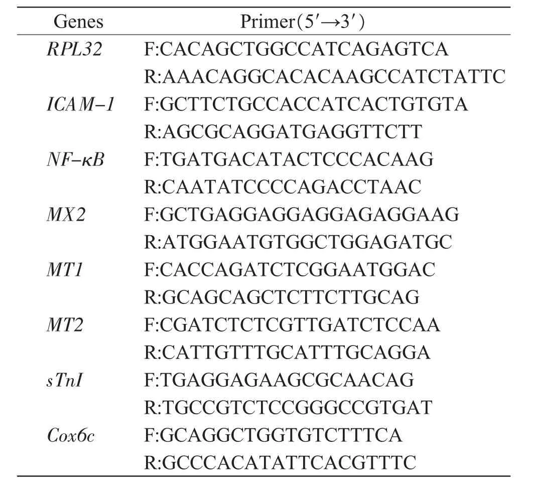
Table1 The primers used for RT-qPCR
The first-strand cDNA was synthesized using a SYBR®Prime Script RT-PCR Kit(TaKaRa Biotechnology,Dalian,China)according to the standard protocols.For reverse transcription,0.4 μg of total RNA was used in a reaction volume of 10 μL.RT-PCR amplification was then performed in a 25 μL reaction mixture containing 12.5 μL SYBR®Premix Ex TaqTM(TaKaRa),9.5μL dH2O,0.5μL of each primer (10 μmol/L),and 2 μL of cDNA according to the instructions provided with the SYBR®Premix Ex TaqTM(TaKaRa).Amplificationwasperformed through one round of pre-denaturation at 95℃for 10 s,and 40 rounds of denaturation at 95℃for 5 s,followed by annealing and extension at 60℃for 20 s.The fluorescence signal was detected at the end of every cycle.All reactions were performedusing aMx3005P real-timePCRSystem (Stratagene,La Jolla,CA).The results were normalized relative to the expression of the ribosomal protein L32(RPL32),which was the most stably expressed gene in contused skeletal muscle[5].The fluorescence curves of the PCR products were evaluated using MxPro software(Stratagene)to determine the expression of ICAM-1,NF-κB,MX2,MT1, MT2,sTnI,and Cox6c mRNA relative to RPL32. The relative gene expression was calculated on Mx-Pro software(Stratagene).
The synthesized cDNA was serially diluted(1, 1∶101,1∶102,1∶103,and 1∶104)in EASY dilution solution(TaKaRa),and 2μL of each dilution was used for amplification in a reaction volume of 25 μL. Sterile purified water was used as a negative control. Statistical analysis
One-wayanalysisofvarianceandtheStudent-Newman-Keuls(SNK)tests was applied to the comparisons of the survival periods of time after muscle contusion.In addition,t test was used to assess statistical significance.In all analyses,P<0.05 was taken to indicate statistical significance.The raw Ct value of each potential gene was normalized based on the Ct value of the reference gene(RPL32). The normalized Ct value distributions of all samples in each group were presented as standard deviations(SD)and calculated by SPSS.
Results
Integrity of total RNA and amplification efficiency of each gene
All RNA samples with RNA integrity number >8.0 and with clearly visible 28S and 18S peaks were employed for RT-qPCR.The dissociation curves of the seven potential candidate genes exhibited signal cusps,and the dissociation temperatures were all>80℃.The amplification efficiency of the seven genes was 80%-120%.
ExpressionofthepotentialmRNAatvarious time points
There were significant differences in the mRNA expression levels of the seven genes between the contusion groups(12,24,and 36 hours later after injury)and control group(0 h)after normalization relative to RPL32(Fig.1).Compared with those of the control group,the levels of ICAM-1,MX2, MT1,and MT2 mRNA expression were significantly increased after contusion(P<0.05).However,the levels of sTnI and Cox6c mRNA expression were significantly decreased(P>0.05)after normalization relative to RPL32.In addition,the expression of NF-κB was attenuated 12 and 24 hours later,respectively,but increased 36 hours later(P>0.05).These genes showed altered expression after the skeletal muscle contusion,suggesting that they were potentially useful for wound age estimation.

Fig.1 Expression of the seven candidate genes at postmortem intervals
Variations in gene expression at various time points
The raw Ct values were all normalized relative to those of RPL32 mRNA,and then converted to normalized Ct values.The SD of normalized Ct value of each gene was calculated to represent the variation in expression after injury.There were significant differences in ICAM-1,NF-κB,and MX2 mRNA expression in the three injury groups when compared with those in the control group(P<0.05).In contrast, there were only slight variations in sTnI and Cox6c expression levels between the three test groups and the control.The extents of variation in MT1 and MT2 expression were found to be inconsistent at the different time intervals(Fig.2).
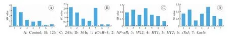
Fig.2 The SD of normalized Ct value of gene expression after contusion
Discussion
Wound age estimation is a critical challenge to forensic pathologists.However,the macroscopic description of a wound is not sufficient enough to determine wound age[6].Gene expression profiling using RT-qPCR is a valuable tool in forensic science for estimating the age of a wound.The most usual solution to this problem is to measure different indicators because many inflammatory cytokines(e.g., TNF-α,TGF-β,fibronectin,IL-6)show variations in expression at different time points after injury[7-10]. However,the degrees of these changes vary with individuals,which is ought to be examined in further studies regarding wound age determination.The primary aim of the current study was to identify the genes which are not only highly correlated with the time interval after injury,but also comparable among individuals.
Seven different function kinds of genes were selected in the study.ICAM-1 is a type of protein present in the membranes of leukocytes and endothelial cells,which produces proinflammatory effects such as inflammatory leukocyte recruitment by signaling through cascades involving a number of kinases[11].NF-κB is a major transcription factor that regulates genes responsible for both the innate and adaptive immune response[12].MX2 is a member of both the dynamin family and the family of large GTPases[13].All three of these genes played important roles in the inflammatory response.Metallothionein (MT)is a family of cysteine-rich,localized to the membrane of the Golgi apparatus,MTs may provide protectionagainstmetaltoxicity[14].Skeletal troponinⅠis part of the troponin complex,which is a major structural protein in skeletal muscle[15].Cox6c is a subunit of cytochrome oxidase,which are a large transmembrane protein complex and the terminal enzyme in the mitochondrial respiratory chain[16].The classes of last two genes were cellular component.
In the current study,the various expressions of seven genes with diverse biological functions in the protection and repair of contused skeletal muscle were compared using the SD of normalized Ct value.A comparison of the relative expression of the genes after contusion indicated that their expression fluctuated at different time intervals(Fig.1),which suggested that all of these genes could be used to estimate wound age.When the variations were compared in gene expression after calculating the SD of normalized Ct value(Fig.2),the levels of variation in sTnI and Cox6c expression were lower than those of ICAM-1,NF-κB,and MX2 mRNA,suggesting that there can be a lower coefficient of variation and greater reliability with sTnI and Cox6c than with other genes.
Conclusion
The current study suggested that genes encoding structural proteins or proteins that serve basic functions could be more suitable as markers to determine wound age.In forensic medicine,it is necessary to select indicator genes that are as stable as possible because they can be applied to any environment.However,definitive conclusions could not be drawn from the results of this experiment;therefore,the results require validation by testing addi-tional indicator genes in future studies.
Acknowledgements
This study was jointly supported by the National Science Foundation for Young Scientists of China(81001347)and the Innovation Foundation of Shanxi Medical University(01201409).
[1]Cecchi R.Estimating wound age:looking into the future[J].Int J Legal Med,2010,124(6):523-536.
[2]Bai R,Wan L,Shi M.The time-dependent expressions of IL-1beta,COX-2,MCP-1 mRNA in skin wounds of rabbits[J].Forensic Sci Int,2008,175(2-3): 193-197.
[3]Kondo T,Ishida Y.Molecular pathology of wound healing[J].Forensic Sci Int,2010,203(1-3):93-98.
[4]Warren GL,Summan M,Gao X,et al.Mechanisms of skeletal muscle injury and repair revealed by gene expression studies in mouse models[J].J Physiol,2007, 582(Pt 2):825-841.
[5]Sun JH,Nan LH,Gao CR,et al.Validation of reference genes for estimating wound age in contused rat skeletal muscle by quantitative real-time PCR[J]. Int J Legal Med,2012,126(1):113-120.
[6]Fronczek J,Lulf R,Korkmaz HI,et al.Analysis of inflammatorycellsandmediatorsinskinwound biopsies to determine wound age in living subjects in forensic medicine[J].Forensic Sci Int,2015,247:7-13.
[7]Yan Z,Sun XL,Hu YL,et al.Different expression of TNF-alpha in brain and peripheral organs after cerebral contusion of rats[J].Fa Yi Xue Za Zhi,2012, 28(4):261-264.
[8]Pan H,Zhang JJ,Xu DD,et al.Expression of CaMKⅡdelta in cerebral cortex following traumatic brain injury[J].Fa Yi Xue Za Zhi,2014,30(3):169-171,177.
[9]Tajiri N,Hernandez D,Acosta S,et al.Suppressed cytokine expression immediately following traumatic brain injury in neonatal rats indicates an expeditious endogenous anti-inflammatory response[J].Brain Res, 2014,1559:65-71.
[10]Kubo H,Hayashi T,Ago K,et al.Temporal expression of wound healing-related genes in skin burn injury[J].Leg Med(Tokyo),2014,16(1):8-13.
[11]Zhang XW,Liu Q,Thorlacius H.Inhibition of selectin function and leukocyte rolling protects against dextran sodium sulfate-induced murine colitis[J].Scand J Gastroenterol,2001,36(3):270-275.
[12]Kim S,Jung JK,Lee HS,et al.Discovery of piperidinyl aminopyrimidine derivatives as IKK-2 inhibitors[J]. Bioorg Med Chem Lett,2011,21(10):3002-3006.
[13]Melén K,Keskinen P,Ronni T,et al.Human MxB protein,aninterferon-alpha-inducibleGTPase,contains a nuclear targeting signal and is localized in the heterochromatin region beneath the nuclear envelope[J].J Biol Chem,1996,271(38):23478-23486.
[14]Kwon A,Jeon SM,Hwang SH,et al.Expression and functional role of metallothioneinsⅠandⅡin the spinal cord in inflammatory and neuropathic pain models[J].Brain Res,2013,1523:37-48.
[15]Zvereva EA,Kovalev LI,Ivanov AV,et al.Enzyme immunoassay and proteomic characterization of troponinⅠas a marker of mammalian muscle compounds in raw meat and some meat products[J].Meat Sci,2015,105:46-52.
[16]Duggan AT,Kocha KM,Monk CT,et al.Coordination of cytochrome c oxidase gene expression in the remodelling of skeletal muscle[J].J Exp Biol,2011, 214(Pt 11):1880-1887.
(Received date:2015-01-28)
(Editor:LIANG Lu)
DF795.1 Document code:A
10.3969/j.issn.1004-5619.2015.02.001
Author:DU Qiu-xiang(1982—),Ph.D,lecturer,major in forensic pathology;E-mail:dqx_2008@126.com
SUN Jun-hong,Ph.D,associate professor,major in forensic pathology;E-mail:junhong.sun@sxmu.edu.cn
Methods The mRNA expression levels of seven candidate genes(ICAM-1,NF-κB,MX2,MT1,MT2, sTnI,and Cox6c)were analyzed in contused rat skeletal muscle at different time points using real-time fluorescent quantitative PCR(RT-qPCR).The raw Ct values were normalized relative to that of RPL32 mRNA,and converted to standard Ct values.At each time point after injury,the standard deviations (SD)of the standard Ct values were calculated by SPSS.Results The expression trends of the seven genes were all found to be related to wound age,but there were lower variation coefficients and greater reliability of sTnI and Cox6c when compared with other genes.Conclusion The genes encoding structural proteins or proteins that perform basic functions can be suitable for wound age estimation.
