下调miR-10a表达对人胰腺癌细胞AsPC-1迁移和侵袭的影响
2013-10-19张恒彭辉勇满昌峰徐娟祁卫东蒋鹏程范钰
张恒 彭辉勇 满昌峰 徐娟 祁卫东 蒋鹏程 范钰
·论著·
下调miR-10a表达对人胰腺癌细胞AsPC-1迁移和侵袭的影响
张恒 彭辉勇 满昌峰 徐娟 祁卫东 蒋鹏程 范钰
目的探讨miR-10a表达对人胰腺癌AsPC-1细胞迁移和侵袭力的影响及其作用机制。方法构建针对miR-10a的小干扰RNA(miR-10a-siRNA),转染处理人胰腺癌AsPC-1细胞,同时设阴性对照siRNA(NC-siRNA)组和空白对照组。采用荧光实时定量PCR法检测3组细胞中miR-10a的表达水平,划痕实验检测细胞迁移,Transwell小室检测细胞侵袭能力,ELISA法检测各组癌细胞培养上清液中基质金属蛋白质酶13(MMP-13)含量。结果对照组、NC-siRNA组和miR-10a-siRNA组AsPC-1细胞miR-10a表达水平分别为1.05±0.08、1.03±0.06、0.02±0.01,穿膜细胞数分别为(150±2.6)、(145±2.2)、(62±1.8)个,培养上清中MMP-13表达量分别为(108.5±2.8)、(107.8±2.5)、(35.8±1.5)pg/ml,miR-10a-siRNA组均显著低于对照组和NC-siRNA组,差异均有统计学意义(P值均<0.01)。 对照组、NC-siRNA组和miR-10a-siRNA组AsPC-1细胞的迁移后间距分别为(385±15)、(395±13) 、(736±18) μm,miR-10a-siRNA组显著大于对照组和NC-siRNA组,差异有统计学意义(P值均<0.01)。结论下调miR-10a表达可抑制胰腺癌AsPC-1细胞迁移和侵袭能力,其机制可能与MMP-13表达下调有关。
胰腺肿瘤; 微RNA; 细胞运动; 肿瘤浸润; 基质金属蛋白质酶-13; miR-10a
MicroRNAs(miRNAs)的异常表达与多种人类癌症的发生密切相关[1-8]。近年的研究发现,miR-10a在人胃癌、甲状腺癌中高表达[9]。Ohuchida等[10]报道,miR-10a在胰腺癌中高表达,并通过抑制HOXA1基因的表达参与细胞的浸润。本研究采用小干扰RNA(small interfering RNA,siRNA)沉默人胰腺癌AsPC-1细胞miR-10a的表达,观察其对细胞迁移、侵袭的影响,探讨其机制。
材料与方法
一、细胞培养及siRNA转染
人胰腺癌AsPC-1细胞系购自南京凯基生物科技发展有限公司,常规培养、传代。靶向miR-10a的siRNA(miR-10a-siRNA)及阴性对照siRNA(NC-siRNA)购自广州锐博公司。转染前一天接种适量细胞至细胞培养板中,每孔加入不含抗生素的培养基。细胞密度达到30%~50%时用50 μl不含血清的培养基Opti-MEM分别稀释1.25 μl 20 μmol/L的siRNA贮存液及1 μl脂质体,轻轻混匀,室温孵育5 min后将稀释的siRNA和脂质体轻轻混匀,室温孵育20 min,加入到细胞的培养液中轻轻混匀。培养4~6 h,更换新鲜培养基,置37℃的CO2培养箱中继续培养48 h。以未转染的AsPC-1细胞作为对照。
二、转染效果鉴定
采用Trizol方法抽提两组转染细胞及对照组细胞总RNA,先逆转录为cDNA,再采用荧光实时定量PCR方法检测细胞miR-10a表达。参照文献[11]方法设计miR-10a引物、PCR 反应条件及计算miR-10a表达量。
三、细胞迁移实验
先用记号笔在6孔板背后用直尺均匀地划横线,大约每隔0.5~1 cm一道,横穿过孔。取各组对数生长期细胞,调整细胞密度为8×10⁵个/ml。每孔加入1 ml细胞悬液,常规培养至细胞铺满单层。用100 μl枪头沿培养板底部呈“一”字形划痕。弃去培养液,用PBS洗细胞3次以去除划下的细胞后继续培养24 h,拍照,计算细胞未爬入的间距。
四、细胞侵袭实验
参照文献[12],应用Transwell小室检测细胞侵袭力。最后在倒置显微镜(×100)下观察穿膜的细胞数。每张膜中央部分和周围部分各随机取3个视野,计数每个视野内的穿膜细胞数,取均值。
五、细胞基质金属蛋白酶13(MMP-13)表达量测定
收集各组细胞培养上清液,采用MMP-13 ELISA试剂盒测定MMP-13含量,按说明书操作。
六、统计学处理
结 果
一、细胞miR-10a表达水平的变化
对照组、NC-siRNA组和miR-10a-siRNA组AsPC-1细胞miR-10a表达量分别为1.05±0.08、1.03±0.06、0.02±0.01(图1),miR-10a-siRNA组显著低于对照组和NC-siRNA组,差异有统计学意义(F=983.280,P=0.000)。

图1 实时PCR的扩增曲线(a)和溶解曲线(b)
二、细胞迁移力的变化
对照组、NC-siRNA组和miR-10a-siRNA组AsPC-1细胞迁移后的间距分别为(385±15)、(395±13)、(736±18) μm(图2)。miR-10a-siRNA组细胞迁移后的间距显著大于对照组和NC-siRNA组,差异有统计学意义(F=883.118,P=0.000)。
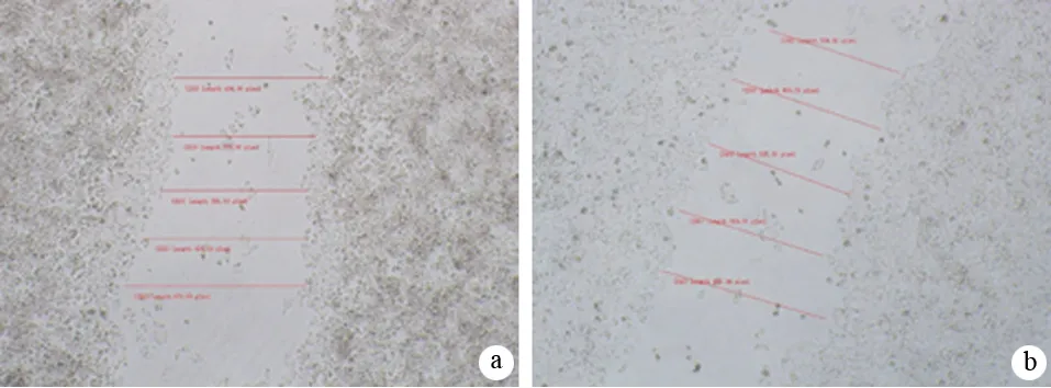

图2对照组(a)、NC-siRNA组(b)、miR-10a-siRNA组(c)AsPC-1细胞的迁移间距
三、细胞侵袭力的变化
对照组、NC-siRNA组和miR-10a-siRNA组AsPC-1细胞的穿膜细胞数分别为(150±2.6)、(145±2.2)、(62±1.8)个(图3),miR-10a-siRNA组明显少于对照组和NC-siRNA组,差异有统计学意义(F=523.500,P=0.000)。


图3对照组(a)、NC-siRNA组(b)、miR-10a-siRNA组(c)的穿膜细胞
四、细胞MMP-13的表达量
对照组、NC-siRNA组和miR-10a-siRNA组AsPC-1细胞的培养上清中MMP-13表达量分别为(108.5±2.8)、(107.8±2.5)、(35.8±1.5)pg/ml,miR-10a-siRNA组显著低于对照组和NC-siRNA组(F=463.853,P=0.000)。
讨 论
大量证据表明,miRNAs的异常表达与多种人类癌症的发生密切相关[1-8]。MiRNAs的发现和应用为胰腺癌等恶性肿瘤的诊断和治疗提供了新的靶点。
胰腺癌细胞转移是一个复杂的过程,迁移和侵袭是重要步骤,也是胰腺癌治疗失败的主要原因之一。积极寻找与胰腺癌侵袭有关的分子机制无疑会有助于胰腺癌的综合诊疗水平。实验研究表明,在许多实体瘤如胃癌、甲状腺癌等瘤组织或细胞中均高表达miRNAs[10],但目前尚不清楚miR-10a在胰腺癌细胞侵袭中的可能作用。
本研究结果显示,与对照组和NC-siRNA组比较,miR-10a组AsPC-1细胞的迁移和侵袭能力均显著减弱,表明miR-10a参与细胞的迁移及侵袭。
肿瘤的浸润与转移的关键是细胞外基质(extracellular matrix, ECM)成分的降解。MMPs能降解ECM。按照底物特异性,人类MMP主要分为明胶酶、间质胶原酶、基质溶解素、机制溶解因子、模型机制金属蛋白质酶及其他等6类。MMP主要功能是可降解一种或多种ECM成分,调控血管形成,又可通过与整合素的相互活化而加强细胞间的粘附作用。MMP-13是MMPs重要成员之一,它在人许多恶性肿瘤包括人胰腺腺癌细胞中高表达[12-15],且与人胰腺癌侵袭有关[16]。本研究结果显示, miR-10a组细胞MMP-13表达量显著低于对照组和NC-siRNA组,提示下调miR-10a表达后可能通过抑制MMP-13的表达而抑制AsPCA-1细胞的迁移和侵袭能力。
[1] Tricoli JV, Jacobson JW. MicroRNA: Potential for Cancer Detection, Diagnosis, and Prognosis. Cancer Res, 2007, 67:4553-4555.
[2] Grammatikakis I, Gorospe M, Abdelmohsen K. Modulation of Cancer Traits by Tumor Suppressor microRNAs. Int J Mol Sc, 2013, 14:1822-1842.
[3] Zabolotneva AA, Zhavoronkov A, Garazha AV, et al. Characteristic patterns of microRNA expression in human bladder cancer. Front Genet, 2013, 3:310.
[4] Pellegrino L, Jacob J, Roca-Alonso L, et al. Altered expression of the miRNA processing endoribonuclease Dicer has prognostic significance in human cancers. Expert Rev Anticancer Ther, 2013, 13:21-27.
[5] Wang Y, Taniguchi T. MicroRNAs and DNA damage response: implications for cancer therapy. Cell Cycle, 2013, 12:32-42.
[6] Wang F, Sun GP, Zou YF, et al. MicroRNAs as promising biomarkers for gastric cancer. Cancer Biomark, 2012, 11:259-267.
[7] Lodewijk L, Prins AM, Kist JW, et al. The value of miRNA in diagnosing thyroid cancer: a systematic review. Cancer Biomark, 2012, 11:229-238.
[8] Zaman MS, Maher DM, Khan S, et al. Current status and implications of microRNAs in ovarian cancer diagnosis and therapy. J Ovarian Res, 2012, 5:44.
[9] Chen W, Tang Z, Sun Y, et al. miRNA expression profile in primary gastric cancers and paired lymph node metastases indicates that miR-10a plays a role in metastasis from primary gastric cancer to lymph nodes. Exp Ther Med, 2012, 3:351-356.
[10] Ohuchida K, Mizumoto K, Lin C, et al. MicroRNA-10a is overexpressed in human pancreatic cancer and involved in its invasiveness partially via suppression of the HOXA1 gene. Ann Surg Oncol, 2012, 19:2394-2402.
[11] Fan Y, Zhang YL, Wu Y, et al. Inhibition of signal transducer and activator of transcription 3 expression by RNA interference suppresses invasion through inducing anoikis in human colon cancer cells. World J Gastroenterol, 2008, 14:428-434.
[12] Boström PJ, Ravanti L, Reunanen N, et al. Expression of collagenase-3 (matrix metalloproteinase-13) in transitional-cell carcinoma of the urinary bladder. Int J Cancer, 2000, 88:417-423.
[13] Leeman MF, McKay JA, Murray GI. Matrix metalloproteinase 13 activity is associated with poor prognosis in colorectal cancer. J Clin Pathol, 2002, 55:758-762.
[14] Lederle W, Hartenstein B, Meides A, et al. MMP13 as a stromal mediator in controlling persistent angiogenesis in skin carcinoma. Carcinogenesis, 2010, 31:1175-1184.
[15] Tan X, Zhou L, Wang W, et al. Genomic analysis of invasion-metastasis-related factors in pancreatic cancer cells. Exp Ther Med, 2010, 1:211-216.
[16] Ohlund D, Ardnor B, Oman M, et al. Expression pattern and circulating levels of endostatin in patients with pancreas cancer. Int J Cancer, 2008, 122:2805-2810.
EffectsofmiR-10adown-regulatedbysiRNAonmigrationandinvasionofhumanpancreaticcancercellAsPC-1
ZHANGHeng,PENGHui-yong,MANChang-feng,XUJuan,QIWei-dong,JIANGPeng-cheng,FANYu.
CancerInstitute,People′sHospitalofZhenjiang,JiangsuUniversity,Zhenjiang212002,China
FANYu,Email:yuf36@sina.com
ObjectiveTo investigate the effects of miR-10a expression on migration and invasion of human pancreatic cancer cells AsPC-1.MethodsSmall interfering RNA targeting at miR-10a(miR-10a-siRNA) was constructed, then it was transfected into pancreatic cancer AsPC-1 cells, and nonsense siRNA (Nc-siRNA) group and blank control group was established. Real time PCR assay was used to detect the expression of miR-10a in the 3 groups, and wound healing assay and Transwell assay were used to determine the migration and invasion abilities of cancer cells. The amount of matrix metalloproteinase-13 (MMP-13) in supernatant of cancer cell culture of each group was examined by ELISA assay.ResultsThe miR-10a levels in control group, NC-siRNA group and miR-10a-siRNA group were 1.05±0.08, 1.03±0.06, 0.02±0.01; and the number of transmembrane cell were (150±2.6), (145±2.2), (62±1.8), the levels of MMP-13 in the supernatant were (108.5±2.8), (107.8±2.5), (35.8±1.5)pg/ml. The values were significantly lower in miR-10a-siRNA group than those in control group and NC-siRNA group (P<0.01). The distance of cultured clone in miR-10a treated cancer cells (736±18 μm) was significantly longer than those in the controls (385±5 μm) and NC-siRNA group (395±13 μm,P<0.01).ConclusionsDown-regulation of miR-10a by siRNA may inhibit migration and invasion of pancreatic cancer AsPC-1 cells, and the down-regulated expression of MMP-13 may be one of the important mechanisms.
Pancreatic neoplasms; MicroRNAs; Cell movement; Neoplasm invasiveness; Matrix metalloproteinase-13; miR-10a
2013-09-29)
(本文编辑:吕芳萍)
10.3760/cma.j.issn.1674-1935.2013.06.004
江苏省普通高校研究生科研创新计划项目(CXLX12_0679);江苏大学临床科技基金(JLY2010007)
212002 镇江,江苏大学附属人民医院肿瘤研究所
范钰,Email:yuf36@sina.com
