Effects of nitric oxide donor N,N'-di-sec-butyl-N,N'-dinitroso-1,4-phenylenediamine on the expression of interferon-gamma in tumor infiltrating lymphocytes
2020-01-07JingFanZhenGaoDePengZhaoPingWangZhengZhongWuXueMeiLi
Jing Fan ,Zhen Gao ,De-Peng ZhaoPing WangZheng-Zhong WuXue-Mei Li
1 Department of Reproductive Medicine Center,Shenzhen Maternity and Child Healthcare Hospital,Shenzhen,Guangdong Province,China
2 The State Key Laboratory of Oncology in South China,Sun Yat-Sen University Cancer Center,Guangzhou,Guangdong Province,China
3 Department of Urology,Shenzhen Second People’s Hospital,First Affiliated Hospital of Shenzhen University,Shenzhen,Guangdong Province,China
Abstract
Key words:nitric oxide donor;tumor infiltrating lymphocytes;N,N'-di-sec-butyl-N,N'-dinitroso-1,4-phenylenediamine;interferon-γ;gas therapy;bladder cancer;autoimmune diseases;controlled release
INTRODUCTION
Nitric oxide (NO) is an important free radical molecule in living organisms.It regulates many biological processes such as vasoconstriction,neurotransmission,and immune activity.1-3The function of NO in the mammalian immune system has been studied intensively.4NO plays a role in the killing response of immune cells to pathogens,and resists pathogen infection as a regulatory molecule.NO regulates acquired immunity and links it to innate immunity.5In the past decade,more and more studies have been conducted to study the role of NO in acquired immunity.Recent studies have shown that NO also plays an important role in the regulation of T lymphocyte differentiation,6,7including regulation of T cell activation and promotion of T cell immune signal transduction.8,9Among them,the functions of CD4+helper T cells and CD8+cytotoxic T cell are regulated by the concentration of NO in endogenous and pericellular environments.
Tumor infiltrating lymphocytes (TILs) are tissue infiltrating lymphocytes isolated from tumor tissues.It is rich in tumorspecific cytotoxic T lymphocytes and natural killer cells.TILs were successfully isolated from fresh tumor tissue by Lotze and Rosenberg10in 1986.In a series of subsequent studies,the activity of TILs was increased and expandedin vitroand reinfused for adoptive immunotherapy.11,12As a new type of immunotherapy strategy,TILs have achieved surprising therapeutic effects in cancers such as melanoma,head and neck cancer,13bladder cancer,14and breast cancer,15but the effects vary greatly from individual to individual.Thus,many patients are not ideally treated.This may be related to the effects of tumor cells and local microenvironment of the tumor,as well as the TILs exhausted16;it may also be related to the state in which TILs are not activated.17Upon activation,TILs release a series of cytokines that regulate their immune functions,such as interferon-γ (IFN-γ),interleukin-2,and tumor necrosis factor-α.18Among them,IFN-γ is an important factor regulating the anti-tumor function of immune cells and plays an important role in TILs-mediated adoptive immunotherapy.It has not only anti-viral and anti-tumor activities but also provides positive feedback to regulate the cellular immune response,which can regulate the phagocytosis,killing,antigen presentation and secretion of cytokines by monocytes-macrophages,which may be resistant to immune system.19
In this study,we introduced the NO small molecule donor N,N'-di-sec-butyl-N,N'-dinitroso-1,4-phenylenediamine(BNN6)that was developed in the past 2 years that was made to release NO by blue-ray (470 nm) irradiation.20It has a great impact on TIL functionality.BNN6 belongs to the cage small molecule NO donor,which has fewer requirements for synthesis experiments,good stability,and can respond to ultraviolet and visible light irradiation.21,22By introducing BNN6,combined with blue-ray short-time irradiation to stimulate NO release,accurate spatio-temporal delivery of NO can be achieved to obtain more scientific research results.The NO released from BNN6 was spread around the isolated and amplified TILs in the medium.Then,the IFN-γ secretion of TILs was investigated to study the functional changes caused by NO.
MATERIALS AND METHODS
Synthesis and characterization of BNN6
BNN6 was synthesized through an addition reaction as reported.20The 4.7 mL (about 20 mmol) N,N'-di-sec-butylamino-p-phenylenediamine (BPA,TCI America Inc.,Portland,OR,USA) is diluted into 25 mL ethanol,then 40 mL of 6 M degassed aqueous solution of NaNO2is added under stirring and nitrogen protection.After 30 min,40 mL of 6 M aqueous solution of HCl was added dropwise using a separating funnel in ice bath.The reaction solution gradually generated beige precipitation.After stirring for 6 hours,the solid product was collected by centrifugation,and washed with water and 50%(v/v) ethanol/water several times to remove residual reactants,and then dried under a freezing vacuum in the dark overnight.The structure of BNN6 was detected by hydrogen nuclear magnetic resonance (1H NMR,Bruker Magnet System,Karlsruhe,Germany,300 MHz/54 mm) and mass spectrum (MS,Waters LC-MS System,Milford,MA,USA) measurements.1H NMR(300 MHz,CDCl3):δ 7.52 (4H),4.94-4.69 (2H),2.00-1.84(2H),1.81-1.69 (2H),1.49 (6H),1.10 (6H).MS (ESI+):calculated for C14H22N4O2,278.2 [M]+;found 279.18 [M]+.
Isolation and culture of TILs
The study was approved by the Medical Ethics Committee of the Shenzhen Second People’s Hospital,the First Affiliated Hospital of Shenzhen University,China (approved No.065)on February 12,2018.The obtained human bladder tumor tissues (preserved in our laboratory) were cut into 5 mm2fragments and disaggregated in RPMI 1640 to obtain single cell suspensions.The cell suspension was filtered through a filter (BD Biosciences,San Jose,CA,USA) and washed by culture medium.The cells were separated by a discontinuous 70% and 100% Isopaque Ficoll gradient (Stemcell Tech.,Mumbai,India) under centrifugation.The targeted TILs were recovered in the 100% gradient interface.Then,the enriched TILs were washed in Dulbecco’s phosphate buffered saline(1% bovine serum albumin) and expanded in TILs culture medium (VIVO XV medium,10% fetal bovine serum,3000 IU/mL interleukin-2).A portion of the cells was stained and analyzed for CD3+,CD4+,and CD8+cells,sorted through flow cytometry (CytoFLEX,Beckman Coulter,Indianapolis,IN,USA).For TILs sorting,TIL cells were dispersed in 20 mL of medium,and cultured in a 90 mm dish for amplification.The 1 mL cells were used for centrifugation at 1500 r/min for 5 minutes.It was then resuspended in 200 μL phosphate buffered saline and albumin buffer (2% bovine serum albumin in phosphate buffered saline),resuspended in 200 μL,with adding 2.5 μL of commercial antibody (blank,CD3,CD4,CD8,CD3+CD4+CD8,Abcam,Cambridge,UK) fluorescein isothiocyanate/P-phycoerythrin (PE)/PECy5.5),incubated at room temperature for 30 minutes,then centrifuged at 1500 r/min for 5 minutes.The supernatant was then discarded,and cell pellet was resuspended in 500 μL of phosphate buffered saline and albumin.Then the threecolor fluorescence flow cytometry detection was employed.
Cell proliferation assay of BNN6 treated TILs
Cell proliferation of TIL cells treated with BNN6 (kept in the dark) and NO released from BNN6 (blue-ray irradiation) was determined by the colorimetric Cell Counting Kit-8 (CCK-8,Beyotime Biotech,Shanghai,China).TILs were seeded in a 96-well plate at a density of 1 × 10⁵cells per well and incubated overnight at 37°C under a 5% CO2atmosphere.After being washed with phosphate buffered saline (pH 7.4),the cells were incubated with 100 μL of BNN6 at different concentrations (0,0.1,0.4,1.2,3.7,11.1,33.3 and 100 μg/mL) at 37°C for 2 hours under the same conditions.Then,the cells were exposed to blue-ray for 45 seconds,and then put back into the incubator.After 24 hours of incubation,the medium was replaced with 100 μL normal medium.After 2 hours,10 μL of CCK-8 was added to each well of the 96-well plates and incubated for 2 hours.The 450 nm absorbance was measured using a plate reader (Bio-Rad,Hercules,CA,USA).Assays were repeated five times.
The IFN-γ secretion in BNN6 treated TILs
To further study the biological effects of BNN6-treated TIL cells,secretion of the T-helper type I cytokine,IFN-γ,was measured by commercial enzyme-linked immunosorbent assay (ELISA) reagents (human IFN-γ ELISA kit,Becton,Dickinson and Company,Franklin Lakes,NJ,USA) and ELISPOT kits (human IFN-γ ELISpot kit,Becton,Dickinson and Company).Cell suspensions at 1 × 10⁶cells/mL density in medium (with 3000 IU/mL of interleukin-2) were prepared and 100 μL was added to each well of the BD ELISPOT plate.The BNN6 was added into the medium with a final concentration of 0,3,10,and 30 μg/mL.The plate was kept under blue-ray irradiation for 45 seconds.Then,the lid was replaced and the cells were incubated at 37°C,5% CO2for 12 hours.The wells were washed 2 times with deionized water and 3 times with prepared wash buffer.Prepared antibody detection solution was added.The cells were incubated for 2 hours at room temperature and then washed 3 times with wash buffer.Prepared streptavidin-horseradish peroxidase solution was added and incubated for 1 hour at room temperature.After washing with wash buffer and phosphate buffered saline,3-amino-9-ethylcarbazole substrate solution was added and monitored for spot development after 10 minutes.The substrate reaction was stopped by deionized water.The plate was air-dried at room temperature overnight until it completely dried.The spots were manually analyzed spots by inspection under the ELISPOT plate reader.
Statistical analysis
The SPSS 21.0 software (IBM,Armonk,NY,USA) was used for statistical analyses.All quantifiable data were evaluated in at least three independent experiments.The pairedt-test was adapted to evaluate the group differences.AP-value <0.05 was deemed to be statistically significant.
RESULTS
Synthesis and characterization of BNN6
BNN6 was synthesized in an aqueous reaction system by BPA and NaNO2in hydrochloric acid solution (Figure1A).After the red oil-like liquid became beige solid products,the BNN6 was purified by washing with ethyl alcohol/water and drying under a vacuum.The characterization of1H NMR and MS showed that the BNN6 synthesized successfully.Each peak of the BNN6 was found in1H NMR (Figure1B) and the 279.18 Da size was in accordance with the molecular structure of BNN6 (Figure1C,301.16 is BNN6 plus one unit of sodium,249.18 is BNN6 minus one unit of NO,and 220.15 is the reactant BPA).We also confirmed the blue-ray responsiveness of the synthesized BNN6 through the color change of the solution.The yellow solution of BNN6 became red after 45 seconds of irradiation by blue ray (Figure1A,digital photo).
Isolation and identification of TILs
We isolated TILs from human bladder tumor tissues by density gradient centrifugation.After amplification of TIL cells,the cell suspension was observed under the light microscope(Figure2A) and identified by flow cytometry.Most of the isolated cells (about 96.80%) were CD3 positive (Figure2B),a surface marker of T lymphocytes.This also showed that the TIL cells were successfully isolated and expanded from tumor tissues.Then,the CD3 positive TILs were further identified for CD4 and CD8 classification by flow cytometry (Figure2C).Most of the TILs were CD4 type T cells (~ 84.99%)while the CD8 type TILs rate was ~10.60%.The CD4 and CD8 double positive cell rate was ~0.37%.

Figure1:Synthesis and characterization of BNN6.
The biological effects of the NO donor BNN6 on TILs
The cytotoxicity of BNN6 to the TILs was measured by a CCK-8 test,and the results indicated a weak cytotoxicity to TILs at concentrations below 33.3 μL/mL of BNN6(Figure3A).Therefore,0,3,10,and 30 μg/mL of BNN6 were selected as the working concentrations to further study the effects on IFN-γ secretion.The ELISA assay of IFN-γ secretion at different BNN6 concentrations show that with increased BNN6 concentrations,the IFN-γ secretion of TILs was decreased.After concentrations higher than 10 μL/mL,there were no significant changes in IFN-γ secretion (Figure3B).The ELISPOT assay was also used to detect the IFN-γ secretion from 0,3,10,and 30 μg/mL BNN6 treated TILs.After analyzing spots by inspection under the ELISPOT plate reader (Figure3C),the number of positive cells was similar to that from the ELISA results (Figure3D).The number of positive spots from the 0,3,10,and 30 μg/mL BNN6 treated TILs was calculated to be 227.3 ± 21.2,193.8 ± 16.8,159.8± 14.6,133.5 ± 17.0,respectively.
DISCUSSION
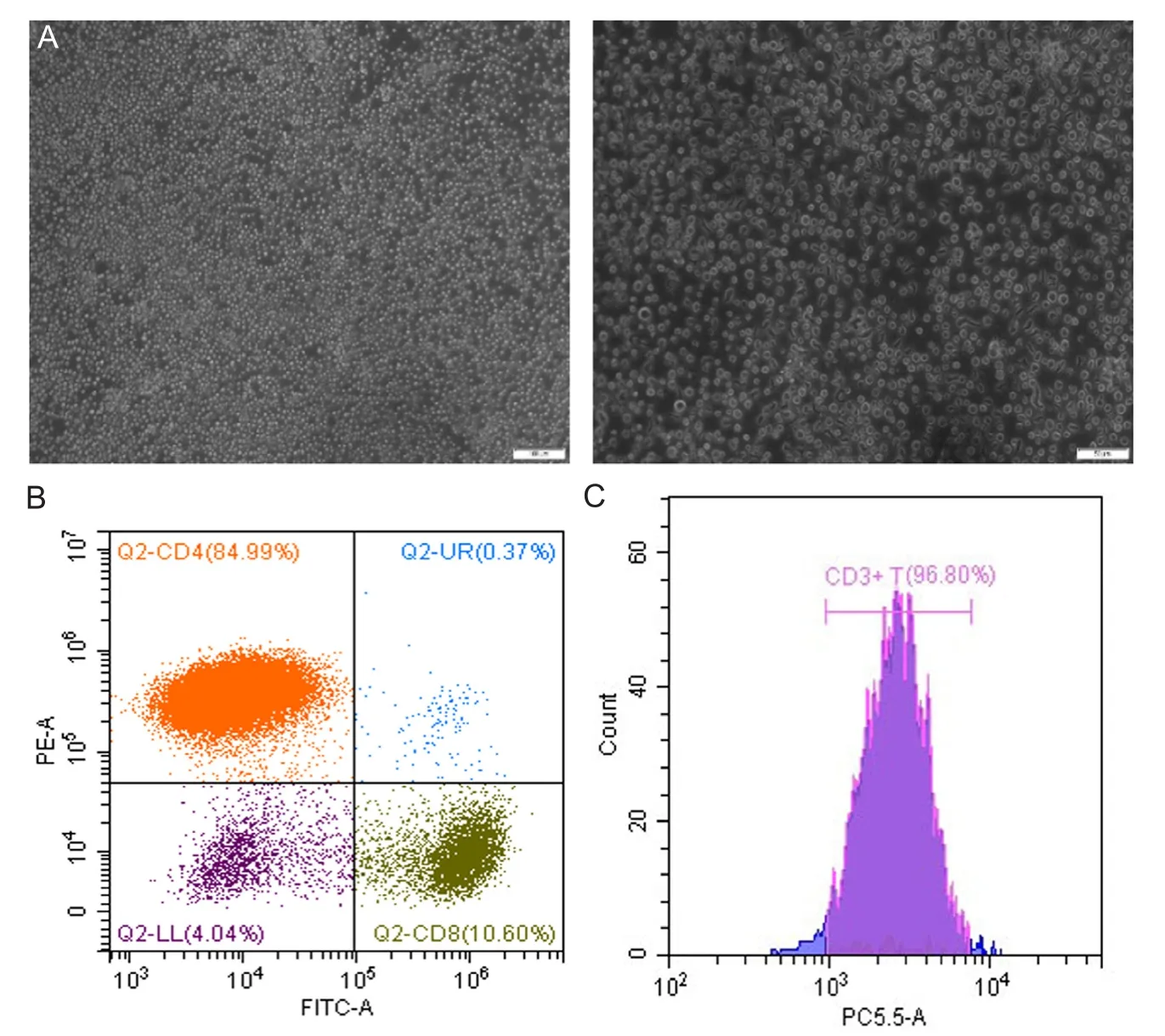
Figure2:lsolation,amplification and identification of tumor infiltrating lymphocyte(TIL) cells.

Figure3:The biological effects of the NO donor BNN6 on TILs.
BNN6 is a novel,caged-structure small molecule NO donor.In this study,BNN6 synthesized by the classical strategy and the phenomenon in the synthesis,characterization and release process were observed.All the phenomena demonstrate the successful synthesis of BNN6 and its ability to release NO in response to blue light.Compared to ultraviolet light,blue light has a relatively long wavelength containing less energy,which may lead to less cytotoxicity.Thus,after confirming that the blue light could stimulate the release of NO from BNN6,we employed it for the subsequent experiments.Then,the TILs which closely related to tumor development and anti-tumor function were isolated from human bladder tumor tissues by density gradient centrifugation.After amplification,TILs were identified by microscope and flow cytometry.These identifications show that the TIL cells were successfully isolated,cultured and expanded successfullyin vitro.After preparation of the TILs,we used BNN6 as the NO donor for biofunctional study on TIL cells.The cytotoxicity of BNN6 to the TILs was studied to confirm the working concentrations.We used both ELISA and ELISPOT assay to detect the IFN-γ secretion at these concentrations of BNN6.The ELISA results show that with increased BNN6 concentrations,the IFN-γ secretion of TILs was decreased.The results of ELISPOT assay are also present a similar trend.In some other similar studies,IFN-γ could induce the production of inducible NO synthase in macrophages and promotes the synthesis of NO.IFN-γ also induces inducible NO synthase production in microglia astrocytes,possibly related with certain parts of the central nervous system.It is related to the occurrence or protection of the disease.Some believe that a small amount of NO has physiological and defensive functions,while a large amount of NO has proinflammatory and damaging effects.23In the current study,NO donors showed inhibition of IFN-γ production,presumably related to BNN6 dosage and NO product concentration.After triggering the exogenous NO donors to release NO,thein vivosystem may sense changes in NO concentration and make corresponding changes,such as regulating the secretion of IFN-γ and inducible NO synthase,thereby reducing endogenous NO production.This may be a mechanism by which NO donors inhibit IFN-γ secretion.Thus,BNN6 or other NO donors may play a role in the treatment of autoimmune diseases.
Author contributions
JF and ZG designed this study.XML conceived the study and supervised this work.JF contributed to major experimental work.DPZ,PW,and ZZW assisted in the experimental and analytic work.JF assisted in chemical synthesis and characterization.JF wrote the paper.All authors were involved in and contributed to the manuscript.
Conflicts of interest
None declared.
Financial support
This work was supported by the Postdoctoral Science Foundation of China (No.2018M643312 to JF,2018M633246 to ZG),National Natural Science Foundation of China (No.81801465 to DPZ,81802537 to ZG),Natural Science Foundation of Guangdong Province,China (No.2018A030310644 to JF),the Sanming Project of Shenzhen Health and Family Planning Commission,China (No.SZSM201512012 to XML).
Institutional review board statement
The study was approved by the Medical Ethics Committee of the Shenzhen Second People’s Hospital,the First Affiliated Hospital of Shenzhen University,China (approved No.065) on February 12,2018.
Copyright license agreement
The Copyright License Agreement has been signed by all authors before publication.
Data sharing statement
Datasets analyzed during the current study are available from the corresponding author on reasonable request.
Plagiarism check
Checked twice by iThenticate.
Peer review
Externally peer reviewed.
Open access statement
This is an open access journal,and articles are distributed under the terms of the Creative Commons Attribution-NonCommercial-ShareAlike 4.0 License,which allows others to remix,tweak,and build upon the work non-commercially,as long as appropriate credit is given and the new creations are licensed under the identical terms.
杂志排行

Medical Gas Research的其它文章
- The role of medical gas in stroke:an updated review
- Revisiting the expanded use of hyperbaric oxygen therapy for treatment of resistant migraines
- Numerical analysis of mechanical ventilation using high concentration medical gas mixtures in newborns
- Micelle-embedded coating with ebselen for nitric oxide generation
- Recent developments in nitric oxide-releasing biomaterials for biomedical applications
- Nitric oxide detection methods in vitro and in vivo