Micelle-embedded coating with ebselen for nitric oxide generation
2020-01-07LiYangLinHuaLiLuJiangJunQiangPanRiFangLuoYunBingWang
Li Yang ,Lin-Hua Li ,Lu JiangJun-Qiang Pan ,Ri-Fang Luo ,Yun-Bing Wang
1 National Engineering Research Center for Biomaterials,Sichuan University,Chengdu,Sichuan Province,China
2 Department of Cardiovascular Medicine,Xi’an Central Hospital,Xi’an,Shaanxi Province,China
Abstract
Key words:micelle;ebselen;nitric oxide;layer-by-layer;cardiovascular implants;anti-coagulation;surface modification;endothelialization
INTRODUCTION
Cardiovascular disease has become the most fatal killer in threatening human health.1,2Coronary stents are devices designed to prevent elastic recoil and intimal hyperplasia associated with percutaneous transluminal coronary angioplasty.3,4With decades of clinical application,coronary stents have already demonstrated the effectiveness and saved millions of lives.Though successful,clinical failures like late stent thrombosis has been treated as a significant risk when using drug-eluting stents,mainly due to the non-selective inhibition of the proliferation of both endothelial cells and smooth muscle cells (SMCs),affected by the released antihyperplasia drug,like sirolimus and paclitaxel.5,6Moreover,delayed re-endothelialization would possibly cause late/very late stent thrombosis.Rainer4discussed the new drug-eluting stent concepts and concluded that the drugs applied should inhibit SMCs proliferation and migration without affecting endothelial regeneration,and reduce the risk of stent thrombosis and anti-inflammation.Besides searching for drugs or a combination of drugs that can provide multifunction,surface coatings which potently address the enhanced antithrombotic and anti-hyperplasia while do no harm to endothelialization are also of special interests.7,8
Nitric oxide (NO) plays a pivotal role as a messenger and signaling molecule in maintaining vascular microenvironment in terms of mediating vascular endothelial function,inhibiting the adhesion of platelets and leukocyte,down-regulating vascular SMC proliferation,and their synthesis of protein and collagen.9,10Healthy endothelial cells could secret a NO flux of 0.5-4 × 10⁻¹⁰mol/(cm2· min).11
Stent implantation usually causes acute blood vessel damage and thus stents fabricated with a coating that mimic the basic function (NO releasing) of endothelial cells would protect the endothelialization process while inhibiting SMCs proliferation and antithrombosis formation.This approached is now well known as “NO-generating” and has been fully developed by researchers,especially represented by Prof.Meyerhoff’s team.12,13Polymers or coatings contain an immobilized catalyst such as glutathione peroxidase-like catalytic mimics(i.e.,selenocystamine,cystamine) and copper nanoparticles or copper (II) ion/ligand complex have been reported by decomposing circulating S-nitrosothiols (RSNO) to release NO at the blood/material interface.14On this basis,Yang et al.15,16had immobilized the NO generation catalyst like copper and selenocystamine on the surface of 316L stainless steels stents and found that due to the potent catalytic effect ofin situgeneration of NOin vivo,both the thrombosis formation and intimal hyperplasia were impressively inhibited.Luo et al.7also grafted cystamine onto heparin backbone and then further prepared a heparin-cystamine/polyethyleneimine nanoparticles functionalized coating to produce the continuous generation of NO.However,somein vivotoxicity studies revealed that there might be the reaction between reduced selenium species and oxygen,which was too fast to produce a significant amount of superoxide that could react with NO to produce peroxynitrite,a toxic species.17And among those selenium catalysts,aromatic organoselenium species have been found to be far less toxic(for example,ebselen (Ebs)).18
As commonly accepted,tuning surface properties is a convenient method for controlling interactions between materials and surrounding microenvironment.Layer-by-layer (LBL)assembly has been treated as a versatile and easy technique for fabrication of multifunctional coatings in biomedical applications.With alternative assembly of selected polyelectrolytes,alone or in combination with bioactive components packaging,desired surface properties are simply achieved.19-21Though widely been investigated,most LBL components are water soluble,which means it is not an ideal platform for loading hydrophobic drug,such as clinically used antihyperplasia drug rapamycin.22,23In order to enforce the local drug administration in the multifunctional LBL coating,the pre-loading of drug into a carrier is feasible.In our previous study,we fabricated a micelle (MIC)-embedded LBL coating with catechol and phenylboronic acid for tunable drug loading and sustained release.24Briefly,a self-healing sandwiched LBL coating was constructed by using chitosan/heparin as the adopted polyelectrolytes with embedding of MICs,in which chitosan backbone was grafted with catechol and the MIC was modified with exposed phenylboronic acid.Moreover,rapamycin and atorvastatin calcium were selected as drug candidates and loaded into MICs,following by drug releasing behavior study.We found that using such coating protocol,the introduction of catechols and boronic acids would endow the coating with enhanced stability by abundant interactions among each coating component (e.g.,boric acid ester bond formation,weak intermolecular cross-linking,π-π interactions and H-bonding).24
Based on above considerations,herein,we used the similar coating approach,and merely changed the preloaded drug to Ebs,a low toxic aromatic organoselenium that has been used as anti-inflammatory drug.18,25Ebs had also been demonstrated as a glutathione peroxidase-like catalyst to decompose RSNOs to release NOin vivo.In this work,we also tested the release profile of Ebs in the MIC-embedded LBL coating,along with the anti-platelet adhesion test and SMCs proliferation test.
MATERIALS AND METHODS
MIC preparation and drug loading
The amphiphilic block MIC molecule is synthesized with three parts,using hyaluronic acid (HA;10 kDa;Sigma-Aldrich,St.Louis,MO,USA) as the backbone,modified with 2-hydroxymethylphenylboronic acid (2-HMPBA;Sigma-Aldrich),and using cholesterol to form the hydrophobic core.In brief,HA (1 g),cholesterol (700 mg;Sigma-Aldrich),2-HMPBA (500 mg)were entirely dissolved in dimethyl sulfoxide (30 mL) at 80°C,following with the adding of N,N′-dicyclohexylcarboimide(500 mg;Sigma-Aldrich) and 4-dimethylaminopyridine (200 mg;Sigma-Aldrich) with continuously stirring for 24 hours at 80°C.Thereafter,the mixture was dialyzed in deionized (DI)water (dialysis membrane bag MWCO = 3000 Da) for 2 days,and the solid phase was separated from the reaction solution by vacuum filtering under vacuum to remove the excessive cholesterol.Finally,the mixture was dialyzed in DI water(dialysis membrane bag MWCO = 3000 Da) for 1 more day.The final product was lyophilized and kept in a moisture-free desiccator for further use.The degree of catechol substitution is determined by proton nuclear magnetic resonance (Bruker Avance,London,UK,400 MHz,D2O).The hydrophobic cholesterol was conjugated to the one end of the hydrophilic HA,and the 2-HMPBA was conjugated to the other endviathe esterification of carboxyl (-COOH) and hydroxyl (-OH).
MIC molecules (40 mg) were completely dissolved into dimethyl sulfoxide (10 mL) at 80°C,and then Ebs was added into the solution.After that,DI water (10 mL) was slowly added into above solution drop by drop under vigorous stirring.Finally,the mixture was dialyzed in DI water for 2 days(dialysis membrane bag MWCO = 500 Da).The concentration of the mixture solution was adjusted to 1 mg/mL by evaporation using rotary evaporators and attenuation using DI water,the pH was adjusted to 6.5 by 1 M hydrogen chloride.Ebs(98%;BioChemPartner,Shanghai,China) loaded MICs were denoted as micelles (MIC)-Ebs and the MICs without drug loaded were labeled as MIC.After that,the size and morphology of MIC and MIC-Ebs were evaluated using transmission electron microscopy (TEM,Hitachi H-600,Tokyo,Japan).
Synthesis of catechol-modified chitosan
Catechol-conjugated chitosan (CS-C) was synthesized using chitosan (Mw100 kDa,80% deacetylated;Sigma-Aldrich)and 3,4-dihydroxybenzaldehyde (3,4-DHB;Sigma-Aldrich)following previously published method by Clifford et al.26The catechol was conjugated to the amine groups of chitosan by Schiff base reaction.Methanol was used to dissolve 3,4-DHB,NaBH4 (Sigma-Aldrich) as a reductant was used to reduce the C=N to C-N,and the degree of catechol substitution on the backbone of chitosan was determined by proton nuclear magnetic resonance.Stock solutions of CS-C (2 mg/mL,pH 6.5) were prepared for further use.
LBL coating construction and characterization
316L stainless steels with the size of 10 mm × 10 mm were mirror-polished as the substrate for biocompatibility test.The MIC-embedded LBL coating fabrication process was shown in Figure1.Briefly,the CS-C,MIC and heparin (185 U/mg;Macklin,Beijing,China) were alternatively and orderly assembled onto the substrate.The deposition pH value for all polyelectrolytes was maintained at pH 6.0 (CS-C and heparin)and pH 7.0 (MIC),respectively.Similar operation could also be found in our previous study,24including the polydopamine coating pre-treatment and subsequent LBL assembly.In this work,the as-prepared coating sample with 10 cycles of LBL assembly was named as LBL10,and the MICs embedded sample with Ebs loading was labeled as LBL10@Ebs.Particularly,ultraviolet-visible light spectrophotometer (UV-2401PC,Shimadzu,Kyoto,Japan) on a quartz plate and the ultraviolet adsorption between 180 and 600 nm was recorded.X-ray photoelectron spectroscopy (XSAM800,Kratos Ltd.,Manchester,UK) was used for further investigations of the surface chemical compositions (Al Kα X-ray source,1486.6 eV).Coatings prepared on silicon wafer were examined by scanning electron microscopy (S-3400N;Hitachi) to study the surface morphology.The Ebs loading capacity,encapsulation efficiency and Ebs release profile were also obtained following the similar protocol.The amount of the released Ebs in the collected medium solution was measuredviahigh performance liquid chromatography (Waters 1525 chromatography,Milford,MA,USA;equipped with a C18 (250 mm × 4.6 mm,5 μm)).
Catalytic ability of NO generation
The NO release catalyzed by sample was examined using Saville-Griess reagent reported before.27,28Briefly,the coated substrate (1 cm × 1 cm) was placed in a 24 well plate and interacted with 1 mL testing solution.The testing solution contained two parts:(1) 0.5 mL donor solution (200 μM ethylenediaminetetraacetic acid,65 μM S-nitroso-N-acetyl-DL-pencillamine (SNAP;Sigma-Aldrich) and 30 μM L-glutathione (Sigma-Aldrich));and (2) 0.5 mL Saville-Griess reagent (2.5 μM).Once interact with the coating surface with catalyst,the released NO would quickly form NO2- and could form diazo complex which would be detected at the absorbance around 540 nm.29The controlled solution is consisted of 0.5 mL Griess reagent and 0.5 mL DI water.The average liberated NO from SNAP was calculated in 30 minutes.
Platelet adhesion
More than five samples were used for statistical count,and each test was done for more than three times.The whole blood was collected from rabbit in negative pressure tubes containing sodium citrate as the anticoagulant.The platelet rich plasma was obtained by centrifuging the obtained whole blood at 1500 r/min for 15 minutes.Briefly,samples were incubated with platelet rich plasma (SNAP containing solution that contained 65 μM SNAP and 30 μM L-glutathione) for 30 minutes at 37°C,and after fixing and dehydration treatment,the platelets morphologies were viewed using scanning electron microscopy.
Smooth muscle cell proliferation
Human umbilical artery SMCs were isolated from newborn umbilical cord (ethics approval was obtained by the Institutional Review Board of the West China Hospital in Sichuan University,approval No.K2018044 on March 3,2018.The enrolled subjects signed the informed consent.) by the explant method as described previously.30SMCs were cultured using Dulbecco’s modified Eagle medium containing 1% antibiotic penicillin-streptomycin and 10% fetal bovine serum).Before seeding,the samples were sterilized by ultraviolet for 1 hour.The samples were then incubated with 1 mL of SMCs suspension solution (with a density of 3 × 10⁴cells/mL) at 37°C under 5% CO2for 4 hours,1 day and 3 days.The cells interacting with NO donors were also carried following our previous study.14The cell viability was tested following the cell counting kit-8 (Dojindo,Kumamoto,Japan) assay.Besides,samples cultured with SMCs were washed completely with saline solution thrice and soaked in 2.5% glutaraldehyde solution for 12 hours,and then stained with rhodamine for 20 minutes.After fully washing,the cells were observedviafluorescence microscope (TE2000;Nikon,Tokyo,Japan).
Statistical analysis
The date was obtained and analyzed using SPSS 11.5 (SPSS,Chicago,IL,USA) and expressed as the mean ± standard deviation (SD).The statistical significance between and within groups was determined using a one-way analysis of variance.
RESULTS
Synthesis of chitosan-catechol and phenylboronic acid modified MICs
It is well known that catechol modified biomolecules will make contribution in strengthening coating stability due to the inherent adhesive properties and enhanced intermolecular interactions.31-33Herein,the catechols were conjugated to chitosan backbones as shown in Figure2A.The value of the catechol proton peaks which appeared from 6.5 to 7.0 ppm indicated successful preparation,and the degree of catechol conjugation was about 31.4%.Besides,the phenylboronic acid modified micellar molecule was also successfully prepared.As shown in Figure2B,the 2-HMPBA peaks appeared from 7.4 to 7.5 ppm,the proton peaks of acetyl group from 7.9 to 8.1 ppm and the cholesterol from 5.5 to 5.75.The value of the degree of 2-HMPBA was 16%,and the value of the degree of cholesterol substitution was 6%.These data indicated successful synthesis of modified molecules.
MICs and MIC-embedded coatings
With the successful synthesis of MIC molecules,which consisted of hydrophilic HA backbone,hydrophobic cholesterol and terminated phenylboronic acid,we further tested the micellular structure and property.The transmission electron microscope results of MIC and MIC-Ebs were shown in Figure3A.The particle size of MIC was around 200 nm,and after drug loading,the size was bigger due to the additional contribution in the hydrophobic core of MICs which resulted in the increasing of particle size (Figure3B).The ultraviolet-visible light absorbance at 280 nm indicated the catechol moieties and the absorbance at 325 nm represented the successful loading of Ebs into the MICs (Figure3C).The averaged particle size,polydispersity index,zeta-potential,drug loading and encapsulation ratio of these MICs are shown in Table1.The polydispersity index value of MIC was 0.125,indicating that the particle size distribution was more concentrated than the drug loaded MICs.Moreover,the zeta-potential value of these MICs was under -20 mV which derived from the carboxyl of HA that were the hydrophilic ends of MIC,which also strongly implied that it could be applied as an anionic polyelectrolyte in constructing LBL coatings.The drug loading efficiency of MIC-Ebs was about 6%,and the encapsulation ratio was more than 30%.The zeta potential value (-27.33 mV) indicated that MICs could be well applied as negatively charged polyelectrolytes,endowing the potential to be applied into the LBL coatings and functionalized as a drug eluting model.
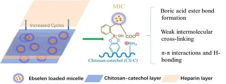
Figure1:The schematic diagram of MIC-embedded layer-by-layer coating construction and the potential intermolecular interactions.

Figure2:The illustration of synthesis of catechol modified chitosan (A) and phenylboronic acid modified MlC molecule(B) with their corresponding proton nuclear magnetic resonance results.

Figure3:The micellular structure and property of MIC and MIC-Ebs.
As shown in Figure1,the existence of catechols and phenylboronic acids would provide more intermolecular interactions among each component.The borate ester bond formation would possibly happen when the pH is higher than 6.34Besides,weak intermolecular crosslinking between adjacent catechols,along with the nucleophilic addition between oxidized quinone and primary amines will also enhance the coating stability.31,35Prior to LBL coating construction,we tested the potential interactions between MICs and catechol-modified chitosan at different pH value.As seen in Figure4,CS-C and MIC were mixed under pH 5.0 and pH 7.0,and the mixture was viewed again after 24 hours.The solution of CS-C and MIC were transparent at pH 5.0.The color of the CS-C solution at pH 7.0 turned to pale yellow,due to the slight oxidation of catechols to quinones.Once mixed together for 24 hours,a faint haze like solution and few precipitates were observed,suggesting weak intermolecular interactions between CS-C and MIC at pH 5.0,mainly are electrostatic interactions.However,at pH 7.0,more epinephelos yellow precipitates were observed,which might be ascribed to the boric acid ester formation between catechols and phenylboronic acid.Thus,more interaction force between CS-C and MIC make the complex denser.On the basis of above phenomenon,the CS-C and MIC would also form similar interactions on the substrate-liquid interface and thus maintain a tunable LBL coating construction process.
We have constructed the MIC embedded LBL coating in our previous work and found that due to the modular assemble of each coating component,the introduced bionic acid ester bond formation and intermolecular crosslinking would help to tunable the coating growth.24In this work,the surface morphology of LBL10and LBL10@Ebs was shown in Figure5A.Obviously,the porosity was maintained and the basic micellar structure with multi-spherical particles near 200 nm in diameter was performed.The coating became denser in the Ebs loaded coatings which might be ascribed to the enriched π-π stacking interactions.Such step-by-step steadily coating growth might make potential contribution in tunable functionality introduction or drug loading.The X-ray photoelectron spectroscopy wide scan indicated successful loading of Ebs with the facile assembly of MICs (Figure5B).The total amount of loaded Ebs on LBL10@Ebs samples was about 52.50 μg/cm2(Figure5C).With the enriched interactions,such coating also presented an inhibition of burst release of Ebs (only 18.6% of released Ebs for the first day),compared to commercial used drug-eluting stents (usually more than 30%).36The coating could also support sustained release of Ebs (more than 1 month) and heparin(more than 20 days).
NO generation and biological effect
As mentioned before,in the presence of reductant L-glutathione,organoselenium would function as a catalyst toin situdecompose RSNOs to release NO.18In this work,thein vitrocatalytic degradation of SNAP was evaluated using Saville-Griess reagent (Figure6).It could be seen that the LBL10@Ebs coating could decompose considerable NO generation level at a speed about 2.0 × 10⁻¹⁰mol/(cm2· min),which was within the level that healthy endothelial cell could produce(0.5-4 × 10⁻¹⁰mol/(cm2· min)).Considering the release profile of Ebs,the sustained release of Ebs would also be expected and give the basic requirement for long-term catalytic effect in NO generation,thus providing anti-thrombotic ability (to be fulfilled in further study).
Following the NO generation ability test,the platelet adhesion to different samples was also tested.As seen in Figure7A and B,the platelets adhered on stainless steel surface were activated with extended pseudopods,suggesting severe platelets activation,which was frequently reported in otherstudy.15,16,37As the coating constructed in our study contains the anti-coagulant heparin,the LBL10coating presented a certain improved anti-platelet adhesion and activation effect.For the catalyst Ebs loaded samples,the LBL10@Ebs could effectively suppress platelet adhesion and activation with the addition of NO donor solution (SNAP containing solution) to mimic the endogenous RSNOs environment in blood.Interestingly,the LBL10@Ebs also showed a slight improved hemocompatibility than LBL10sample,which still needed investigation.The drug Ebs was also found to present to upregulate the level of CD62P on platelet membrane.38Briefly,the synergetic effect of heparin and NO generation ability of Ebs could effectively enhance the coating hemocompatibility and thus could be well allied as a safe blood-contacting material.

Figure4:The interactions between CS-C and MIC under pH 5.0 and pH 7.0,respectively.

Table1:The properties of micelles

Figure5:The surface morphology and Ebs release of LBL and LBL@Ebs.

Figure6:The nitric oxide (NO) generation ability of the ebselen loaded coating.
Excessive SMCs proliferation after stents implantation has been treated as a key factor for in-stent restenosis.1And at that condition,without endothelial cell protection,the SMCs proliferation could also further cause thrombus deposition and microphages invasion.2Thus,fabricating a surface that functions well to suppress SMCs proliferation is of special significance.As reported,NO could inhibit SMC proliferation through the cyclic guanosine monophosphate-dependent pathway.10We have also evaluated the SMCs proliferation behavior on different samples,which were presented in Figure8A and B.The controlled stainless steel samples presented nice affinity for SMCs proliferation,which agreed with other studies.7,15,16The introduced heparin and the generated NO indicate a synergistic effect to suppress the growth of SMCs.Heparin could suppress SMCs proliferation in a dose dependent manner.Based on our previous findings,heparin release content at the early stage could reach up to 10 μg/cm,24which is higher than the reported inhibitory density (3.5-5 μg/cm).39The strongest inhibitory effect could be seen on LBL10@Ebs samples with the addition of SNAP,due to the additional effect ofin situreleasing of NO.Though we have demonstrated the effect of MIC-embedded LBL coating with Ebs loading on inhibit platelet activation and SMCs proliferation,the antiinflammatory ability of Ebs was not sufficiently evaluated.Besides,furtherin vivoimplantation of such coating is also needed to better verify their biological function,especially the subcutaneous experiment (to evaluate inflammation response)and stent implantation (to assess the in-stent restenosis and thrombus deposition).Nevertheless,the current study has already proved the potential of MIC-embedded coating with Ebs loading for modifying cardiovascular implants.
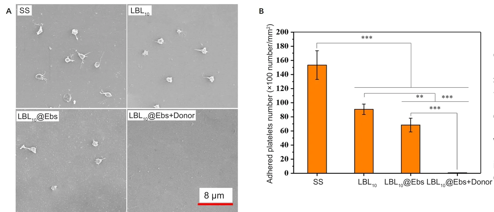
Figure7:The morphology (A) and number of the adhered platelets (B)on different samples.

Figure8:SMCs proliferation behavior of LBL10 and LBL10@Ebs.
DISCUSSION
The key function of native endothelium is to secrete functional agent,including nitric oxide (NO),prostacyclin (PGI2),antithrombin III and membrane-bound species (heparan sulfate).These functional components can effectively inhibit thrombus formation,and suppress excessive intima hyperplasia.Thus,developing multifunctional coatings that can mimic the endothelium function would make sense in the whole reendothelialization process.In this study,heparin,a clinically used anti-coagulant was adopted as a derivate of endothelium secreted heparan sulfate.The nitric oxide generation,which also means thein situcontinuous release of nitric oxide,was simulated.How to combine those two functional mimics are also the main purpose of this work.
As LBL assembly is a versatile and facile technique to address desired surfaces properties,it can be recommended to fabricate multifunctional coatings,with the assembly of polyelectrolytes,alone or in combination with drug loading.Herein,a sandwich-like LBL coating was prepared to achieve the combination of functional polyelectrolytes and loaded drugs.The polyelectrolytes were consisted of catechol-modified chitosan,phenylboronic acid modified MICs and heparin.The drug Ebs was pre-loaded into a MIC,which was functionalized with phenylboronic acid groups.The phenylboronic acid can form covalent bonding with catechol-modified chitosan at pH 7.4,so as to stabilize the LBL coating,resulting sustained release of loaded components.Within the abundant interactions inside the coating components,the loaded Ebs could present inhibited burst release and maintain sustained release till 1 month.The stability of the coating would be a potential barrier for protecting the loaded components.Therefore,the Ebs in the coating couldin-situcatalyze and decompose RSNOs to NO in the blood/material interface.The Ebs loaded coating could effectively suppress platelet adhesion/activation and SMCs proliferation,due to the synergetic effect of both heparin andin situgeneration of safe level NO.With the sustained release of Ebs,the catalytic effect of Ebs on decomposing RSNOs to continuous release NO could be expected and such MIC-embedded coating with Ebs loading showed potential for vascular material modification.
Author contributions
Concepts,design,funding support,manuscript review and guarantor:RFL,JQP and YBW;definition of intellectual content,data analysis,manuscript editing,literature research:LHL and LJ;experimental studies:LY,LHL,LJ;data acquisition:LHL,LJ;statistical analysis:LJ;manuscript preparation:LY,LJ and RFL.All authors read and approved the final version of manuscript for publication.
Conflicts of interest
None declared.
Financial support
This work was supported by the National Key Research and Development Program of China (No.2017YFB0702503,2016YFC1102200),National Natural Science Foundation of China (No.11802190),Sichuan Science and Technology Major Project of China (No.2014SZ0128,2018SZDZX0011),the 111 Project (The Program of Introducing Talents of Discipline to Universities) of China (No.B16033)and Clinical Research Award of the First Affiliated Hospital of Xi’an Jiaotong University of China (No.XJTU1AF-CRF-2015-007).
Institutional review board statement
The approval was obtained by the Institutional Review Board of the West China Hospital in Sichuan University,approval No.K2018044 on March 3,2018.
Copyright license agreement
The Copyright License Agreement has been signed by all authors before publication.
Data sharing statement
Datasets analyzed during the current study are available from the corresponding author on reasonable request.
Plagiarism check
Checked twice by iThenticate.
Peer review
Externally peer reviewed.
Open access statement
This is an open access journal,and articles are distributed under the terms of the Creative Commons Attribution-NonCommercial-ShareAlike 4.0 License,which allows others to remix,tweak,and build upon the work non-commercially,as long as appropriate credit is given and the new creations are licensed under the identical terms.
杂志排行

Medical Gas Research的其它文章
- The role of medical gas in stroke:an updated review
- Revisiting the expanded use of hyperbaric oxygen therapy for treatment of resistant migraines
- Numerical analysis of mechanical ventilation using high concentration medical gas mixtures in newborns
- Effects of nitric oxide donor N,N'-di-sec-butyl-N,N'-dinitroso-1,4-phenylenediamine on the expression of interferon-gamma in tumor infiltrating lymphocytes
- Recent developments in nitric oxide-releasing biomaterials for biomedical applications
- Nitric oxide detection methods in vitro and in vivo