Microstructures,mechanical properties,corrosion,and biocompatibility of extruded Mg-Zr-Sr-Ho alloys for biodegradable implant applications
2023-03-24FislKiniJixingLinAlirezVhidKhurrmMunirCuieWenYunngLi
Fisl Kini ,Jixing Lin ,Alirez Vhid ,Khurrm Munir ,Cuie Wen ,Yunng Li,∗
a School of Engineering,RMIT University,Melbourne,Victoria 3001,Australia
b School and Hospital of Stomatology,Wenzhou Medical University,Wenzhou 325027,China
cInstitute for Frontier Materials,Deakin University,Geelong,Victoria 3217,Australia
Abstract In this study,the microstructures,mechanical properties,corrosion behaviors,and biocompatibility of extruded magnesium-zirconiumstrontium-holmium (Mg-Zr-Sr-Ho) alloys were comprehensively investigated.The effect of different concentrations of Ho on the microstructural characteristics,tensile and compressive properties,corrosion resistance,and biocompatibility were investigated.The microstructures of the extruded Mg-1Zr-0.5Sr-xHo (x=0.5,1.5,and 4 wt.%) alloys consisted of α-Mg matrix,fin α-Zr particles,and intermetallic phase particles of Mg17Sr2 and Ho2Mg mainly distributed at the grain boundaries.Extensive {10¯12} tensile twins were observed in the partially recrystallized samples of Mg-1Zr-0.5Sr-0.5Ho and Mg-1Zr-0.5Sr-1.5Ho.Further addition of Ho to 4 wt.% resulted in a complete recrystallization due to activation of the particle stimulated nucleation around the Mg17Sr2 particles.The evolution of a rare earth (RE) texture was observed with the Ho addition,which resulted in the weakened basal and prismatic textures.Furthermore,a drastic increase of 200% in tensile elongation and 89% in compressive strain was observed with Ho addition increased from 0.5 to 4 wt%,respectively.The tension-compression yield asymmetry was significantl decreased from 0.62 for Mg-1Zr-0.5Sr-0.5Ho to 0.98 for Mg-1Zr-0.5Sr-4Ho due to the weakening of textures.Corrosion analysis of the extruded Mg-Zr-Sr-Ho alloys revealed the presence of pitting corrosion.A minimum corrosion rate of 4.98 mm y−1 was observed in Mg-1Zr-0.5Sr-0.5Ho alloy.The enhanced corrosion resistance is observed due to the presence of Ho2O3 in the surface fil which reduced galvanic effect.The formation of a stabilized surface fil due to the Ho2O3 was confirme through the electrical impedance spectroscopy and XPS analysis.An in vitro cytotoxicity assessment revealed good biocompatibility and cell adhesion in relation to SaOS2 cells.
Keywords: Mg-Zr-Sr-Ho alloy;Mechanical properties;Corrosion;Cytotoxicity;EBSD;Electrical impedance spectroscopy;Potentiodynamic polarization.
1.Introduction
High-purity magnesium (Mg) has been known for its biomedical properties and its potential as a biodegradable implant material since the late 19th century.The major shortcomings experienced during those early studies were its inferior mechanical properties and high corrosion rate (CR)bothin vitroandin vivo[1].Based on the biocompatibility,mechanical,and corrosion property requirements,a limited number of the alloying elements were recommended [2].Consequently,Mg-zirconium-strontium-holmium (Mg-Zr-Sr-Ho) and Mg-Zr-Sr alloys were developed which exhibited superior mechanical and corrosion properties along with excellent biocompatibility [3,4].
Sr is considered an osteoconductive element and showed improved bone mineralization,formation of Sr-substituted hydroxyapatite (HA),and enhanced bone growth around the implant after implantation in mice femurs and in dog femoral arteries [5,6].Sr also promoted osteoblast maturation and improved vertebral bone density [7].Formation of new bone directly adjacent to an Mg-1Zr-2Sr implant was reported 3 months after implantation in rabbits [3] and radiography of the newly formed bone showed high mineral density due to the osteointegration properties of Sr.
The addition of rare earth elements (REEs) to Mg alloys has been found to improve their mechanical and corrosion properties [8–10].In vivotests of RE-containing Mg alloy(LAE442) showed an acceptable host response and low CR[11].The cytotoxicity assessment of Mg-1Ho alloy indicated a cell viability of 96% in relation to murine calvarial preosteoblast cell line (MC3T3-E1) [12] and least hemolysis rate among the Mg-heavy REE alloys [13].Zambanini et al.[14] reported that Ho addition up to 5 wt.% to bioactive glass showed an appreciable improvement in the cell viability of osteoblast MC3T3-E1 cells and strong cytotoxicity in relation to osteosarcoma MG63 cells.Similarly,an enhanced bioactivity and a reduced cytotoxicity were observed in the Hocontaining 58S bioactive glass [15].Jiao et al.[16] confirme the cytocompatibility of the extruded Mg-6Ho-xZn (x=0,0.5 and 1.5 wt.%) alloys via bothin vivoandin vitroassessments;and the extruded Mg-Ho-Zn alloys showed higherin vivobiocompatibility than pure Mg after implantation in rabbits for 10 d.Feyerabend et al.[17] reported good biocompatibility of Mg alloys that contain heavy REEs afterin vitrotests using SaOS2 cells.Furthermore,holmium oxide nanoparticles (Ho2O3) were used in the magnetic resonance imaging and showed no toxicity at concentration ≤16 μg/mL[18].Moreover,the half-lethal dose LD50(LD50is define as the concentration capable of killing half of the seeded cells[17]) for rats is reported as 560 mg kg−1[13,19].In addition,Ho addition was reported to improve the strength and elongation of Mg-Ho-Zn alloys [20].
A recent study on extrusion of Mg-Zr-Sr showed substantial increases of 26.9% in ultimate compressive stress (σUCS)and 100% in compressive yield stress (σCYS) as compared to the as-cast conditions.Moreover,the CR significantl reduces due to favorable accumulation of the Mg17Sr2phase at the grain boundaries (GBs) [21].To examine the benefit of extrusion,the as-cast Mg-Zr-Sr-Ho alloys were extruded and their mechanical,corrosion,and biocompatibility properties were investigated in this study.Their mechanical properties were evaluated by tensile and compression tests,and the dependence of these properties on the microstructure and crystallographic texture was determined.Hydrogen evolution(HE)testing,potentiodynamic polarization (PDP) testing,and electrical impedance spectroscopy (EIS) were used to determine the corrosion behavior,while human osteoblast-like SaOS2 cells were used to assess the biocompatibility of the Mg-Zr-Sr-Ho alloys.Based on the results,the effects of extrusion and alloying elements on the microstructural characteristics and material properties have been determined.
2.Materials and methods
The actual chemical compositions of the Mg-Zr-Sr-Ho alloys were measured by inductively coupled plasma atomic emission spectroscopy (ICP-AES) (Service provided by Spectrometer Services Pty.Ltd.,Australia).Composition analysis was conducted on three samples for each alloy.Required lengths of the testing samples for microstructural characterization,and mechanical,corrosion,and cytotoxicity tests were cut from the extruded rods using electrical discharge machining (EDM).
2.1.Extrusion of Mg-Zr-Sr-Ho alloys
As-cast Mg-Zr-Sr-Ho samples were prepared under the similar conditions reported in the previous study [22].Briefl,pure Mg,Mg-30Zr,Mg-30Sr and Mg-10Ho alloys (Hunan Rare Earth Metal and Material Institute,China) were used as the master alloys in this study.To prevent iron (Fe) contamination in the molten Mg,a coated steel crucible was used during the casting.The Mg alloys were melted under an atmosphere of high purity argon.The melt was stirred for 30 min then casted at 700 °C into cylindrical steel dies with an inner diameter of 22 mm,which were preheated to 250 °C.The 22 mm diameter Mg alloy cylindrical ingots were then machined to remove the out layer to obtain cylindrical bars with a diameter of 20 mm and a length of 40 mm.The as-cast samples were not subjected to any post-heat treatment before extrusion.
A schematic of the extrusion apparatus used in this study is shown in Fig.S1 (see Supplementary information).The ram was powered using an Instron 500 kN machine under compression.A fla die with a half-angle of 90° was used for the extrusion [23].The bore of the container was 30 mm in diameter.The as-cast cylindrical bars of 20 mm in diameter and 40 mm in length were used as billets.The temperature of the billet was measured using a dummy sample,having the same composition and size as that of the billet and embedded with a thermocouple.Initially,the container was heated to the extrusion temperature of 400 °C at a heating rate of∼60 °C/min and held for approximately 3 h Subsequently,the die and spacer were inserted and maintained at 400 °C for 30 min.The billet was then placed inside the container,on top of the die and held for 10 min to homogenize the temperature.Extrusion was conducted at a constant temperature of 400 °C,a stem speed of 1 mm/s,and an extrusion ratio(ER) of 36:1.Finally,the extruded rods of 5 mm in diameter were obtained.After extrusion,the samples were air-cooled to room temperature (water quenching was not used) and were not subjected to any post-heat treatment.An equivalent plastic strain (∈eqv) of 4.181 was calculated using the formula derived from the incremental changes in configuratio of a body,during the deformation process (see supplementary information) [24–27].
2.2.Microstructural characterization
Samples for microstructural characterization were prepared with their surfaces perpendicular to the extrusion direction(ED).The phase constitutes of the Mg-Zr-Sr-Ho alloys were characterized by X-ray diffractometry(XRD,Bruker AXS D4 Endeavor).For XRD analysis,samples were scanned over the angular range of 5–90° at a step size of 0.02° using Cu-Kαradiation(λ=0.154 nm).The International center for Diffraction Data (ICDD PDF4) database was used for indexing the phases.For electron backscatter diffraction (EBSD) analysis,Mg alloy samples were cold mounted in epoxy resin and the exposed surfaces were ground up to 4000 grit SiC papers and polished using 0.04 μm colloidal silica-based oxide polishing suspension (OP-U NonDry).The EBSD data was analyzed using the software package AZtec crystal 5.1 (Oxford Instruments).For microstructural observation,the polished samples were sonicated in ethanol and then etched using a picral reagent (formulation: 3.0 ml picric acid,5 ml acetic acid,50 ml ethanol,and 10 ml distilled water).Optical microscopy (Leica DM2500M with 3.1 MP CCD) and scanning electron microscopy coupled with energy-dispersive X-ray spectroscopy (SEM-EDS) (JEOL 7200F Schottky Field Emission)were carried out to study the microstructures of the etched samples and the fractured surfaces of the samples after tensile testing.The grain size (GS) was measured using the linear-intercept method according to ASTM E122-12[28].The average GS was calculated by counting the number of grains intercepted by fi e straight lines long enough to yield at least 50 intercepts.SEM was also used to study the corroded surfaces of samples after immersion in simulated body flui (SBF) for 24 h
2.3.Mechanical testing
Cylindrical samples with a length/diameter (L/D) ratio of 2 and dogbone shaped samples with a 4 mm gauge diameter and 18 mm gauge length were cut from the extruded rods by EDM for compression and tensile tests,respectively.All samples prepared for mechanical testing were polished with SiC paper up to 800 grit to remove the surface defects.
Tensile and compression tests were undertaken in accordance with ASTM B557M-15 [29],ASTM E8/E8M-16a [30],ASTM E9-09 [31] and ASTM E09-19 [32].Both compression and tensile testing were conducted at a loading rate of 1 mm/min at room temperature using a uniaxial testing machine (Instron 5569).The stress-strain curves obtained from compression and tensile testing were used to calculate the respective yield strength,ultimate strength,and elongation and maximum strain at failure.The yield strength was calculated using the 0.2% offset method.Five samples from each alloy composition were tested to determine the averaged values.
2.4.Corrosion behavior characterization
Disc samples with 5 mm in diameter and 3 mm in thickness were prepared for corrosion testing.Four samples from each alloy composition were used for corrosion testing.The sample surfaces were mechanically ground up to 1200 grit SiC paper,polished using a 9 μm diamond suspension with felt nap mats,washed with acetone,ethanol,and distilled water,and dried in a cool airstream.Corrosion behavior of the Mg alloys was assessed using potentiodynamic polarization(PDP),electrical impedance spectroscopy(EIS),and hydrogen evolution (HE) tests in SBF at 37 °C.For corrosion testing,the SBF volume to sample’s surface area ratio was maintained as 381.97 ml/cm2in accordance to ASTM G31-72 [33].The preparation of SBF involved the addition of reagents in 1 L of pure water in the following order: NaCl (5.403 g),NaHCO3(0.504 g),Na2CO3(0.426 g),KCl (0.225 g),K2HPO43H2O(0.230 g),MgCl26H2O (0.311 g),0.2 M NaOH (100 ml),HEPES (17.892 g),Na2SO4(0.072 g),and 1.0 M NaOH(15 ml).For immersion test,the disc samples were immersed in SBF for 24 h.The corrosion rate (CR) was determined through PDP,EIS,and HE tests.Three samples for each group of alloys were tested.The PDP and EIS testing were carried out using a three-electrode cell system with a saturated calomel electrode (SCE) as the reference electrode,a platinum electrode (1.5×1.5 cm2) as the counter electrode,and the alloy samples as the working electrode.The samples were connected to a copper wire and then cold-mounted in epoxy resin leaving an exposed area of 0.785 cm2.The PDP curves were obtained with a scan range between ±5 V at a scan rate of 1.5 mV s−1using an electrochemical workstation(VSP-300,BioLogic,France).The requirement of measuring current with change in potential from the corrosion potential to +1.6 V SCE was also complied [34].The EIS was measured at a frequency range from 10 MHz to 100 kHz at 10 mV in amplitude using ZsimpWin software.The samples were immersed in SBF for 1 h in order to stabilize the OCP.The corrosion rate determined from PDP is denoted as CRi,which is calculated from the PDP curves using the equation given in ASTM G102-89 [35]:
where CRiis the corrosion rate(mm y−1),icorris the corrosion current density (μAcm−2),ρis the density of the sample(gcm−3),K1is a constant (3.27×10⁻³),and EW is the alloy equivalent weight (dimensionless).
All HE tests were conducted in SBF at 37 °C using an inverted funnel as described in previous studies [3,36].The instantaneous hydrogen evolution rateVH(ml cm−2d−1) is determined by the slope of the volume of evolved hydrogen versus time.The corrosion rate determined by HE tested is denoted CRh,which is related to the hydrogen evolution rate given by [37]:
2.5.Biocompatibility assessment
The biocompatibility of the Mg alloys was assessedin vitrousing osteoblast-like cells,i.e.the human SaOS2 osteosarcoma cell line exhibiting osteoblastic properties [38].The SaOS2 cells were cultured in modifie minimal essential medium (MMEM) at 37 °C in a humidifie atmosphere composed of 5% CO2in air for 72 h.Biocompatibility tests were carried out in accordance with the indirect contact method as described in previous studies [3,39].Briefl,extracts were prepared for each Mg alloy specimen using MMEM,with a surface area/MMEM ratio of 0.8 cm2ml−1in a humidifieatmosphere of 5% CO2at 37 °C for 72 h After that,the extracts were collected and filte-sterilized with a 0.22 μm filte (Falcon,BD Biosciences,USA).The control was prepared in the same way by incubation of MMEM without the addition of the Mg alloy samples.All extracts from the Mg alloys and the control were supplemented with 10% fetal bovine serum (FBS) (Bovogen Biologicals,Australia) and 1%penicillin-streptomycin (Gibco,Life Technologies,Australia).The SaOS2 cells were seeded into a 48-well plate at a density of 10,000 cells per well with 400 μL of supplemented MMEM and then incubated for 24 h to allow cell attachment on the plate.Then the supplemented MMEM was replaced with 400 μL of the respective specimen extracts and control supplemented with 10% FBS and 1% penicillin-streptomycin.After cell culturing for 3 and 5 d,the cytotoxicity of the Mg alloys was assessed using 3-(4,5-dimethylthiazol-2-yl)−5-(3-carboxymethoxyphenyl)−2-(4-sulfophenyl)−2H-tetrazolium(MTS) (CellTiter® Aqueous One Solution,Wisconsin,UA).In brief,the supplemented extracts and control in each well were removed and the wells were washed three times with phosphate buffered saline (PBS).Then 300 μL phenol red free RPMI 1640 medium (Gibco,Life Technologies,Australia) was added to each well.After that,100 μL MTS solution was added to each well and the plate was incubated for 1 h at 37 °C.After incubation,100 μL of solution from each well was transferred into a 96-well plate.The optical absorbance was measured using a microplate reader(FlexStation 3 Microplate Reader,Waltham,CA,USA) at 490 nm.The viable cell counts were calculated according to the optical absorbance.Three samples from each alloy were tested for cytotoxicity assessment.
For cell adhesion studies,Mg alloy discs were placed in 48-well plates and each well was seeded with 200 μL of a SaOS2 suspension in MMEM (10,000 cells.ml−1) and incubated at 37 °C in humidifie air with 5% CO2for 3 d After incubation,the Mg alloy discs with the attached cells were fi ed with 3.9% (w/v) glutaraldehyde solution (Sigma Aldrich,Australia) for 2 h at room temperature and then rinsed three times with PBS,followed by progressive dehydration in ethanol/distilled water gradients (50,60,70,80,90,95 and 100%) for 10 min each.The Mg alloy discs were then chemically dried in hexamethyldisilazane (HMDS)reagent (Sigma Aldrich,Australia) for 1 h.Finally,the disc samples were sputter-coated with iridium(approx.6 nm thickness) using a sputter coater (LEICA EM ACE600) to make the samples ready for SEM observation.Adherence of SaOS2 cells on the surfaces of Mg alloy discs were imaged using SEM (FEI Nova NanoSEM).
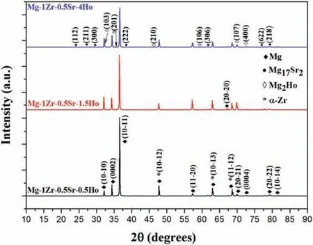
Fig.1.XRD spectra of the extruded Mg-Zr-Sr-Ho alloys.
2.6.Statistical analysis
Experimental data are expressed as mean ± standard deviation.A one-way analysis of variance (SPSS Inc.,USA)was used to determine the statistical significanc of the differences observed between groups.P <0.05 was accepted as statistically significant
3.Results and discussion
3.1.Chemical compositions and XRD spectra of extruded Mg-Zr-Sr-Ho alloys
The chemical compositions of the extruded Mg-1Zr-0.5SrxHo (x=0.5,1.5 and 4 wt.%) alloys measured by ICP-AES is given in Table 1.The iron (Fe) concentration of maximum 30 ppm (ppm=wt.%×10,000) [40] was observed in Mg-1Zr-0.5Sr-0.5Ho alloy,which was well below the tolerance limit of 170 ppm,above which precipitation of the Fe-rich body-centered cubic (bcc) phase was observed [41,42].Furthermore,removal of Fe content from the Mg melt through Zr addition is also well reported [41,43].Based on the corrosion resistance,the tolerance limit of impurities in Mg was suggested by Cramer et al.[44].The concentrations of aluminum(Al),manganese (Mn),zinc (Zn) and copper (Cu) shown in Table 1 were found well below the proposed threshold values,as beyond these limits,a deleterious effect on the CRs was observed in binary Mg alloys [45].Zr and Fe exhibited a mutual solubility in the molten Mg and readily form Fe-Zr intermetallic phases which generally settle down in the melt if allowed [46].Nonetheless,complete removal of the intermetallic phases and undissolved Zr was unlikely,as stirring was conducted in a coated steel crucible during casting,in order to break down the undissolved Zr particles suspended in the melt and to attain a homogenous distribution [47,48].
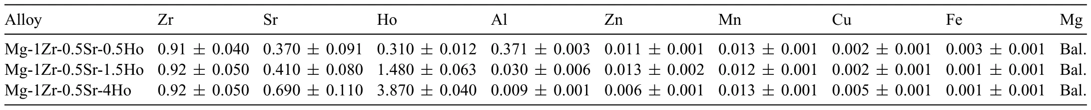
Table 1 Chemical composition of extruded Mg-Zr-Sr-Ho alloys measured from ICP-AES.All compositions are given in wt.%.
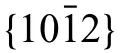
Table 2 Corrosion properties of extruded Mg-Zr-Sr-Ho alloys obtained from PDP curves.
The XRD spectra of extruded Mg-Zr-Sr-Ho alloys is given in Fig.1.The spectra revealed the presence ofα-Mg,Mg17Sr2,Mg2Ho andα-Zr.Low intensity peaks of Mg2Ho were observed especially in Mg-1Zr-0.5Sr-4Ho.Moreover,with an increase in Ho content to 4 wt.%,an increase in the peak intensities of Mg17Sr2was noted (Mg-1Zr-0.5Sr-4Ho).This was accompanied with an overall decrease in the intensities of Mg peaks.
The solubility limits of Sr (bcc),Zr (hexagonal closepacked) (hcp) and Ho (hcp) inα-Mg (hcp) were reported as 0.11,3.8 and 28.08 wt.%,respectively [49].Furthermore,the atomic radii calculated from the self-consistent fiel method were reported as 1.7435,1.6421,1.5803 and 1.4400[50] and the electronegativity values calculated with Iodine(I) were reported as 0.95,1.26,1.23 and 1.30 for Sr,Zr,Ho,and Mg,respectively [51].The solubilities of REEs in Mg showed a general correspondence with their crystal structures,atomic/ionic radii,and electronegativity values [52,53].Briefl,a similar crystal structure and atomic radius close to the Mg,large interplane distance of the Mg-RE solid solution,and a small electronegativity difference,enhance the solubility of the REEs in Mg.Consequently,the larger solubility of Ho in Mg could be attributed to its more suitable chemical properties than Sr and Zr.
As per the thermodynamics and binary phase diagrams,α-Mg,Mg17Sr2(hcp),α-Zr (hcp) and Mg24Ho5(bcc) were the expected phases in the Mg-1Zr-0.5Sr-xHo (x=0.5 to 4 wt.%) alloys [54–56].Both Mg and Zr have hcp structure and therefore exhibit overlapping of peaks in the XRD patterns [57].However,instead of the expected Mg24Ho5phase,Mg2Ho was detected in the XRD spectra.The Mg2Ho phase forms with a peritectic reaction at 695 °C and exhibit hexagonal,MgZn2-type structure.It is worth noting that the casting of the Mg-Zr-Sr-Ho alloys was conducted at 700 °C.Furthermore,in case of heavy REEs,i.e.yttrium (Y) subgroup -Y to lutetium (Lu),the formation of Mg2RE compounds was reported [56].A comparison of the standard Gibbs free energies of phase formation calculated for Mg-REs at 700 °C revealed Mg2RE as a more favorable phase than Mg24RE5,mainly due to its low entropy of formation [58].On the other hand,the most negative heat of formation among all the secondary phases in Mg-Zr-Sr-Ho was reported as −5.72 kJ/atom for Mg17Sr2[59].Consequently,formation of the Mg17Sr2in Mg-Zr-Sr-Ho alloys was anticipated because of its unfavorable thermodynamic potential and the low solubility of Sr in Mg.
3.2.Optical microstructures of extruded Mg-Zr-Sr-Ho alloys
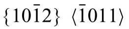
Optical micrographs of extruded Mg-Zr-Sr-Ho alloys are shown in Fig.2.Extensive twinning and heavily deformed grains with interdendritic regions were observed in Mg-1Zr-0.5Sr-0.5Ho and Mg-1Zr-0.5Sr-1.5Ho,thereby,clearly depicting a lack of recrystallization in these alloys.Moreover,most of the twins were contained within the grains and terminated at the GBs or at second phase particles as indicated with red arrows in Fig.2(a-2) and (b-2).On the other hand,the Mg-1Zr-0.5Sr-4Ho alloy exhibited equiaxed grains without presence of any twins,thus,signifying the manifestation of a complete recrystallization.An increase in the secondary phase particles at GBs was observed with Ho addition in Mg-1Zr-0.5Sr-xHo (x from 0.5 to 4 wt.%).Resultantly,the GBs in Mg-1Zr-0.5Sr-4Ho alloy were comparatively thickened and heavily occupied by the secondary phase particles(Fig.2c).The secondary phases observed in the micrographs were mainly comprised of dark-colored and relatively lightcolored particles,as indicated with black and white arrows,respectively,in Fig.2(c-2).
As evident from the optical micrographs in Fig.2,an increase in volume fraction of the recrystallized grains was observed with the Ho addition to 4 wt.%.This behavior is generally in contrast to the reported observations that REE addition in Mg alloys suppressed the dynamic recrystallization(DRX)process due to the solute drag effect[74].Nonetheless,Hadron et al.[75] suggested that the Y addition caused an alteration from the basal slip to a prismatic cross-slip in Mg-Y alloy.The transition to a non-basal slip mechanism promoted the continuous dynamic recrystallization (CDRX) which induced a change in the texture.On the other hand,DRX induced by Sr and RE-rich particles in Mg alloys is also well reported [76,77].Furthermore,it was noted that enthalpy of mixing at the respective compositions for Mg-Ho alloys was observed more negative than that of Mg-Sr and Mg-Zr alloys[54,61,78].Therefore,it can be deduced that addition of Ho in Mg-Zr-Sr-Ho alloy would promote segregation of the Sr during casting to form an intermetallic Mg17Sr2phase due to its low solubility and unfavorable thermodynamic potential.This was substantiated with an increase in the dark-colored Mg17Sr2and light-colored Mg2Ho particles in the extruded Mg-1Zr-0.5Sr-4Ho alloy (Fig.2(c-2)).Hence,an increase in the Sr and RE-rich particles could promote particle-stimulated nucleation (PSN),which led to an enhanced DRX.
3.3.SEM microstructures and EDS composition maps of extruded Mg-Zr-Sr-Ho alloys

Fig.3.SEM micrographs and EDS maps of extruded Mg-Zr-Sr-Ho alloys: (a) Mg-1Zr-0.5Sr-0.5Ho;(b) Mg-1Zr-0.5Sr-1.5Ho;(c-1) Mg-1Zr-0.5Sr-4Ho;and(c-2) SEM image and EDS of the magnifie region of Mg-1Zr-0.5Sr-4Ho in (c-1).Respective elemental compositions in wt.% calculated from the EDS are given in the maps.Images were taken from surfaces perpendicular to ED.Images collected using backscatter electron imaging technique are indicated as BSE image.
SEM images and EDS composition maps of extruded Mg-Zr-Sr-Ho alloys are shown in Fig.3.Similar to the observations of optical microscopy (Fig.2),SEM micrographs of the extruded Mg-1Zr-0.5Sr-0.5Ho and Mg-1Zr-0.5Sr-1.5Ho alloys also revealed the presence of irregular-shaped and deformed grains (Fig.3a and b),whereas DRX grains with thickened GBs and a bimodal grain-size distribution were observed in the extruded Mg-1Zr-0.5Sr-4Ho (Fig.3(c-1)).The size of the large grains ranged from 4.5 to 10.5 μm,while the smallsized grains were measured around 1.5 ± 1.0 μm (the size of a few selected grains measured using SEM software is shown in Fig.3(c-1)).In addition,presence of small-sized grains around the Mg17Sr2particles were observed,which could be attributed to the PSN (regions highlighted with blue circles in Fig.3(c-1)).The distribution of the alloying elements investigated via EDS mapping revealed inhomogeneous dispersedα-Zr particles in Mg matrix,while Mg17Sr2phase was observed mainly accumulated at the GBs.Moreover,the number ofα-Zr and Mg17Sr2particles were found increased with the addition of Ho in Mg-1Zr-0.5Sr-xHo (x from 0.5 to 4 wt.%).The highly soluble Ho element was found homogeneously distributed within the Mg matrix;hence,only a very few Ho-rich particles were observed.Furthermore,a slight segregation of Ho within the matrix was observed in Mg-1Zr-0.5Sr-4Ho alloy (EDS map of Ho in Fig.3c).
The increase in DRX with addition of REEs in Mg could be attributed either to the RE segregation/RE solute clustering [79,80] or from PSN [81,82].The segregation of REEs at GBs and solute clustering within the matrix is well reported in Mg-RE alloys [79,83].A retarding force exerted by the secondary phase particles on the migrating GBs,strongly influence the recovery,grain growth and recrystallization kinetics,as described by the Zener drag effect [84].The boundary drag produced due to the segregation of REEs in Mg-Gd alloy showed a direct effect on recrystallization and grain growth behavior [85].In addition,the RE clusters were observed to interact with the dislocation slip and altered the basal slip to a prismatic cross-slip mechanism,thereby,modifying the texture in Mg-Y alloy [75].However,at extrusion temperatures of 400 °C for Mg-Zr-Sr-Ho alloys,the Zener drag would be reduced due to a higher GB mobility and dissolution of the fin RE-rich precipitates,thus,enhancing the DRX [86].
Notwithstanding the above,large-sized Sr and RE-rich particles (≥1 μm) are well reported for promoting the DRX through PSN [81].However,a low growth kinetics of the PSN grains was explained on the basis of an early impingement of the growing grains within a PSN cluster,more severe effect of the precipitates leading to a higher critical Gibbs-Thompson diameter and their random orientation [87].Thus,difference between the growth kinetics of conventional DRX and PSN induced recrystallized grains could produce a bimodal recrystallized microstructure [88,89],as observed in Mg-1Zr-0.5Sr-4Ho (Fig.3(c-1)).
3.4.EBSD crystallographic characteristics of extruded Mg-Zr-Sr-Ho alloys


Fig.4.Orientation maps,PFs,IPFs,and in situ EDS maps collected during EBSD analysis of extruded alloys: (a) Mg-1Zr-0.5Sr-0.5Ho;(b) Mg-1Zr-0.5Sr-1.5Ho;and (c) Mg-1Zr-0.5Sr-4Ho.The special boundaries having a rotation angle of 86° about 1¯210 axis and representing the tensile twins are delineated with the red color in Fig.4a and b [91].The boundaries in IPF maps marked with white color indicates low-angle grain boundaries (LAGBs) (2°–15°),whereas black colored boundaries signify high-angle grain boundaries (HAGBs) ≥15°.Selected DDRX and PSN locations are marked with black,and white circles,respectively.The IPF maps of the surfaces shown are perpendicular to the ED.Nb: Due to the cylindrical symmetry of the specimen,both RD and ND are geometrically equivalent and are perpendicular to ED (i.e.,the normal in the PFs).
The DDRX mechanism in Mg alloys is facilitated by the low SFE of basal planes that restricts the dynamic recovery due to limited cross-slip and thus promotes the nucleation of the DDRX grains [110].The low SFE implies a large stacking fault width which inhibit the cross-slip or dislocation climb,thus increases the stored strain energy (SSE) and promotes the DDRX [111–113].Furthermore,the influenc of the extrusion parameters on DDRX is also well studied,as the processing temperatures ≥300 °C and low strain rates were reported to promote DDRX in Mg alloys [114].Meanwhile,PSN mainly depends upon the particle size and volume fraction,and the strain gradient across the particle-matrix interface,due to the dislocation density.Initially,the subgrains having LAGBs are formed in the deformation zone,which then readily transforms to HAGBs under the deformation strain.The nucleated PSN grain are randomly oriented and grows till the deformation zone is consumed [115].The role of the intermetallic Mg-Sr in PSN leading to texture weakening is also well-reported [116,117].Furthermore,as applicable to this study,at extrusion temperature of 400 °C and Sr content of 0.5 wt.%,the PSN was suggested as a favorable mechanism in AZ31 alloy [118].Consequently,it can be deduced that the RE addition caused an increase in Mg17Sr2phase,which augmented the PSN in Mg-1Zr-0.5Sr-4Ho and contributed to the observed weakening of the texture.
The broadening of the basal pole orientations around the RD/ND directions(also termed as‘basal pole split’)manifests the evolution of a RE texture by addition of the REEs in Mg alloys[119].Furthermore,the basal pole splitting is characterized by the reorientation of the{0001}poles between 35nd 45° from ND towards TD [120–122] and was observed to be more influence by the heavy REEs due to their higher solute drag on the GB mobility [120,123].The basal pole splitting observed at 45.27° from RD towards ND in the extruded Mg-1Zr-0.5Sr-0.5Ho is indicated in the PF shown in Fig.4a.Correspondingly,a fully developed RE texture in the IPFs showed theparallel to the ED,whereas the peak intensity was located at 27° fromand exhibited a weakened intensity of ∼3 MRD from the edge of the IPF [98].The evolution of the RE texture that promotes the weakening of an overall texture,was found to be highly influence by the REE addition in Mg alloys [124].Depending upon the REE addition,a minimum content was required to initiate the formation of the RE texture,which weakened the overall texture intensity with further REE addition [125].The RE texture evolution was attributed to the changes in SFEs,and interaction of the RE solutes with the dislocations and boundaries[126,127].As the mobility of the dislocations and GBs was retarded by a large-sized REE and a high solute segregation at the GBs,respectively [125].The increase in the solute segregation which led to the development of the RE textures in the Mg-Zr-Sr-Ho alloys is apparent in the EDS maps of Ho shown in Fig.3.It could be further corroborated with an observed decrease in the texture intensities with an evolving RE texture (PFs and IPFs in Fig.4).Overall,with an addition of Ho,the basal fibe was maintained with the formation of a RE component,that led to the texture weakening by dispersion of {0001} poles between the RD/ND,while tilting towards the ED.Nonetheless,the contribution from PSN in randomizing the texture was also significant
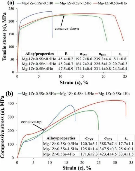
Fig.5.Tensile and compressive properties of extruded Mg-Zr-Sr-Ho alloys:(a) tensile stress-strain curves and tabulated Elastic Modulus (E,GPa),ultimate tensile strength (σUTS,MPa),tensile yield strength (σTYS,MPa),and tensile elongation (εt,%));and (b) compressive stress-strain curves and tabulated ultimate compressive strength (σUCS,MPa),compressive yield strength(σCYS,MPa),and compressive strain (εc,%).NB: ED and loading axis coincide with specimen longitudinal axis.The axis of loading is aligned with ED.
3.5.Mechanical properties of extruded Mg-Zr-Sr-Ho alloys
The tensile and compressive stress-strain curves along with the tabulated mechanical properties of extruded Mg-Zr-Sr-Ho alloys are shown in Fig.5.The average elastic modulus(E)of the extruded Mg-Zr-Sr-Ho alloys calculated from the stressstrain curves was 46.1 ± 1.1 GPa,which is comparable to that of the pure Mg and its alloys ≈45 GPa [128].The Mg alloys showed elastic modulus closer to that of natural bone(3–20 GPa) and are therefore more suitable in mitigating the stress shielding effect as compared to the conventional permanent metal implants (such as stainless steels and titanium alloys) [129].Firstly,with an increase in Ho content from 0.5 to 4 wt.%,similar trends in the tensile elongationεtand compressive strainεcwere observed;as both properties showed an increase of 200% and 89%,respectively.Furthermore,a substantial increase in ductility was achieved at the expense of a marginal 3.3% decrease in the ultimate tensile strength (σUTS) and 9.6% in the tensile yield strength (σTYS)(Fig.5a).Conversely,the improvement in malleability was accompanied by a significan increase of 8.9% in ultimate compressive strength (σUCS) and 42.6% in compressive yield strength (σCYS) (Fig.5b).
Secondly,the tension-compression yield asymmetry(σCYS/σTYS) was significantl reduced from 0.62 for Mg-1Zr-0.5Sr-0.5Ho to 0.98 for Mg-1Zr-0.5Sr-4Ho.This anisotropic behavior between theσCYSandσTYSwas found to be highly influence by the texture evolution with the REE addition[130].Work hardening during the compression was observed to be more pronounced than in the tension loading.Overall,Mg-1Zr-0.5Sr-4Ho exhibited superior mechanical properties among the Mg-1Zr-0.5Sr-xHo(x=0.5–4.0 wt.%)alloys,with the largestεtandεcof 24.3 and 33.4%,and highestσUCSandσCYSof 423.4 and 171.6 MPa,respectively.Besides,Mg-1Zr-0.5Sr-4Ho also showed a decentσUTSof 231.1 MPa andσTYSof 174.1 MPa[131],which were slightly lower than the maximumσUTSof 239.2 andσTYSof 192.7 MPa in Mg-1Zr-0.5Sr-0.5Ho alloy(Fig.5a).No apparent correlation between the GS andσTYSwas observed,as the largest GS 12.46 ± 1.33 μm observed in Mg-1Zr-0.5Sr-0.5Ho exhibited the highestσTYSof 192.7 MPa,thus undermining the Hall-Petch relationship[21].Nevertheless,the strong influenc of the texture onσTYSalso cannot be disregarded [132].
The improvement in the mechanical properties from RE addition in Mg-RE alloys is dependent upon the grain size,texture,second phase particles,and strain hardening [133].A weakened texture was reported to enhance theεin extruded Mg-0.2Ce alloy [134].Extruded Mg-0.5Ca exhibited increasedσTYSandσUTSdue to grain refinemen and precipitation strengthening,and a largerεdue to the weakened texture;however,a further increase in Ca addition to 2.0 wt.%showed an insignifican effect on the strength but caused a drastic decrease inεdue to the re-strengthening of the texture and possible cracking at the increased secondary particles/matrix interfaces [133].A large increase in theεtandεcwith increasing Ho addition suggested a strong effect of the RE texture on the plastic deformation,as the highestεtof 24.3% andεcof 33.4% was observed in Mg-1Zr-0.5Sr-4Ho,which exhibited a fully developed RE texture as shown in the PFs and IPFs (Fig.4c).The orientation spread of the basal poles in a RE-textured microstructure is between the RD and ND,which increased the presence of {0001} planes,oriented such that the basal 〈a〉 slip is promoted [135,136].The shape of the curves in the tension (concave-down) and compression (concave-up) signifie that the deformation through slip was dominant in tension,while twinning was the prevalent deformation mode in compression [137].A tilt of thecaxis between 70° and 90° from the compression direction favored the extension twinning and enhanced the strain-hardening rate in Mg-RE alloys [138,139].
Furthermore,with Ho addition,the activation of the basal slip with the evolution of the RE texture is corroborated with the decrease in average Schmidt Factor (m) for the extension twins[140],i.e.,from 0.46 in Mg-1Zr-0.5Sr-0.5Ho to 0.41 in Mg-1Zr-0.5Sr-4Ho,respectively.Conversely,mfor basal planes remained unchanged at 0.42.This illustrates that with the reorientation of the basal poles,due to the RE texture,tensile loading in the ED is unfavorable for the extension twinning [98].Hence,the competing basal slip or the additional pyramidal slip systems,could be activated and significantl enhanced the ductility in extruded Mg-Zr-Sr-Ho alloys [140,141].Moreover,a decrease in the tension-compression yield asymmetry with the Ho addition,is attributed to the increase inσCYS,as increase in the extension twinning would enhance the strain hardening,during the compressive loading along the ED [103,139].
The individual contributions and combined effect of GS,precipitates,texture,and solid strengthening on the tensile and compressive strengths of Mg alloys were evaluated.The GS showed no correlation with theσTYS,as the decrease in GS with Ho addition from 0.5 to 1.5 wt.% showed a decreasedσTYS,whereas further Ho addition to 4 wt.% slightly increased theσTYS.However,in case ofσCYS,the Hall-Petch relationship was found valid.It may be noted that theσoandkin the Hall-Petch relation,i.e.,σTYS=σo+kd−1/2manifests the CRSS for easy slip system in the grain volume and difficul slip/twinning systems near the GBs,respectively[142].This implies that the Hall-Petch relation is texture dependent,as activation of the deformation modes based on the CRSS is influence by the crystallographic orientation of the sample.The non-dependence of theσTYSon GS is attributed to the easy activation of the twinning during tensile loading,as with an increase in Ho addition,the basal poles were observed moving/tilting towards the ED (PFs in Fig.4).Thus,in spite of the decrease in GS,reduction in theσTYSwas observed.On contrary,generation of the extension twins with Ho addition became unfavorable during the compression loading,thusσCYSwas observed obeying the Hall-Petch relation[143].
To evaluate the solid solution strengthening from Ho addition,the microstructures and yield strengths were compared with the previously studied extruded Mg-1Zr-0.5Sr alloy [21].The extruded Mg-1Zr-0.5Sr and Mg-1Zr-0.5Sr-0.5Ho showed GS of 3.42 and 12.46 μm,respectively.However,both alloys exhibited similar microstructures,i.e.,heavily deformed grains with Zr precipitates.In addition,Ho was mainly observed as a solid solution in the Mg-1Zr-0.5Sr-0.5Ho,as negligible Ho precipitates were observed due to its high solubility and low content (see EDS map of Ho in Fig.3a).The extruded Mg-1Zr-0.5Sr alloy exhibited a Hall-Petch relation with a linear fi ofσTYS=112.8+198.8d−1/2[21],thusσTYSof 169.1 MPa was calculated for the GS of 12.46 μm.On the other hand,the extruded Mg-1Zr-0.5Sr-0.5Ho showed aσTYSof 192.7 MPa,thereby confirmin the contribution of 23.6 MPa from 0.5 wt.% Ho addition to the solid solution strengthening.Similarly,significan effects of REEs on the solid solution strengthening of Mg alloys were also reported for Gd and Y[144].Furthermore,theσCYSof extruded Mg-1Zr-0.5Sr and Mg-1Zr-0.5Sr-0.5Ho alloys were noted as 145.5 and 120.3 MPa,respectively.The extruded Mg-1Zr-0.5Sr-0.5Ho alloy exhibited the Hall-Petch relation with a linear fi ofσCYS=38.6+274.1d−1/2,thusσCYSof 186.7 MPa was calculated for the GS of 3.42 μm.This implies that with the same GS,0.5 wt.% Ho addition resulted in a solid solution strengthening of 41.2 MPa.
On the other hand,the EDS maps of Ho in Figs.3 and 4 showed only minor precipitation of Ho-rich particles,mainly due to the high solubility of Ho in the Mg matrix.Therefore,it is deduced that the contribution from Ho addition to the precipitation strengthening remained insignifican in the extruded Mg-1Zr-0.5Sr-xHo (x=0.5,1.5 and 4 wt.%).
A comparison of the tensile properties with those of the extruded pure Mg revealed an increase of 29.3% inσTYS,24.2%inσUTSand 61.0% inεt;similarly,a comparison of the compressive properties revealed an increase of 35.8% inσCYSand 58.6% inσUCS,with an insignifican change inεc[90].Furthermore,a comparison with the previously studied as-cast Mg-Zr-Sr-Ho alloy [145] revealed an increase of 32.3% inσUCSand 58.0% inσCYSwith negligible effect onεc.A comparison of the microstructural characteristics reveals that the GS of the as-cast Mg-Zr-Sr-Ho alloys ranged between 20 and 80 μm.In contrast,the extruded alloys exhibited a significan fine GS of 4 to 12 μm.Furthermore,the as-cast Mg-Zr-Sr-Ho showed non-equiaxed grains with a dendritic structure,whereas equiaxed grain morphology was observed in the extruded Mg-1Zr-0.5Sr-4Ho alloy (Figs.3c and 4c).Thus,a decrease in the GS and formation of a uniform equiaxed grains in the extruded Mg-Zr-Sr-Ho alloys caused a significan increase in the strength and ductility.This clearly demonstrates that the extrusion indeed enhanced the mechanical properties of Mg-Zr-Sr-Ho alloys.A further comparison of the compressive properties between the extruded Mg-1Zr-0.5Sr [21] and Mg-1Zr-0.5Sr-4Ho showed an increase of 46.5% inσUCS,22.5% inσCYS,and an elongation of 147.4%.Moreover,comparison of the tensile properties showed an increase of 292.9% in ductility at the expense of a marginal decrease of 10.7% inσUTS.Consequently,it is deduced that the Ho addition significantl enhances the compressive strength and ductility of extruded Mg-Zr-Sr alloys.
3.6.Tensile fractography of extruded Mg-Zr-Sr-Ho alloys
The SEM micrographs of the tensile fractured surfaces of extruded Mg-Zr-Sr-Ho alloys is shown in Fig.6.The Mg-1Zr-0.5Sr-0.5Ho having minimumεtof 8.1% exhibited limited number of dimples,which is a characteristic feature of the ductility [146].A large number of microcracks were observed at the GBs and/or particle-matrix interface,as indicated with the blue ellipses in Fig.6(a-2).In addition,the particles exhibited cleavage facets at a few locations.With an increase in Ho addition,Mg-1Zr-0.5Sr-1.5Ho and Mg-1Zr-0.5Sr-4Ho showed increased number of dimples,which manifests the largeεtof 20.77 and 24.3%,respectively (Fig.6b and c).Furthermore,large-sized cracks were observed in regions enriched with the Sr content,as also revealed by the insert of EDS map in Fig.6(b-1).All the microvoid sites,i.e.,dimples were embedded with Ho-rich particles,thereby signifying the contribution of the Ho addition in enhancing the ductility of Mg-1Zr-0.5Sr-1.5Ho and Mg-1Zr-0.5Sr-4Ho alloys (Fig.6(b-2) and (c-2)).Moreover,the microvoids were found deepened and the cleavage facets as observed in the Mg-1Zr-0.5Sr-0.5Ho alloy were not present.
3.7.HE of extruded Mg-Zr-Sr-Ho alloys
HE curves and their corresponding average CRh(mm y−1)of extruded Mg-Zr-Sr-Ho alloys are given in Fig.7.The CRhof 5.70±0.97,10.91 ±0.35 and 12.36 ±0.46 mm y−1were measured forx=0.5,1.5,and 4.0 wt.% in Mg-1Zr-0.5SrxHo,respectively.Both HE curves of Mg-1Zr-0.5Sr-0.5Ho and Mg-1Zr-0.5Sr-4Ho showed similar trends after 25 h immersion in SBF,along with the distinct pit initiation point at around 150 h (indicated with black arrows in Fig.7).However,a drastic increase in H2production was observed for Mg-1Zr-0.5Sr-4Ho between 10 and 25 h immersion in SBF(highlighted with a circle in Fig.7).Whereas,the HE curve of Mg-1Zr-0.5Sr-1.5Ho alloy initially showed a sharp rise in the H2production,thereafter the slope of the curve was consistently decreased till completion of the experiment.Consequently,an inflectio point of pit initiation is not apparent in the HE curve of Mg-1Zr-0.5Sr-1.5Ho alloy (Fig.7).
Fig.6.SEM images of tensile fractured surfaces of extruded Mg-Zr-Sr-Ho alloys: (a) Mg-1ZSr-0.5Sr-0.5Ho;(b) Mg-1ZSr-0.5Sr-1.5Ho,and (c) Mg-1ZSr-0.5Sr-4Ho.
The SEM micrographs of the corroded sample surfaces after 24 h immersion in SBF are shown in Fig.8.Pitting was observed in all samples,as indicated with the red arrows in Fig.8.The maximum CRhof 12.36 mm y−1was observed in Mg-1Zr-0.5Sr-4Ho,which is also depicted by a drastic increase in dissolution between 10 and 25 h immersion and a large slope after the pitting initiation (Fig.7),conforming to the increased number of pit holes shown in Fig.8c.The drastic increase in H2production after 10 h immersion implies the initial breakdown of the newly developed surface fil and is attributed to the high galvanic effect in the Mg-1Zr-0.5Sr-4Ho alloy.However,as the corrosion proceeded,a stabilized Ho-rich surface fil formed till its breakdown at 150 h immersion in SBF.The crater at location X in Fig.8a,was probably formed by the unintended removal of the corroded surface during the sample handling.Nonetheless,the surface condition at a depth of approximately 133 μm (measured via SEM software),revealed a robust surface in a corrosion-free condition.This indicates that the Mg-1Zr-0.5Sr-0.5Ho alloy exhibiting the least CRhof 5.7 mm y−1is intrinsically more corrosion resistant than the other two compositions (Fig.7).Lastly,the Mg-1Zr-0.5Sr-1.5Ho alloy showed a more compact surface fil with less cracking.However,the island-like structures developed from the pitting corrosion were observed in the Mg-1Zr-0.5Sr-1.5Ho alloy,as indicated with ‘Y’ at a few selected locations in Fig.8b.
Fig.7.HE curves of extruded Mg-Zr-Sr-Ho alloys in SBF at 37 °C.Average CRh of each alloy,calculated from the volume of hydrogen gas evolved,are also shown.
The effect of the REE addition on the corrosion of Mg alloys was mainly attributed to the formation of the intermetallic Mg-RE phases,which increased the galvanic corrosion[9,152].Conversely,Chang et al.[153] reported a decrease in CR with an RE addition until the precipitation occurred in the Mg-RE alloys i.e.,within the solid solution range.Furthermore,the formation of RE-oxides in the inner layers of the corrosion fil also reduced the CRs in Mg-RE alloys mainly due to the formation of the more stabilized passive layers [152,154].An increase in the CRhwith the Ho addition to 4 wt.% in Mg-1Zr-0.5Sr-4Ho is considered due to the increase in the non-homogenously distributed secondary phase particles,i.e.,Mg17Sr2,α-Zr and Mg2Ho phases (EDS maps in Fig.3c) [152,155].
Fig.8.SEM micrographs of the corroded surfaces after immersion in SBF for 24 h at 37 °C: (a) Mg-1Zr-0.5Sr-0.5Ho,(b) Mg-1Zr-0.5Sr-1.5Ho,and (c)Mg-1Zr-0.5Sr-4Ho.Selected pitting locations are indicated with the red arrows.
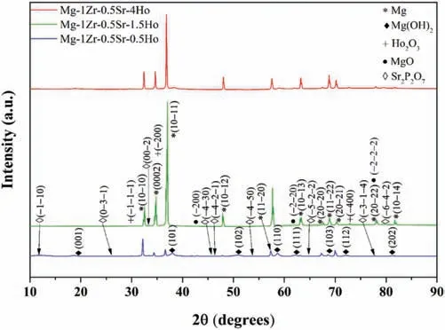
Fig.9.XRD spectra of corroded surfaces of extruded Mg-Zr-Sr-Ho alloys after immersion in SBF for 24 h at 37 °C.
The formation of a stabilized protection fil of the RE oxides at 500 °C was reported in Mg-3Y and Mg-3Sc [154].Furthermore,the outer layers of the film were mainly comprised of the RE oxides,which enhanced the corrosion resistance and were thermodynamically more favorable to form than the MgO at 500 °C [156].Nonetheless,the deterioration in corrosion resistance with REE addition cannot be overlooked,as with an increase in the intermetallic Mg-RE phase,contribution from the galvanic corrosion is enhanced[8,9].Alternately,an RE addition within the solid solution range is suggested to be beneficia [153,157].Consequently,it is deduced that a strong fil passivation in Mg-1Zr-0.5Sr-0.5Ho was formed,which comprised of Ho oxides/hydroxides near the outer layers and hence resulted in the minimum CRh.Further increase in Ho addition in Mg-1Zr-0.5Sr-4Ho worsened its corrosion resistance due to the increased intermetallic Mg2Ho phase.
3.8.Characterization of corrosion products
The XRD spectra of the corroded surfaces of extruded Mg-Zr-Sr-Ho alloys is given in Fig.9.The formation of Ho2O3and MgO were observed in Mg-1Zr-0.5Sr-1.5Ho and Mg-1Zr-0.5Sr-4Ho,while Mg,Mg(OH)2and Sr2P2O7were detected in all the three compositions.The presence of the Mg(OH)2and MgO in the outer layers of the surface fil was observed in the binary Mg-RE alloys [158].Furthermore,the inner layers contained RE-oxides which were found to stabilize the passive layer [159].
The corrosion morphology and elemental distribution of extruded Mg-Zr-Sr-Ho alloy surfaces were further investigated using SEM and EDS,and the corresponding images and composition percentages are shown in Fig.10.The surface of the Mg-1Zr-0.5Sr-0.5Ho sample was thoroughly covered with a corrosion film which showed low Mg content (EDS at locations 1 and 2 in Fig.10a) and was mainly comprised of O,along with Ho and HA (Ca10(PO4)6OH2) [160].Conversely,regions around the clusters of secondary particles were found containing high concentration of Mg,indicating an increased percentage of Mg(OH)2in the surface fil at these sites (location 4 in Fig.10a).Whereas clusters ofα-Zr were heterogeneously distributed in the surface film It is worth noting that theα-Zr did not appear in the XRD spectra in Fig.9.This is because both Mg and Zr have similar crystal structure and lattice parameters,thus leading to the overlapping of peaks in the XRD patterns [57].
On the other hand,the corroded surfaces of Mg-1Zr-0.5Sr-1.5Ho and Mg-1Zr-0.5Sr-4Ho alloys also showed Mg-rich regions with island-like structures (Fig.10b and c).Furthermore,these regions contained a high number of secondary phase particles and pitting beneath the surface [161].In addition,an oxide-rich surface fil with no apparent pitting was noted in these alloys.The EDS results of the oxide-rich regions revealed the presence of Ho-rich particles;in addition,the corrosion fil was found enriched with Ho and HA (locations 3 and 4 in Fig.10b).
The corrosion precipitates forming the island-like structures are the pit sites.Moreover,the number and size of the pit holes were found dependent upon the extent and size of the secondary phase particles,which acted as the cathodic sites in theα-Mg matrix [162,163].Consequently,an increase in the CRs with increasing Ho addition was attributed to the increase in the number of secondary phase particles,which enhanced the galvanic effect.This is also evident from the large number of secondary particles at/around the pit sites (Fig.10b and c).Furthermore,the pit-free regions of the corrosion fil showed low Mg concentration and were enriched with Ho in the form of Ho2O3,as indicated in the XRD pattern (Fig.9).The formation of the RE oxides was found thermodynamically viable and was mainly observed inside the Mg(OH)2layers or formed RE(OH)x[154].In Mg-RE alloys,with the presence of the RE-oxides in the inner layers of the surface,the corrosion film were found more stabilized [158].Nonetheless,the MgO was also observed in the inner layers of the surface fil in Mg-Y-Nd-Zr alloys [159].The improvement in the surface protection fil with an RE addition was attributed to the subsequent increase in the Pilling-Bedworth ratio(PBR) [164].A PBR of 1.23 was calculated for the Ho2O3,which was well above that of the MgO (0.81) and closer to the Mg(OH)2of 1.74 [165–169].An oxide fil having a PBR ≥1 was reported to form a complete coverage on the surface,although a difference in PBR between Ho2O3and Mg(OH)2would generate some internal stresses within the growing interface[167,169].Furthermore,the surface film in the extruded Mg-Zr-Sr-Ho alloys were found enriched with Ca and P,which indicated the presence of the Sr-substituted HA [6].
For further assessment of the corrosion film the XPS profile showing the oxidation states of the Mg and Ho in Mg-1Zr-0.5Sr-0.5Ho alloys are given in Fig.11.The survey spectrum revealed the presence of Ho in the corrosion film as indicated with the red arrows in Fig.11a.The deconvoluted peak of O 1s in Fig.11b showed contribution from the characteristic H2O3peak at 529.8 eV [170].Whereas,the peak at 531.6 eV was observed only less by 0.2 eV than the binding energy (BE) of 531.8 eV reported for Mg(OH)2[171].Nonetheless,the Mg hydrates have BEs reported between 530.0 and 533.2 eV [172].The peak component at 530.6 eV may be the contribution from MgO and Sr(OH)2[173,174].The presence of the Mg2+peak at 1303.8 eV in the Mg 1s spectrum depicts the presence of the MgO or Mg(OH)2(Fig.11d) [175].Furthermore,the H2O3was confirme with the presence of its characteristic peak component at 161.8 eV,in the Ho 4d scan (Fig.11c) [170,176].The presence of Cl implies that the process of surface pitting may have occurred in the Mg-1Zr-0.5Sr-0.5Ho alloy [36,177].
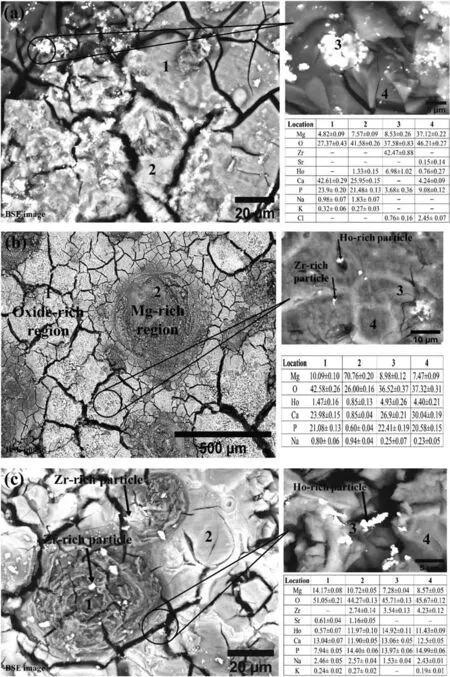
Fig.10.SEM images and EDS compositions of the corroded surfaces of extruded Mg-Zr-Sr-Ho alloys after immersion in SBF for 24 h at 37 °C: (a)Mg-1Zr-0.5Sr-0.5Ho,(b) Mg-1Zr-0.5Sr-1.5Ho,and (c) Mg-1Zr-0.5Sr-4Ho.All compositions are given in wt.%.
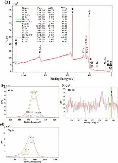
Fig.11.XPS spectra of corroded surface of extruded Mg-1Zr-0.5Sr-0.5Ho alloy: (a) survey scan of the entire binding energy;(b) O 1s;(b) Ho 4d;and (c)Mg 1s.The deconvoluted peaks are also indicated with the peak components in O 1s and Mg 1s spectra.
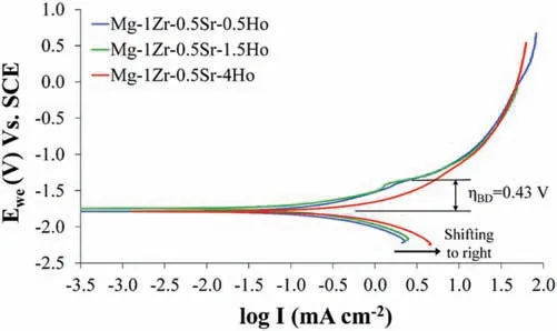
Fig.12.PDP curves of extruded Mg-Zr-Sr-Ho alloys.The maximum breakdown potential (ηBD) measured for Mg-1Zr-0.5Sr-1.5Ho is shown.The black arrow indicates shifting of the cathodic curves towards right.
3.9.PDP curves of extruded Mg-Zr-Sr-Ho alloys
PDP curves and relevant Tafel parameters of extruded Mg-Zr-Sr-Ho alloys are given in Fig.12 and Table 2,respectively.The CRiobtained from the PDP tests are 4.98 ± 0.62,7.95 ± 0.63 and 9.37 ± 0.48 mm y−1for Mg-1Zr-0.5Sr-0.5Ho,Mg-1Zr-0.5Sr-1.5Ho and Mg-1Zr-0.5Sr-4Ho,respectively.No significan change in the corrosion potential (Ecorr)was observed with the Ho additions.However,an increase in the cathodic activity,i.e.,shifting of the PDP curves toward the right is evident in Fig.12.Furthermore,Mg-1Zr-0.5Sr-1.5Ho and Mg-1Zr-0.5Sr-4Ho alloys showed similar cathodic kinetics in their PDP curves.Whereas,the anodic curve of Mg-1Zr-0.5Sr-1.5Ho showed a distinct breakdown potential (ηBD),i.e.,the pitting initiation at 0.43 V.A further increase in the overpotential exhibited similar anodic behavior,although the curve merged in Mg-1Zr-0.5Sr-4Ho at −0.78 V,which suggests an insignifican contribution from the corrosion products during the pitting,as also depicted by the HE curve and SEM image of Mg-1Zr-0.5Sr-1.5Ho alloy in Fig.7 and Fig.8b,respectively.The Mg-1Zr-0.5Sr-4Ho showed the maximum anodic and cathodic current densities which resulted in the highest CRiof 9.37 mm y−1(Table 2).
The corrosion potentials (Ecorr) of REEs are similar to Mg,thus a smaller difference between the intermetallic phase and Mg is expected within the solid solution range,which can reduce the micro galvanic corrosion [178].A shift in the Ecorrto ennobled potentials with an RE addition was reported in the Mg-RE alloys [179,180].Furthermore,increasing the RE content in the respective binary Mg-RE alloy also ennobled the Ecorr[8].The increase in the cathodic activity is attributed to the increase in the intermetallic Mg2Ho phase that enhanced the galvanic corrosion [8,181,182].An improvement in the fil passivation due to the incorporated RE-oxides in the corrosion products has been reported for the Mg-RE alloys [183,184].TheηBDof 430 mV observed in Mg-1Zr-0.5Sr-1.5Ho (Fig.12) indicated the presence of a protection fil against the corrosion attack [185].Conversely,the absence of such an inflectio point in the PDP curve of the Mg-1Zr-0.5Sr-4Ho alloy manifests the initiation of a rapid dissolution at relatively low overpotentials,i.e,ηBDis closer to Ecorr[186,187].This is also corroborated with the HE curve of the Mg-1Zr-0.5Sr-4Ho alloy shown in Fig.7,indicating a drastic increase between 10 and 25 h immersion in SBF.The merging of Mg-1Zr-0.5Sr-1.5Ho curve into Mg-1Zr-0.5Sr-4Ho anodic branch at a high potential (Fig.12) is attributed to the formation of a more stabilized corrosion product due to the increased presence of Ho2O3[184].
It was noted that with addition of Ho in Mg-Zr-Sr-Ho alloys,both the anodic and cathodic current densities were increased (Fig.12).Moreover,any improvement in the anodic kinetics beyond the pitting potential,may not be sufficien to counteract the enhanced cathodic activity from RE addition [8].Consequently,an increase in icorrand corresponding CRiwas observed with increasing Ho addition from 0.5 to 4 wt.% in the Mg-1Zr-0.5Sr-xHo alloys (Table 2).The contribution of the REEs in the corrosion resistance of Mg-RE alloys is mainly to form a more stabilized passive layer via formation of the RE-oxides which hinders the penetration of the Cl−anions by their interaction with the RE3+cations in Mg(OH)2lattice [152].Furthermore,the corrosion potentials and content of the Mg-RE intermetallic phases determines the cathodic current densities.It was reported that depletion of the eutectic Mg12Nd phase with the T4 temper heat treatment in Mg-4Nd alloy caused an improvement in the CR due to shifting the Ecorrto a value closer to that of Mg (−1.84 V) vs SCE [152].Consequently,the minimum CRiof 4.98 mm y−1is attributed to the formation of a more stabilized passive layer from Ho2O3and the low content of the intermetallic Mg2Ho phase in Mg-1Zr-0.5Sr-0.5Ho,which resulted in the reduced cathodic activity in Fig.12.

Fig.13.EIS plots of extruded Mg-Zr-Sr-Ho alloys in SBF at 37 °C: (a) Nyquist plot;(b) and (c) Bode plots.
3.10.EIS of extruded Mg-Zr-Sr-Ho alloys
To further unravel the corrosion mechanisms involved in the extruded Mg-Zr-Sr-Ho alloys,an EIS investigation was conducted,and the EIS plots along with the equivalent electric circuit used for fittin the curves is given in Fig.13.In addition,Table 3 tabulates the relevant parameters and CReiscalculated from the Stern-Geary equation,which included the Tafel slopes(βaandβc)and polarization resistance(Rp)obtained from the PDP and EIS experiments,respectively[37,188]:
The Nyquist plot of Mg-1Zr-0.5Sr-0.5Ho alloy in Fig.13a revealed the presence of two discrete capacitive semicircles,which were modeled with the two parallel resistor-capacitor(RC) circuits connected in series,as given in the equivalent electric circuit.Furthermore,at frequency ≤0.1 Hz,an inductive loop was observed in all samples and are indicated inFig.13.The presence of the two capacitive loops in Mg-1Zr-0.5Sr-0.5Ho is also confirme by the two humps observed in Fig.13c.The high frequency capacitive arc is assigned to the charge transfer resistance (Rct),whereas the medium frequency to the corrosion fil [189–191].Finally,the low frequency inductive loop represents the relaxation process of adsorbed species,such as adsorbed Mg(OH)+or Mg(OH)2[192].Consequently,absence of the second semicircle in Mg-1Zr-0.5Sr-1.5Ho and Mg-1Zr-0.5Sr-4Ho alloys signifie a limited protection and minimal protection contributed from the surface film Furthermore,the minimum CReisof 9.43 mm y−1in Mg-1Zr-0.5Sr-0.5Ho is corroborated with the large diameter of the high frequency semicircle and the increasing slope in Fig.13a and b,respectively.Correspondingly,the maximum Rctof 152.8 and Rfof 37.6Ωcm2were measured in Mg-1Zr-0.5Sr-0.5Ho alloy (Table 3).The large Rctvalues depicts the presence of an enhanced corrosion product fil covering the surface,i.e.,Mg(OH)2or MgO layer formed upon immersion in the electrolyte[193,194].Nonetheless,the Mg-1Zr-0.5Sr-0.5Ho exhibited maximum inductance,which indicates that the Cl−adsorption on the surface was pronounced,as the same is reported to increase the oxidation of metal [194].
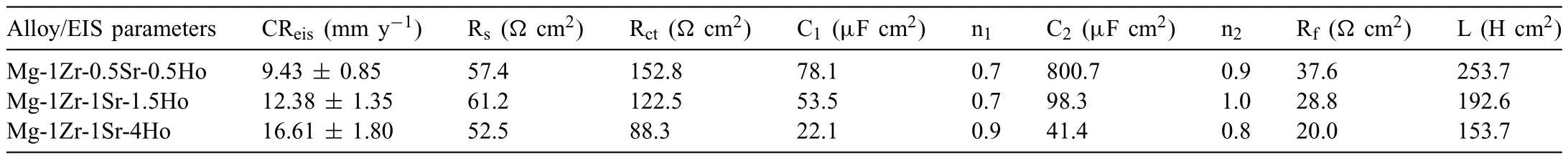
Table 3 EIS parameters of extruded Mg-Zr-Sr-Ho samples.
As per the circuit configuratio of the equivalent model,CReisis governed by the net impedance of the circuit in series,which depicts that both faradic reaction kinetics(Rct)and surface protection film (Rf) contributed to the overall corrosion resistance.A strong surface protection in Mg-1Zr-0.5Sr-0.5Ho is evident from the presence of the second capacitive loop at 0.46 Hz in Fig.13c.Moreover,the constant-phase element(C1) in Table 3,showed values close to that of the doublelayer capacitance of 50 μF cm−2[195,196].A decrease in the Rctand Rfwas reported with an increase in the intermetallic phase which enhanced the galvanic corrosion in Mg-Zn alloy[197].Similar behavior is observed with the increasing Ho addition in Mg-Zr-Sr-Ho alloys (Table 3).The same is also depicted in the PDP curves that showed an increased cathodic activity with the increase in Ho addition (Fig.12).The contribution of the protection fil in Mg-1Zr-0.5Sr-0.5Ho could be estimated with an apparent increase in the impedance of approximately 100Ωcm2in Fig.13b.Further addition of the Ho increased the galvanic corrosion due to the enhanced Mg2Ho and Mg17Sr2phases,which were evident in the EDS maps of Sr and Ho in Fig.3.The absence of the island-like structures,which are also indicative of the pitting,further substantiates the minimal galvanic effect in Mg-1Zr-0.5Sr-0.5Ho(Fig.10a).

Fig.14.Comparison of CRs obtained from HE,PDP,and EIS methods in extruded Mg-Zr-Sr-Ho alloys.
3.11.Corrosion mechanism in extruded Mg-Zr-Sr-Ho alloys
A comparison of the CRs obtained from the HE,PDP,and EIS methods of extruded Mg-Zr-Sr-Ho alloys is given in Fig.14.The CRs calculated from the respective techniques showed a similar trend in each composition.The reasons for the differences in CRs among the investigating techniques were discussed in various studies [188,198–200].The extruded Mg-Zr-Sr-Ho showed minimum CRiof 4.98 mm y−1in SBF,compared to 2.6 mm y−1for extruded pure Mg[201].However,a comparison of the minimum CRiwith the ascast Mg-Zr-Sr-Ho alloys [145] revealed a decrease of 19.5%,which conforms to the fact that extrusion of Mg-Zr-Sr-Ho alloys enhanced their corrosion resistance.From the microstructural point of view,a significan segregation of the intermetallic Ho-containing phase was observed at the GBs with the Ho addition in the as cast Mg-Zr-Sr-Ho alloys.Whereas the extruded Mg-1Zr-0.5Sr-4Ho exhibited a significantl refine GS with only minor Ho segregation at the GBs (EDS map in Fig.3c).This implies that the galvanic corrosion was more pronounced in the as-cast condition due to the relatively high quantity of the intermetallic phases at the GBs.Similarly,a comparison between extruded Mg-1Zr-0.5Sr [21] and Mg-1Zr-0.5Sr-0.5Ho showed a significan reduction of 53.4% in CRi.Therefore,it is deduced that Ho addition substantially reduces the CR in extruded Mg-Zr-Sr alloys.
It is worth noting that the high-purity (HP) Mg showed a very low CR of 0.3–0.4 mm y−1for biodegradable implant applications [202,203].A further improvement in CR was achieved in the ultra-high-purity (UHP) Mg which exhibited a CR of 0.25 mm y−1[204].However,the inferior mechanical strength of HP and UHP Mg is the major concern for biodegradable orthopedic implants [204,205].Consequently,to achieve optimized corrosion and mechanical properties,Mg-Zr-Sr-Ho alloys were developed in this study.Nevertheless,Mg alloys with satisfying corrosion,mechanical,and biocompatibility properties are still limited [206].Furthermore,discrepancies betweenin vitroandin vivodegradation rates (i.e.,in vivotests showing significantl lower CRs)have also been widely reported [207,208].
Mg is a naturally passive metal that undergoes pitting corrosion at its free corrosion potential (Ecorr) when exposed to chloride ions in a non-oxidizing medium.As a result,the corrosion of Mg alloys in neutral or alkaline salt solutions typically takes the form of pitting [42].Kirkland et al.[209] reported pitting corrosion in most of the biocompatible Mg alloys including the HP Mg and a minimum CR of 0.5 mm y−1was observed for AZ91,which does not exhibit good biocompatibility [210],whereas the majority of Mg alloys showed CRs between 2 and 10 mm y−1in minimum essential medium [209].Although,the pitting corrosion was also observed in this study,a CR of 4.98 mm y−1implies that the extruded Mg-Zr-Sr-Ho alloys may exhibit a lower CRin vivo.
In view of the finding above,the corrosion mechanism observed in the extruded Mg-Zr-Sr-Ho alloys can be proposed.The influenc of the Ho addition on the Ecorrwithin a solid solution range,i.e.,containing minimal intermetallic Mg2Ho,was insignifican due to the similar electrode potentials of Ho and Mg [178,211].However,with an increase in the intermetallic Mg2Ho phase at the GBs and the segregation of the Ho within theα-Mg matrix,the micro-galvanic effect became more pronounced [212].This led to an increase in the cathodic activity,as evident from the shifting of the cathodic curves and the increased pitting in Fig.12 and Fig.10c,respectively.Furthermore,the presence of a stabilized protection fil was formed in the Mg-1Zr-0.5Sr-0.5Ho alloy due to the presence of Ho2O3in the corroded surface layers (XPS in Fig.11b).However,with an increase in the secondary phases,pitting due to the galvanic effect is enhanced in Mg-1Zr-0.5Sr-1.5Ho and Mg-1Zr-0.5Sr-4Ho alloys,which deteriorated the growing surface film This is further corroborated with the absence of the second capacitive arcs in Fig.13a.
Upon immersion in the SBF,the extruded Mg-1Zr-0.5Sr-0.5Ho alloys underwent an anodic dissolution ofα-Mg to form a porous fil of Mg(OH)2,incorporated with the H2O3.The presence of the H2O3stabilized the fil by displacing the Mg2+with the Ho3+ions,which could retard the Cl−penetration [152].The enhanced stability can be further confirme from the presence of the MgO in the XRD and XPS spectra (Figs.9 and 11b),which was reported to augment the formation of a stabilized fil in the Mg-RE alloys [199,212].Furthermore,an improvement in the fil stability is also attributed to the reduction in the internal stresses due to the formation of H2O3[167].In addition,the EDS investigations of the corrosion products revealed the presence of the HA,which enhanced the corrosion resistance of the Mg alloys.It was reported that the formation of Sr-substituted HA in SBF is assisted by the presence of the Sr in Mg alloys[6].
3.12. In vitro cytotoxicity and cell adhesion of extruded Mg-Zr-Sr-Ho alloys
Thein vitrocytotoxicity of the extruded Mg-Zr-Sr-Ho alloys was evaluated by measuring the viability of the SaOS2 cells after culturing for 3 d in MMEM.Subsequently,the cell adhesion on the sample surfaces was investigated using SEM.A graph of the cell viability (CV) and corresponding SEM images of the cell morphologies are given in Fig.15.The CV of all the Mg-Zr-Sr-Ho samples was observed around 80% and were statistically insignifican with apvalue ≥0.05[213].This implies that increasing of Ho addition in the extruded Mg-Zr-Sr-Ho alloys has no significan effect on their cytotoxicity.It may be noted that as per ISO 10993–5,a material with CV ≥70% is considered non-cytotoxic [214].The SaSO2 cell adhesion on the Mg-Zr-Sr-Ho samples revealed well-spread lamellae (flattene cells) with extended lamellipodia and filopodia thereby exhibiting good cell adhesion[215–217],as indicated in Fig.15(b-2) and Fig.15(c-2),respectively.Moreover,at a few locations an aggregation of the cells was observed (Fig.15(b-2)).
In vitroandin vivostudies have shown direct correlation between the CR and the biocompatibility of Mg alloys [1,208,218].Thus,a decrease in CR with RE addition showed enhanced biocompatibility as the Mg-RE alloys possessed significantl better capability to sustain endothelial cell attachment and growth with no adverse reaction in human aorta endothelial cells for 7 d [219].It was reported that the Dy,Gd,Y,Eu,Nd and Pr exhibited non-cytotoxicity in human osteosarcoma cell line MG63 [17],whereas La and Ce showed anti-carcinogenic properties and inhibited leukemic cells,namely HL-60 and NB4 cells [220].Furthermore,cytotoxic effects of the REEs were observed at concentrations where localized corrosion occurred [221].Similarly,the role of other alloying elements is also critical,as Sr addition in Mg has been observed to increase the alkaline phosphatase activity of MG63 cells and enhanced the bone density around implants [3,5,222,223].Li et al.[224] demonstrated that the addition of Sr at 2 wt.% to Mg-1Ca-xSr (x=0.2 to 2 wt.%)induced higher CV,protein adsorption,alkaline phosphatase activity,extracellular mineralization,and osteogenesis via the extracellular signal-regulated kinase pathway.Furthermore,the Ho-containing bioactive glasses exhibited an increase in cell proliferation of osteoblast-like MC3T3-E1 cells [225].The released ion concentration of Ho was calculated to be 0.156 mg/L based on the highest CRiof 9.37 mm y−1measured for Mg-1Zr-0.5Sr-4Ho with a sample surface area of 0.785 cm2in 300 ml of SBF and assuming that the released ion concentration in 72 h (time period of the hydrogen evolution test) [226].The half-lethal dose LD50of Ho in rats was reported as 560 mg kg−1[13].Thus,it can be seen that the released Ho ion concentration is well below the LD50limit even at the highest CR of the Mg-1Zr-0.5Sr-4Ho alloy.It may be noted that the cytotoxicity of REEs was found dependent on the ionic radii and solubility in the Mg [17].Resultantly,the heavy REEs such as Dy and Gd were found to be least toxic in the Mg alloys.

Fig.15.Biocompatibility of extruded Mg-Zr-Sr-Ho alloys: (a) cell viability of SaOS2 cells after culturing for 3 d and SEM micrographs of SaOS2 cells on:(b) Mg-1Zr-0.5Sr-0.5Ho;(c) Mg-1Zr-0.5Sr-1.5Ho;and (d) Mg-1Zr-0.5Sr-4Ho.
The cytotoxicity tests of the extruded pure Mg showed a CV of 95% with respect to the negative control in relation to SaOS2 cells [227].Nevertheless,the cytotoxicity results of the extruded Mg alloys were compared with the earlier studied as-cast Mg-Ho and Mg-Zr-Sr-Ho alloys [13,145].Firstly,an increase in Ho addition from 1 to 10 wt.% in the as-cast Mg-Ho alloys showed a slight increase from 96 to 98% in CV after 3 d of culturing,although the CR was increased from 0.5 to 6 mm y−1for Mg-1Ho and Mg-10Ho,respectively.This implies that the toxicity is not affected by the Ho addition within the solubility range.The effect of corrosion on the cytotoxicity is related to the effect of the trapped H2gas,pH,metal ion concentration,and corrosion products in the surrounding tissues/cells [228,229].Therefore,it is suggested that the cytotoxicity caused from corrosion is counteracted by the improvement in cell growth and proliferation due to the inherent biofunctionality (e.g.,osteogenesis) of the alloying elements.Secondly,an increase in CV after 5 d of cell culturing was also observed with Ho addition up to 3 wt.% in the as-cast Mg-1Zr-2Sr-xHo (x=1,3 and 5 wt.%) alloys[4].Consequently,the flattene cells with extended filopodi on the extruded Mg-Zr-Sr-Ho alloys indicated good cell adhesion and proliferation;moreover,in spite of an increase in the CRs with the Ho addition,no significan change in CV was observed;this may be attributed to the competing effects on CV between the corrosion and Ho addition [4].
4.Conclusions
The mechanical,corrosion,and biocompatibility properties of the extruded Mg-Zr-Sr-Ho alloys was comprehensively investigated.The effects of REE addition and extrusion on the textures,tensile and compressive properties,corrosion behavior,and biocompatibility were evaluated.Based on the results,a corrosion mechanism was proposed.The main conclusions are as follows:
(1) A decrease in grain size was observed with increasing Ho addition in the extruded Mg-Zr-Sr-Ho alloys.Minimum grain size of 4.35±1.04 μm was measured in the extruded Mg-1Zr-0.5Sr-4Ho alloys.
(2) The intermetallic Mg17Sr2and Mg2Ho phases were mainly distributed at the grain boundaries.EDS mapping results revealed an increase in the secondary phases with increasing Ho addition.
(4) The extruded Mg-Zr-Sr-Ho alloys exhibited the basal texture with the {0001} planes aligned parallel to ED.Evolution of RE texture was observed with increasing Ho addition.Consequently,the Mg-1Zr-0.5Sr-4Ho showed a completely developed RE texture,which resulted in weakening of the basal and prismatic textures.
(5) An increase in Ho content from 0.5 to 4 wt.% caused 200% improvement in the tensile elongation and 89% improvement in compressive strain.The large elongation is attributed to the weakened textures and were accompanied by only a marginal decrease of 3.3% in tensile strength but a significan increase of 8.9% in compressive strength.
(6) An increase of Ho content from 0.5 to 4 wt.% in Mg-1Zr-0.5Sr-xHo resulted in a decrease in the tensioncompression yield asymmetry from 0.62 to 0.98 due to weakening of the textures.
(7) Tensile fracture analysis revealed microcracks at the grain boundaries and particle-matrix interface in Mg-1Zr-0.5Sr-0.5Ho.An increase in the Ho content transformed the quasi-brittle intergranular fracture to a more ductile transgranular fracture.
(8) A comparison of the maximum compressive properties with the previously studied as-cast Mg-Zr-Sr-Ho alloy demonstrates that the extrusion indeed enhanced the mechanical properties of Mg-Zr-Sr-Ho alloys.Further comparison of the compressive properties between the extruded Mg-1Zr-0.5Sr and Mg-1Zr-0.5Sr-4Ho also confirme that Ho addition significantl improves the compressive strength and ductility of extruded Mg-Zr-Sr-based alloys.
(9) Corrosion analysis of extruded Mg-Zr-Sr-Ho alloys revealed the presence of pitting corrosion.A minimum corrosion rate of 4.98 mm y−1was calculated from the PDP curves of Mg-1Zr-0.5Sr-0.5Ho alloy.The enhanced corrosion resistance is attributed to the presence of Ho2O3in the surface fil and reduced galvanic corrosion due to less intermetallic phases.The maximum corrosion rate of 9.37 mm y−1was observed for Mg-1Zr-0.5Sr-4Ho due to the increased galvanic effect.
(10) The formation of a stabilized corrosion fil due to the presence of Ho2O3was confirme by EIS and XPS.The maximum fil resistance of 37.6Ωcm2and charge transfer resistance of 152.8Ωcm2were measured in Mg-1Zr-0.5Sr-0.5Ho alloy.
(11) A comparison of the minimum CRiwith the previously studied as-cast Mg-Zr-Sr-Ho alloys revealed that extrusion of Mg-Zr-Sr-Ho enhances the corrosion resistance.Similarly,a comparison between extruded Mg-1Zr-0.5Sr and Mg-1Zr-0.5Sr-0.5Ho showed that Ho addition substantially reduces the CR of extruded Mg-Zr-Sr-based alloys.
(12) The corrosion mechanism included the formation of a corrosion fil that is stabilized by Ho2O3,which retards the ingress of the Cl−ions from the corrosion environment.However,with increasing Ho addition,the galvanic corrosion increased,leading to pitting and subsequent damage to the surface film
(13) Extruded Mg-Zr-Sr-Ho alloys exhibits non-cytotoxicity and good SaOS2 cell adhesion.Furthermore,increasing Ho addition in the Mg-Zr-Sr-Ho alloys showed insignificant effect on their cell viability.
Declaration of Competing InterestThe authors declare that they have no known competing financia interests or personal relationships that could have appeared to influenc the work reported in this paper.
Acknowledgments
The authors acknowledge the financia support for this research by the Australian Research Council(ARC)through the Future Fellowship (FT160100252) and the Discovery Project(DP170102557).The authors also acknowledge the scientifi and technical assistance of RMIT University’s Microscopy and Microanalysis Facility,a linked laboratory of the Australian Microscopy &Microanalysis Research Facility.
Supplementary materials
Supplementary material associated with this article can be found,in the online version,at doi:10.1016/j.jma.2022.10.002.
杂志排行

Journal of Magnesium and Alloys的其它文章
- Development of high-strength magnesium alloys with excellent ignition-proof performance based on the oxidation and ignition mechanisms: A review
- Development and application of magnesium alloy parts for automotive OEMs: A review
- Recent advances in surface endothelialization of the magnesium alloy stent materials
- Recent developments in high-pressure die-cast magnesium alloys for automotive and future applications
- Exploring the contribution of oxygen reduction reaction to Mg corrosion by modeling assisted local analysis
- Fabrication of a model specimen for understanding micro-galvanic corrosion at the boundary of α-Mg and β-Mg17Al12