Electroacupuncture preconditioning alleviates myocardial ischemiareperfusion injury through the hypothalamic paraventricular nucleusinterposed nucleus nerve pathway
2022-07-20WEIXiaotongLILiaoyuanZHANGYatingSHUQiWANGShuaiyaCHENPianpianHULingYUQingCAIRonglin
WEI Xiaotong,LI Liaoyuan,ZHANG Yating,SHU Qi,WANG Shuaiya,CHEN Pianpian,HU Ling,YU Qing,CAI Ronglin
WEI Xiaotong,LI Liaoyuan,ZHANG Yating,SHU Qi,Wang Shuaiya,CHEN Pianpian,Graduate School of Anhui University of Chinese Medicine,Hefei 230012,China
HU Ling,YU Qing,CAI Ronglin,Institute of Acupuncture and Meridian,Anhui University of Chinese Medicine,Hefei 230012,China;Key Laboratory of Acupuncture and Moxibustion Fundamentals and Techniques of Anhui Province,Anhui University of Chinese Medicine,Hefei 230038,China
Abstract OBJECTIVE:To explore whether the paraventricular nucleus (PVN) participates in regulation of the antimyocardial ischemia-reperfusion injury (MIRI) effect of electroacupuncture (EA) and whether this is achieved through the PVN-interposed nucleus (IN) neural pathway.METHODS:The modeling method of myocardial ischemia reperfusion injury was achieved by ligating the left anterior descending coronary artery in Sprague-Dawley rats.We used the Powerlab multi-channel physiological recorder system to record electrocardiograms and analyze the changes in ST segment displacement;2,3,5-Triphenyltetrazolium chloride staining was used to observe the percentage of myocardial infarction areas.Detecting cardiac troponin I(cTnI),lactate dehydrogenase (LDH) in serum was done with an enzyme-linked immunosorbent assay kit.Morphological changes in the myocardium were detected in each group with hematoxylin-eosin staining of paraffin sections.Detection of c-fos protein expression in the PVN of the hypothalamus was done with the immuneofluorescence method.The Plexon multi-channel acquisition system recorded PVN neuron discharges and local field potentials in each group of rats.Offline Sorter software was used for cluster analysis.Neuro Explorer software was used to perform autocorrelation,raster and frequency characteristics and spectral energy analysis of neuron signals in each group.RESULTS:Compared with the MIRI model group,the areas of myocardial infarction in the EA group were significantly reduced;the expression of cTnI,LDH in serum was decreased significantly.The firing frequency of pyramidal cells in the PVN was significantly increased and the spectrum energy map showed energy was reduced,c-fos expression in PVN was reduced,this indicated that neuronal activity in the PVN participates in the effect of EA improving myocardial injury.In addition,we used the kainic acid method to lesion the IN and observed that the effect of EA was weakened.For example,the area of myocardial infarction of lesion IN +EA group in rats was significantly increased compared with that resulting from EA group,the expression of cTnI,LDH in serum was significantly increased,the firing frequency of pyramidal cells in the PVN was significantly reduced.A spectral energy diagram shows that the energy after damage was higher than that of EA group.At the same time,the expression of c-fos in the PVN increased again.CONCLUSION:Our results indicated that the PVN-IN nerve pathway may participate as an effective pathway of EA to improve the effect of myocardial injury.
Keywords:acupuncture;myocardial reperfusion injury;L-lactate dehydrogenase;troponin I;paraventricular hypothalamic nucleus;interposed nucleus
1.INTRODUCTION
Electroacupuncture preconditioning has been shown to have a protective effect on the heart in rodent models and patients undergoing heart valve replacement surgery,1,2but the main mechanism is not understood.In recent years,the relationship between the brain and meridians and viscera has been given increasing attention by scholars,and the central integration mechanism of the acupuncture effect is still largely unclear.Janget al3explored the relationship between meridians,acupoints and viscera by injecting virus tracer,and found out that it may be related to the central nervous system of the brain and spinal cord.The myocardial ischemiareperfusion injury (MIRI) mechanism is complicated,which further aggravates myocardial ischemia injury.4Therefore,acupuncture pretreatment is of great significance to improve MIRI.
As an important nucleus of the hypothalamus,the PVN is the central integration area and the most important central part of the sympathetic efferent activity,and it regulates cardiovascular function through afferent and efferent fibers.5,6It is a key nucleus for regulating the cardiovascular system.Decreased excitability of PVN neurons can reduce the degree of MIRI,and vice versa,can aggravate the damage.7In the early stage of the research,by implanting the PVN in the rat myocardial ischemia model with multi-electrode microarray recording technologyin vivo,it was found that the activity of pyramidal cells was increased in the PVN and the activity of the interneurons was inhibited.Therefore,the PVN participates in this process.8These results indicate that PVN is one of the important nuclei in improving cardiac function by acupuncture.Studies have shown that damage to the medial part of the cerebellum can change the behavioral response caused by damage to the hypothalamus.9There is a direct and two-way fiber connection between the hypothalamus and the interposed nucleus (IN).10But whether the PVN -IN participates in acupuncture preconditioning of MIRI has not been confirmed.Therefore,it is believed that the PVN-IN neural pathway may be one of the important pathways involved in visceral activity.Based on this,we suggest that the PVN participates in the acupuncture effect and may be achieved through the PVN-IN pathway.Therefore,in this study,the characteristics of PVN electrical activity were recorded by multi-channel microarray recording technology,the temporal and spatial connections of nuclear neurons in different brain regions were observed,and the role of PVN-IN neural pathway in the mechanism of acupuncture effect was verified by destroying IN.In order to explore the regulation mechanism of PVN-IN,we studied the neural pathway involved in acupuncture treatment of MIRI,which laid an experimental foundation for further clarifying the role of hypothalamus cerebellum in anti MIRI.
2.MATERIALS AND METHODS
2.1.Experimental animals
Adult male Sprague-Dawley rats,weighing (220 ± 30) g,were obtained from Jinan Pengyue Experimental Animal Breeding Co.,Ltd.(Jinan,China),license number:SCXK (Lu) 20190003.Animals were reared under the same conditions as in Suzhou Feng's IVC-Ⅱ (intelligent)independent air supply isolation cage,the temperature in the cage was (24 ± 1) ℃,and the light/dark cycle was 12 h.Food and water were supplied freely and they were reared adaptively for one week.
2.2.Experimental grouping
Rats were randomly divided into the sham group,MIRI model group (MIRI group),acupuncture at the heart meridian points group+MIRI model group(electroacupuncture group,EA group),and lesion IN +acupuncture at the heart meridian points group+MIRI model group (lesion+EA group).All experimental operations strictly abided by the“Guiding Opinions on the Treatment of Experimental Animals”(Ministry of Science and Technology of the People's Republic of China,2006) This study was reviewed and approved by the Experimental Animal Ethics Committee of Anhui University of Chinese Medicine (No.2020028).
2.3.Experimental reagent and instruments
The reagents and instruments used in the experiments included:isoflurane anesthetic purchased from Shenzhen Reward Life Technology Co.,Ltd.(Reward,Shenzhen,China);kainic acid purchased from MCE China;Acupuncture needles Purchased from Suzhou Tianxie Acupuncture Instrument Co.,Ltd.(specification:0.25 mm × 25 mm,Suzhou,China);a Huatuo brand SDZ-Ⅱ electronic acupuncture instrument purchased from Suzhou Medical Products Factory Co.,Ltd.(Suzhou,China);2,3,5-Triphenyltetrazolium chloride (TTC) solution was purchased from Beijing Solaibao Technology Co.,Ltd.(Beijing,China);Powerlab Multichannel Physiological recorder (AD Instruments,Shanghai,China);Microfilament electrode (Dallas,USA);Neuro Explorer software (version 5.029);C-fos primary antibody application Santa (Dallas,USA);Second antibody was purchased from Abcam (Abcam,UK);DAPI purchased from Beyotime (Shanghai,China);Anti-fluorescence quenching was purchased from Beyotime (Shanghai,China);cTnI kit used in Shanghai Future Industry Co.,Ltd.(Shanghai,China);LDH kit used in Nanjing Jiancheng Institute of Bioengineering (Nanjing,China).
2.4.Production of the MIRI animal model
We improved coronary artery ligation to establish a rat MIRI model.The rats were fasted for 12 h before the operation.After weighing,the rats were anesthetized by intraperitoneal injection of 2% pentobarbital sodium.11The limbs and heads were fixed on a test board in the supine position,and lead II was recorded.Signs of successful MIRI modeling included the local myocardial tissue being pale or cyanotic after 30 min of ischemia ligation,the ST segment of the electrocardiogram being elevated or the T wave shown to be towering;the myocardial tissue in the ischemic area turned red at 2 h of reperfusion,and a significantly elevated ST segment was reduced by 50% of the above.The chest cavity was closed and the muscle skin was sutured layer by layer.In the sham group,the coronary arteries were not ligated after thoracotomy,and the needle was used to pass through the corresponding part once.The other operations were the same as those in the model group.Before ligation in each group of experimental animals,if the ECG was abnormal,if they died without reaching the end of observation,and if they were unsuccessful in modeling,they were eliminated.
2.5.Lesioning of the IN region
The destruction procedure involved putting the rat in the induction box of the gas anesthesia machine and maintaining anesthesia for 2-3 min.After anesthesia,the head and nose from the induction box were placed into the stereotaxic anesthesia mask and fixed.The anesthesia concentration was 1.5%-2.0%.According to the coordinates of the rat Paxions brain in the atlas for IN(AP:11.1-11.4 mm,RL:2.0-2.1 mm,H:6.2-6.4 mm).12The position was determined and the skull was drilled with a skull drill.Slowly a micro-injector that had been injected with kainic acid was placed into the predetermined site,and bilateral injection of 0.4 μL of 1 g/L kainic acid solution into the IN.The needle was left in for 5 min after the injection and then slowly removed to suture the wound.Penicillin sodium was injected intramuscularly to prevent infection,and the animals were kept warm while waiting for recovery after anesthesia.After three days of administration,EA pretreatment was carried out for 7 d.13After data collection,Nissl staining was performed to detect the morphological changes of neurons in the damaged nucleus.
2.6.EA pretreatment
The rats of each group were fixed on a cylindrical rat fixator,the forelimbs of the rats were exposed,and the heart meridian acupoint group was acupunctured using the acupoints Shenmen (HT7) and Tongli (HT5) on both sides.Disposable sterile acupuncture needles were used and sites were pierced 2-3 mm.The needle handle was connected to a Huatuo brand SDZ-Ⅱ electronic acupuncture instrument.The two needles on the same side were connected to positive and negative poles.A continuous wave,stimulation voltage of 1 V,frequency of 2 Hz,and rat slight shaking of the limbs in degrees was carried out for 20 min/time per day for seven consecutive days.Acupoint locations were chosen according to the "Experimental Acupuncture and Moxibustion" test.MIRI replication was performed within 1 h after the last EA.14No EA was given to the sham group and MIRI group.
2.7.ECG record of ST segment displacement value changes
Using a PowerLab multichannel physiological recorder,the standard Ⅱ lead electrocardiogram of rats was recorded.Needle-shaped electrodes were inserted into the rats' limbs (right upper limbs and both lower limbs)and the rat's ECG signal was continuously monitored.The built-in Chart software was used to analyze the changes of ST segment of rats in each group before modeling,30 min of ligation,and 2 h of reperfusion.
2.8.TTC staining method to detect myocardial infarction area
Four anesthetized rats from each group were taken and their hearts were extracted.They were then washed in 4 ℃ phosphate buffer and frozen at -20 ℃ for 30 min.The heart was cut into five pieces below the ligature line from the apex to the bottom of the heart perpendicular to the long axis of the heart.The thickness was about 1-2 mm.The heart was then immediately immersed in TTC solution,incubated at 37 °C in the dark for 20 min,fixed with 4% paraformaldehyde,and photographed under a microscope.The hearts were analyzed with ImageJ software;the white area was the ischemic infarction area,and the red area was the non-ischemic area.The percentage of myocardial infarction area=left ventricular tissue slice infarct area/ total area of left ventricular tissue section × 100%.
2.9.Serum biochemical indices were determined with an enzyme-linked immunosorbent assay
After 2 h reperfusion,5 mL of the abdominal aorta was taken,and the supernatant was centrifuged for detection according to the cTnI,LDH kit operation instructions.
2.10.Hematoxylin-eosin (HE) staining
The heart was fixed in paraformaldehyde for 24 h,dehydrated and embedded conventionally,and then sectionalized with HE staining (5 μm).
2.11.Recording of neuronal discharges in the PVN
Except for the sham group,the models were replicated in the remaining groups.After the animals recovered for 24 h,the animals underwent a craniotomy according to the rat brain atlas under anesthesia.Microwire electrodes were implanted unilaterally at 1.4 mm behind the fontanelle and 0.4 mm to the right of the median line.A manual micro-thruster was used to slowly push the electrode tip to the PVN at a pace of 10 μm/s.Signals were recorded for 5 min after a stable neuron discharge was observed.All data were stored digitally.Neurons were sorted with Offline Sorter3 software and the final neuron classification was exported in SPK and FP .pl2 file formats.Then,Neuro Explorer software was used to further analyze the data of the .pl2 files of SPK and local field potential (LFP).For Spike,the autocorrelation analysis mainly used the relationship between neuron firing and the time to obtain firing times and patterns.Neurons can be divided into interneurons and pyramidal neurons according to the time course of action potentials,total firing times,and autocorrelation maps.15,16Choosing 0-40 Hz,the time was the FP of 300 times the window to calculate the power of its corresponding frequency band.Then,by comparing the difference between the number of neuron firings and LFP in each group,the changes between the groups were observed.At the end of the experiment,Nissl staining was used to morphologically identify the electrode positions.
2.12.Determination of c-fos expression with an immunofluorescence method
Rats in each group were perfused for 2 h after myocardial ischemia and reperfusion and brains were taken.After the perfusion,the rat brains were fixed in paraformaldehyde for 24 h,dehydrated,and embedded in paraffin sections with a thickness of 5 μm.Then brain slices were stained by immunofluorescence.We observed and took pictures under a fluorescence microscope,and the proportion of c-fos labeled positive cells in PVN was counted.
2.13.Statistical analysis
SPSS 23.0 (Chicago,IL,USA) software was used for statistical analyses,and the data were expressed as the mean ± standard deviation ().Differences between groups were compared with a one-way analysis of variance.The homogeneity of variance test was performed before pairwise comparisons between groups.The least significant difference test was used for uniform variance,and Tamhane’sT2 test was used for uneven variance.P <0.05 indicates that the difference is statistically significant.
3.RESULTS
3.1.Evaluation of ECG model replication and comparison of ST segment potential displacement in each group of rats
According to the changes in the ST segment of the ECG,judgements were made about whether the MIRI rat model was successfully replicated.The following figure shows the ECG changes of each group.The picture shows that the ST segment of the ECG has significant changes before and after ligation,which decreasing by more than 50% (Figure 1).This indicated that the MIRI model of rats in this study was successfully replicated (n=6 for each group).
Before the MIRI model was copied,the ST segment displacement values of each group were basically the same (P >0.05).Except for the sham group,the ST segment displacement values of the rats in each group were increased 30 min after ligation compared with those before model replication (P <0.01).The displacement value of ST segment in rats after reperfusion for 2 h was less than half of that of ligation for 30 min (P <0.05,<0.01),therefore the model was successfully established.The ST segment of the MIRI group was higher than that of the sham group at 30 min after ligation and 2 h of reperfusion (P <0.01),and the ST segment displacement value of the EA group was lower than that of the MIRI group and the lesion+EA group (P <0.05,< 0.01,Table 1).
Table 1 Statistical analysis of electrocardiogram of rats in each group ()

Table 1 Statistical analysis of electrocardiogram of rats in each group ()
Notes:The MIRI group refers to the myocardial ischemia reperfusion injury model group.EA group refers to the electroacupuncture of the heart meridians group+myocardial ischemia-reperfusion injury group.The lesion+EA group refers to lesion of the intercerebellar nucleus +electroacupuncture pretreatment+MIRI group.PVN:paraventricular nucleus;MIRI:myocardial ischemia-reperfusion injury;EA:electroacupuncture.Results except for sham group,the ST segment displacement values of rats in each group increased 30 min after ligation compared with that before ligation,and decreased reperfusion 2 h compared with 30 min after ligation.Compared with before ligation,aP < 0.01;compared with 30 min after ligation,bP < 0.05,cP < 0.01;compared with the sham group,dP < 0.01;compared with the MIRI group,eP < 0.01;compared with the lesion+EA group,fP < 0.05.
3.2.Comparison of the areas of myocardial infarction in each group of rats
Compared with the sham group,the myocardial infarction area in the MIRI group was significantly increased (P <0.01).Compared with the MIRI group and lesion+EA group,the myocardial infarction area in the EA group was significantly reduced (P <0.01,< 0.05)(n=4;Figure 2A,2B).
3.3.cTnI and LDH expression statistics in serum
Compared with the sham group,cTnI,LDH expression in the MIRI group was increased significantly (P <0.01);compared with the MIRI group and the lesion+EA group,the EA group was significantly decreased (P <0.05,< 0.01) (n=6,Figure 2C,2D).
3.4.HE staining results
In the cross section of heart,myocardial cells in sham group were neatly arranged with clear cross section lines and a few vacuoles.The MIRI group and lesion+EA group the myocardial cell injury had irregular arrangement,unclear transverse lines,and the EA group still had enlarged spacers and vacuoles,but the orderly arrangement of myocardial cells was significantly better than the MIRI group and the lesion+EA group(Figure 3).
3.5.Autocorrelation analysis of electrical activity of PVN neurons in each group of rats
Multi-channelin vivorecording technology was used to record the stable signal in the PVN for 5 min;first,through Offline Sorter software analysis,and then using Neuro Explorer software for autocorrelation analysis of neuron firing sequences.The results showed that there were two types of pyramidal cells and interneurons in each group (n=6 per group;Figure 4).

Figure 1 Changes of ST segment in each group at different time periods
3.6.Comparison of two types of PVN neuron firing raster images of rats in each group
Neuro Explorer software can display the recorded spike signal with a raster image (0-100 s).Pyramidal cell discharge results showed that,compared with the sham group,the MIRI group had significantly sparser discharges;compared with the MIRI group and the lesion+EA group,the EA group was significantly denser;and the Interneurons discharge continued to be dense after the model was copied (n=6 per group,Figure 5).

Figure 2 Statistical analysis of myocardial infarction size,cTnI and LDH between groups
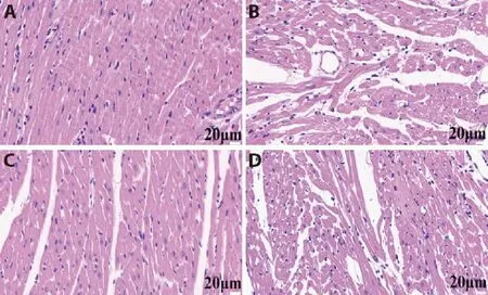
Figure 3 Hematoxylin-eosin staining results in each group
3.7.Comparison of the firing numbers of PVN neurons in each group of rats
After converting the spike signal emission sequence(time=300 s) into a discharge frequency histogram.The Pyramidal cell discharge of the MIRI group was significantly reduced compared with the sham group (P <0.01).
Compared with the MIRI group and the lesion+EA group,the discharge of the EA group was significantly higher (P <0.01).The Interneurons all increased after the model was copied (P <0.05) (n=6 per group,Figure 6).
3.8.Analysis of PVN energy spectrum of rats in each group
LFP reflects the cluster activity of neurons;from blue to red indicates that the energy is from small to large.The results showed that the MIRI group had the strongest LFP spectrum energy,the lesion+EA group was strong,the EA group was lower,and the sham group was the lowest (n=6 per group,Figure 7).
3.9.c-fos expression statistics in the PVN
The c-fos expression changes were observed in each group and statistical analyses were performed.The results showed that the expression of c-fos in the PVN of the MIRI group (9.43 ± 0.73) was significantly higher than that of the sham group (3.10 ± 1.40) (P <0.01);compared with the MIRI group and lesion+EA group(8.56 ± 2.50),the expression of c-fos in the PVN of the EA group (3.93 ± 0.55) was significantly reduced (P <0.01,n=3 per group;Figure 8).

Figure 5 Comparison of raster images of PVN neuron electrical activity in rats from each group

Figure 6 PVN neuron spike counts in each group.

Figure 7 Comparison of spectral energy diagrams between the groups (time=300 s;frequency=40 Hz).

Figure 8 c-fos expression in the PVN of each group (immunofluorescence staining,partial image was 200 times,scale was 50 μm)
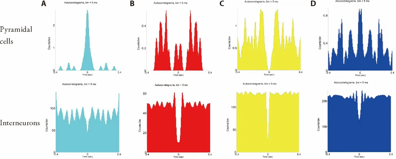
Figures 4 Autocorrelation analysis of PVN neuron electrical activity in each group of rats
4.DISCUSSION
Acupuncture at the points of the heart meridian can significantly improve heart disease according to animal and clinical studies.Acupuncture is safe and feasible for the treatment of cardiovascular diseases.17It effectively relieves angina pectoris and improves heart function by exciting the vagus nerve and regulating the autonomic nervous system.18-20In traditional Chinese medicine,if there are diseases in the viscera,the Yuan acupoint should be selected.It is believed that the Yuan acupoint is the place with the richest vitality in the meridian of the human body.There is also a saying that collateral points allow communication between the meridians of the outside and inside.Shenmen (HT7) is the Yuan acupoint and Tongli (HT5) is the Luo acupoint of the heart meridian.It is a commonly used acupuncture point for improving heart function.21Electroacupuncture can activate the discharge activity of pyramidal cell in the PVN,inhibit interneuron and produce an anti-myocardial ischemia effect.8Acupuncture can adjust the excitability of PVN neurons through the nerve fibers between the hippocampus and PVN,thereby transmitting to the sympathetic nerves to produce protection of the heart.22Zhanget al23showed that EA can inhibit the reduction of Apelin expression in the PVN area of traumatic rats.Therefore,it can improve the cardiac function of rats with thoracic surgery trauma.Another study pointed out that a damaged PVN at different time periods will have a significant impact on the heart rate and blood pressure of hypertensive rats.24Therefore,the PVN is considered to be an important nucleus for regulating the cardiovascular system.25
Current studies have demonstrated that the cerebellum not only regulates body movements,but also participates in the regulation of visceral activities.26Having study showed that the application of HRP retrograde axon technology and fMRI confirmed mutual projection between the cerebellum and hypothalamus,and the projection between the two was through the direct projection of many single synapses and the indirect projection of multiple synapses.27-29Previous research of our group showed that damage to the fastigial nucleus can affect changes in DA,5-HT,and other neurotransmitters and the number of nerve discharges in the lateral hypothalamus,which is significantly different from the effect of EA preconditioning for improving the myocardium.30Recently,there have been many studies on the visceral function of the fastigial nucleus of the cerebellum,but little is known about the IN.Studies have found that the afferent signals in the IN have an effect on PVN neurons,and it is believed that the IN can actively regulate cardiovascular diseases through the pathway of PVN-IN.31Therefore,we speculate that the effect of acupuncture preconditioning on improving myocardial effects is not only due to the involvement of a single nucleus in the PVN,but may be related to the PVN-IN pathway.
Multi-channel microarray technology can be used to monitor the activity of neurons by recording the potential values outside the cell.Observing neuronal firing between the brain nuclei can occur in time and space and they can be distinguished as int and pyramidal cell by their discharge frequency,mode,and waveform.Multichannel electrophysiology technology is used for extensive research of neurons.After being stimulated and depolarized,c-fos is expressed in neurons and is commonly used as an indicator of neuron activity in neurobiology.32The LFP reflects the synthesis of electrical activity of the neuron cluster in a certain spatial range,and the changes in the neuron activity in the nucleus are observed through the changes in color.cTnI,LDH are the gold standard for diagnosing myocardial infarctions,and they are good indicators of the state of the heart muscle.
In this study,we used multi-channel microarray electrode technology to observe the electrical activity of neurons in the PVN,TTC staining to observe the area of myocardial infarction,and immunofluorescence to detect the changes in c-fos expression in the PVN.In addition,we detected expressions of LDH and cTnI in serum.It was found that there were two types of neurons;namely,interneuron and Pyramidal cell.It is generally believed that interneuron is an inhibitory neuron and Pyramidal cell is an excitatory neuron.Compared with the sham group,the number of interneuron firings in the other groups was continuously in a higher state,indicating that the MIRI model was successfully replicated,causing abnormal excitation of the Interneuron in the PVN.However,after EA,it was observed that the area of myocardial infarction in the EA group was reduced compared to the MIRI group.HE staining showed that the morphology of myocardial cells was significantly improved after EA.The expression of the cTnI and LDH in serum was significantly decreased in EA group.The number of Pyramidal cell discharges decreased significantly after the model was copied,but Pyramidal cell discharges increased significantly after EA,which indicates that EA caused the activity of Pyramidal cell in the PVN.The LFP showed that the local energy after EA pretreatment was significantly lower than that of the MIRI group.The c-fos protein in the EA group was also lower than in the MIRI group.It suggested that the activation of Pyramidal cell in PVN by acupuncture relatively inhibits interneuron and regulates downstream neuron firing to improve heart function.
In addition,kainic acid treatment of lesions in the nucleus is a method to promote degeneration and death of neurons in the nucleus without affecting the nerve fibers passing through.33In this study,lesions in the IN were used to observe changes myocardial infarction area,cTnI,LDH in serum;in the number of neuronal firings,and c-fos protein expression in the PVN.It was found that after the IN was damaged,the area of myocardial infarction was larger than that of the EA group,cardiomyocytes returned to a severe state;the expression of cTnI and LDH in the lesion+EA group increased significantly again;the number of Pyramidal cell discharges in the PVN was significantly lower than that of the EA group;the LFP energy in the PVN was higher than that in the EA group,the expression of c-fos in the lesion+EA group was higher than that of the EA group,suggesting that after the IN is damaged,the effect of acupuncture on inhibiting abnormally excited neurons and improving the myocardium was weakened.Therefore,the fiber projection of the PVN-IN may be involved in acupuncture to improve the cardiovascular effect.In summary,acupuncture at the heart meridian points can significantly inhibit the activity of abnormally excited neurons during myocardial reperfusion by regulating the activity of neurons in the PVN.It is worth noting that after the IN was damaged,activity of Pyramidal cell in the PVN of the lesion+EA group was decreased,the area of myocardial infarction was increased,the expression of cTnI and LDH in the lesion+EA group increased significantly again,the expression of c-fos was increased.This showed that the projection in the PVNIN pathway may be a potential way for acupuncture preconditioning to treat MIRI.Therefore,heart activity is regulated while myocardial function is improved through acupuncture at the Shenmen (HT7) and Tongli(HT5) points and the PVN-IN nerve pathway plays a key role in this process.
Based on the neural circuit between the hypothalamus and cerebellum,this research considered the PVN to deeply study the relationship between the PVN-IN pathway and the heart.We provided an experimental basis for the central integration mechanism of the acupuncture heart meridian to improve the myocardial effect in the next step.The next step will be to clarify the involvement of specific neuron types in the PVN,and more clearly the hypothalamic-cerebellar nerve pathway,which is involved in the anti-myocardial effect of acupuncture.
5.REFERENCES
1.Hao F,Cai RL,Yu Q,et al.Effect of electroacupuncture preconditioning on the expressions of NF-κB p65,IκBα and IKKβ in myocardial tissue of the rats with acute myocardial ischemiareperfusion injury.Zhong Guo Zhen Jiu 2020;40:1103-7.
2.Yang L,Yang J,Wang Q,et al.Cardioprotective effects of electroacupuncture pretreatment on patients undergoing heart valve replacement surgery:a randomized controlled trial.Ann Thorac Surg 2010;89:781-6.
3.Jang I,Cho K,Moon S,et al.A study on the central neural pathway of the heart,Nei-Kuan (EH-6) and Shen-Men (He-7) with neural tracer in rats.Am J Chin Med 2003;31:591-609.
4.Chi HJ,Chen ML,Yang XC,et al.Progress in therapies for myocardial ischemia reperfusion injury.Curr Drug Targets 2017;18:1712-21.
5.Zhong MK,Duan YC,Chen AD,et al.Paraventricular nucleus is involved in the central pathway of cardiac sympathetic afferent reflex in rats.Exp Physiol 2008;93:746-53.
6.Chen WW,Xiong XQ,Chen Q,et al.Cardiac sympathetic afferent reflex and its implications for sympathetic activation in chronic heart failure and hypertension.Acta Physiol (Oxf) 2015;213:778-94.
7.Chen C,Zhang Y,Cheng XY,et al.Central nervous system regulation mechanism of myocardial ischemia-reperfusion injury in rats:neuronal excitability in the paraventricular nucleus of the hypothalamus.Zhong Hua Ma Zui Xue Za Zhi 2018;38:1293-7.
8.Cai RL,Cui S,Wu ZJ,et al.Effect of electroacupuncture at“Shenmen”(HT7)-“Tongli”(HT5) of heart meridian on neuronal activities in paraventricular nucleus of hypothalamus in myocardial ischemia rats.Zhen Ci Yan Jiu 2018;43:406-13.
9.Zhu JN,Yung WH,Kwok-Chong Chow B,et al.The cerebellarhypothalamic circuits:potential pathways underlying cerebellar involvement in somatic-visceral integration.Brain Res Rev 2006;52:93-106.
10.Lu JH,Mao HN,Cao BB,et al.Effect of cerebellohypothalamic glutamatergic projections on immune function.Cerebellum 2012;11:905-16.
11.Han J,Xuan JL,Hu HR,et al.Protective effect against myocardial ischemia reperfusion injuries induced by hyperoside preconditioning and its relationship with PI3K/Akt signaling pathway in rats.Zhong Guo Zhong Yao Za Zhi 2015;40:118-23.
12.Paxinos G,Watson C.The rat brain in stereotaxic coordinates.People's Medical Publishing House,2005,in press.
13.Liu F,Li BM.Brain function damage and inactivation methods commonly used in learning and memory research.Zhong Guo Xing Wei Yi Xue Ke Xue 2006;222-3.
14.Lin WZ,Wang P.Experimental acupuncture and moxibustion.Shanghai Science and Technology Press,1999,in press.
15.Barthó P,Hirase H,Monconduit L,et al.Characterization of neocortical principal cells and interneurons by network inter-actions and extracellular features.J Neurophysiol 2004;92:600-8.
16.Xu JM,Wang CQ,Lin LN.Multi-channelin vivorecording techniques:signal processing of action potentials and local field potentials.Sheng Li Xue Bao 2014;66:349-57.
17.Painovich J,Longhurst J.Integrating acupuncture into the cardiology clinic:can it play a role? Sheng Li Xue Bao 2015;67:19-31.
18.Wang JS,Yu XD,Deng S,et al.Acupuncture on treating angina pectoris:a systematic review.Medicine 2020;99:e18548.
19.Cui S,Xu J,Wang J,et al.Effect of electroacupuncture stimulation of heart meridian on autonomic nervous activities in acute myocardial ischemia rats.Zhen Ci Yan Jiu 2016;41:515-20.
20.Armstrong K,Gokal R,Todorsky W.Neuromodulating influence of two electroacupuncture treatments on heart rate variability,stress,and vagal activity.J Altern Complement Med 2020;26:928-36.
21.Sun YZ,Yao J,Zhou L.Research progress in the ancient and modern methods of acupuncture method of matching points of the original collaterals.Liaoning Zhong Yi Yao Da Xue Xue Bao 2019;21:5-9.
22.Cui S,Wang K,Wu SB,et al.Electroacupuncture modulates the activity of the hippocampus-nucleus tractus solitarius-vagus nerve pathway to reduce myocardial ischemic injury.Neural Regen Res 2018;13:1609-18.
23.Zhang HH,Wang YJ,Zheng C,et al.Apelin in the hypothalamic paraventricular nucleus improves cardiac function in surgical trauma rats.Sheng Li Xue Bao 2018;70:99-105.
24.Ciriello J,Kline RL,Zhang TX,et al.Lesions of the paraventricular nucleus alter the development of spontaneous hypertension in the rat.Brain Res 1984;310:355-9.
25.Xu B,Zheng H,Patel KP.Relative contributions of the thalamus and the paraventricular nucleus of the hypothalamus to the cardiac sympathetic afferent reflex.Am J Physiol Regul Integr Comp Physiol 2013;305:R50-9.
26.Saab CY,Willis WD.Cerebellar stimulation modulates the intensity of a visceral nociceptive reflex in the rat.Exp Brain Res 2002;146:117-21.
27.Onat F,Cavdar S.Cerebellar connections:hypothalamus.Cerebellum 2003;2:263-9.
28.Kullmann S,Veit R.Resting-state functional connectivity of the human hypothalamus.Handb Clin Neurol 2021;179:113-24.
29.Dietrichs E,Haines DE.Interconnections between hypothalamus and cerebellum.Anat Embryol 1989;179:207-20.
30.Yu Q,Cai RL,Shao XF,et al.Effect of electroacupuncture preconditioning on the contents of dopamine and 5-hydroxytryptamine in lateral hypothalamus area and cerebellar fastigial nucleus of rats with myocardial ischemia-reperfusion injury.Zhong Guo Zhen Jiu 2021;41:525-30.
31.Wen YQ,Zhu JN,Zhang YP,et al.Cerebellar interpositus nuclear inputs impinge on paraventricular neurons of the hypothalamus in rats.Neurosci Lett 2004;370:25-9.
32.van den Hoogen NJ,Kwok CHT,Trang T.Identifying the neurodevelopmental differences of opioid withdrawal.Cell Mol Neurobiol 2021;41:1145-55.
33.Coyle JT,Molliver ME,Kuhar MJ.In situ injection of kainic acid:a new method for selectively lesioning neural cell bodies while sparing axons of passage.J Comp Neurol 1978;180:301-23.
杂志排行
Journal of Traditional Chinese Medicine的其它文章
- Efficacy of meridian massage for motor function after a stroke:a systematic review and Meta-analysis
- Antiviral Activity of Medicinal Plants against Human Coronavirus:a systematic scoping review of in vitro and in vivo experimentations
- Fuzheng Kang' ai decoction (扶正抗癌方) inhibits cell proliferation,migration and invasion by modulating mir-21-5p/human phosphatase and tensin homology deleted on chromosome ten in lung cancer cells
- Correlation between slow transit constipation and spleen Qi deficiency,and gut microbiota:a pilot study
- Efficacy of Kushen decoction (苦参汤) on high-fat-diet-induced hyperlipidemia in rats
- Hyperpolarization-activated cyclic nucleotide-gated 2 contributes to electroacupuncture analgesia on lumbar disc herniation-induced radicular pain through activation of microglia in spinal dorsal horn