Furostanol Saponins from Asparagus cochinchinensis and Their Cytotoxicity
2021-12-16RuoSongZhangYangYangLiuPeiFengZhuQiongJinZhiDaiXiaoDongLuo
Ruo-Song Zhang ·Yang-Yang Liu·Pei-Feng Zhu ·Qiong Jin ·Zhi Dai·Xiao-Dong Luo ,
Abstract Phytochemical investigation on the roots of Asparagus cochinchinensis led to the isolation of one new furostanol saponin, named 26- O- β- D-glucopyranosyl-22 α-hydroxyl-(25 R)-Δ 5(6) -furost-3 β,26-diol-3- O- α- L-rhamnopyranosyl-(1 → 2)-[β- Dglucopyranosyl-(1 → 4)- α- L-rhamnopyranosyl-(1 → 4)]- β- D-glucopyranoside ( 1), along with three known congeners ( 2 ).The structure of new saponin was elucidated via comprehensive inspection of its HRMS and NMR spectral data as well as chemical technology, whereas those of known ones were identifi ed by comparison of their NMR and MS spectral data with those reported in literatures.All isolated saponins were evaluated for their cytotoxic eff ects on two human liver (MHCC97H) and lung adenocarcinoma (H1299) cancer cells in vitro.Among them, both 1 and 2 showed signifi cant cytotoxicity against above mentioned cell lines.Further studies revealed that these two saponins could signifi cantly inhibit their proliferation of MHCC97H and H1299 cells.
Keywords Steroid saponins · Asparagus cochinchinensis · Cytotoxicity · Structural elucidation
1 Introduction
Steroid saponins, whose aglycones were usually a spirostanol or its derivatives [1], were commonly found from roots, tubers, leaves, blooms or seeds in more than 100 families of plants [2,3].Compared with other glycosides, the strong foam-forming property in aqueous solution of steroidal saponins was their main feature [2,4].Previous researches revealed steroidal saponins possessed various pharmacological activities, such as antifungal [5], hypocholesterolemic [6], antimitotic [7] and cAMP phosphodiesterase inhibitory [8] eff ects.Among them, a large number of publications have revealed steroid saponins shared diff erent cytotoxic properties that promoted their potential as anticancer drugs or adjuvants [9,10].
Asparagus cochinchinensis, belonging to the genusAsparagus(Liliaceae), is well-known as “Tianmendong” in China.Its roots have been historically used in Chinese folk medicine for the treatment of cough, acute and chronic bronchitis, chronic pharyngitis, hemorrhoids, and tumors for thousands of years [11].Apart from steroidal saponins [12], phenolic compounds [13], norlignans [14] and alkaloids [15] have been isolated from this plant as revealed by previous phytochemical studies.However, steroidal saponins obtained from title species were proved to be its major and bioactive components responsible for its cytotoxic [16], antiinfl ammatory [17], hepatotoxic and nephrotoxic [18], and anti-neuroinfl ammatory [11] properties.In continuation of a search for bioactive constituents from plants of the Yunnan province [19], a chemical investigation was performed on the roots ofA.cochinchinens.As a result, a total of steroidal saponins ( 1 -4) were isolated and identifi ed including one new and three previously described furostan-type steroidal saponins.Their cytotoxic eff ects on two human cancer cells MHCC97H and H1299 were also evaluated (Fig. 1).
2 Results and Discussion
Saponin 1 was obtained as a white amorphous powder.It had a molecular formula of C57H94O27as determined by the observed (+)-HRESIMS protonated ion peak atm/z1233.5879 [M + Na] + (calcd for C57H94O27Na, 1233.5875).It showed a positive reaction to the Ehrlich’s reagent (red color), suggesting a furostanol skeleton [20].The 13 C NMR spectrum (Table 1) displayed 57 carbons, of which 27 were assigned to the aglycone part and the remaining 30 were attributed to fi ve hexose units.With the aid of the HSQC experiment, the 1 H and 13 C NMR spectrum (Table 1) attributable to the aglycone moiety showed resonances for four characteristic steroidal methyls atδH0.83 (3H, s,CH3-19), 0.93 (3H, d,J= 6.6 Hz,CH3-27), 1.00 (3H, s,CH3-18), and 1.26 (3H, d,J= 6.7 Hz,CH3-21), together with their corresponding carbons atδC 16.3 (CH 3 -19), 17.3 (CH 3 -27), 19.2 (CH 3 -18), 16.3 (CH 3 -21); two oxygenated methines atδH3.82 (1H, m) and 4.88 (1H, m), along with their corresponding carbons atδC77.8 (CH-3) and 80.8 (CH-16); an olefi nic group atδH5.26 (1H, brs) as well asδC121.6 (CH-6) and 140.6 (C-5); and a ketal carbon atδC110.4 (C-22).The abovementioned data indicated that the aglycone of 1 should be a furostanol one as that of protodioscin ( 2) [21].Moreover, the aglycone of 1 was further confi rmed a by the following diagnostic 1 H1H COSY, HMBC, and ROESY correlations (Figs. 2 and 3).The 1 H1H COSY experiment revealed three structural fragments including CH 2 -1CH 2 -2-CH-3CH 2 -4, CH-6CH 2 -7CH-8/(CH-9CH 2 -11CH 2 -12)/CH-14CH 2 -15CH-16CH-17CH-20CH3-21, and CH2-23CH2-24CH-25/(CH3-27)/CH2-26.Moreover, the observed HMBC fromδH1.00 (CH3-18) toδC39.7 (CH2-12), 40.4 (C-13), 56.4 (CH-14), and 63.6 (CH-17), fromδH0.83 (CH3-19) toδC37.3 (CH2-1), 140.6 (C-5), 50.1 (CH-9), and 36.9 (C-10), and from bothδH1.26 (CH3-21) andδH2.00 (H-23a) toδC110.4 (C-22) established the aglycone of 1 to be 22α-hydroxyl-(25R)-furost-Δ 5(6) -3β,26-diol.The ROESY correlations ofδH1.00 (Me-18) with 1.51 (H-8)/2.17 (H-20)/1.94 (H-23b) and ofδH0.83 (Me-19) with 1.51 (H-8) and 1.68 (H-1a) verifi ed these protons were placed at the same side, whereas the observed ROESY correlations ofδH0.94 (H-1b) with 3.82 (H-3)/0.86 (H-9), ofδH1.02 (H-14) with 0.86 (H-9)/1.87 (H-17), and ofδH1.87 (H-17) with 4.88 (H-16) indicated these protons were located at the other side.Additionally, the 25Rconfi guration of 1 was assigned according to the small chemical shift diff erence between Ha-26 and Hb-26 at Δab = 0.34 ppm (Δab > 0.57 ppm for 25S, and Δab < 0.48 ppm for 25R) [22].In view of aforementioned evidence, the aglycone of 1 was thus elucidated as 22α-hydroxyl-(25R)-furost-Δ 5(6) -3β,26-diol.
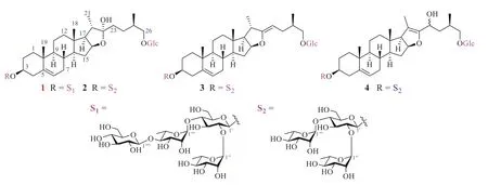
Fig.1 Structures of 1-4

Table 1 1 H and 13 C NMR spectral data of 1 (600 and 150 MHz, pyridine- d 5 )

Fig.2 Key 1 H- 1 H COSY and HMBC correlations of 1
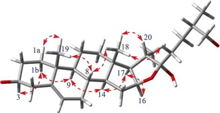
Fig.3 Key ROESY correlations for the aglycone moiety of 1
As for the sugar units of 1, its 1 H NMR spectrum (Table 1) displayed the presence of fi ve anomeric proton signals atδH 4.73 (1H, d,J= 7.8 Hz, H-1″), 4.88 (1H, d,J= 7.7 Hz, H-1′), 5.14 (1H, d,J= 7.7 Hz, H-1″″), 5.74 (1H, brs, H-1) and 6.27 (1H, brs, H-1″), which showed correlations in the HSQC spectrum with fi ve anomeric carbons atδC104.6 (CH-1″), 100.1 (CH-1′), 106.4 (CH-1″″), 101.8 (CH-1), and 101.6 (CH-1″).With the assistance of MS spectrum, the sugar moiety of 1 was preliminary determined.Specifi cally, the [MH]ion (m/z1209.6) displayed 1 had a molecular weight (MW) of 1210.6 Da in the negative ion mode of ESIMS n .The observed ions withm/zvalues of 1047.5, 901.5, and 755.4 indicated the sequential cleavage of two rhamnopyranosyl units followed by the cleavage of a glucopyranosyl moiety from the parent [MH]ion (m/z1209.6), respectively.Likewise, the MS 2 spectrum also aff ordedm/zvalue of 593.4 that was indicative of the loss of one glucopyranosyl group from the C-3 position or the C-26 position (Scheme 1).Also, acid hydrolysis of 1 also gave D-glucoses and L-rhamnoses as the sugar residue, which was confi rmed by HPLC analysis of their corresponding PMP derived adducts.All the anomeric protons of D-glucose possessedβ-confi gurations due to their 3JH1,H2coupling constants (7.8, 7.7, and 7.7 Hz), and both anomeric protons of L-rhamnoses sharedα-confi gurations due to the chemical shifts of C-3 (δC72.5 and 72.2) and C-5 (δC69.3 and 68.3), respectively.In the HMBC spectrum, the long-range correlations fromδH4.88 (H-1′) toδC77.8 (CH-3), fromδH4.73 (H-1″) toδC74.9 (CH2-26), fromδH6.27 (H-1″) toδC77.6 (CH-2′), fromδH5.74 (H-1) toδC77.1 (CH-4′), and fromδH5.14 (H-1″″) toδC 84.9 (CH-4) established the sequence for 3-O-sugar chain as an [α- L-rhamnopyranosyl-(1 → 2)]-[β- Dglucopyranosyl-(1 → 4)-α- L-rhamnopyranosyl-(1 → 4)]-β- Dglucopyranosyl moiety and for 26-O-sugar chain asβ- Dglucopyranosyl moiety, respectively.Based on the above information presented, the structure of 1 was thus elucidated to be 26-O-β- D-glucopyranosyl-22α-hydroxyl-(25R)-Δ 5(6) -furost-3β,26-diol-3-O-α- L-rhamnopyranosyl-(1 → 2)-[β- Dglucopyranosyl-(1 → 4)-α- L-rhamnopyranosyl-(1 → 4)]-β- Dglucopyranoside.
Additionally, three known steroidal glycosides were identified as protodioscin ( 2) [21], (25R)-26-O-β- D-glucopyranosyl-3β,20α,26-trihydroxyfurostan-5, 22-diene-3-O-α- L-rhamnopyranosyl-(1 → 2)-[α-Lrhamnopyranosyl-(1 → 4)]-O-β- D-glucopyranoside ( 3) [23], and dioscoreside H ( 4) [24] by comparison of their spectroscopic data with those reported in the literatures.
The steroid saponins obtained from species of Liliaceae have shown the potential to signifi cantly inhibit the proliferations of various human tumor cell lines in vitro [25– 29].Therefore, all isolated compounds were evaluated for their cytotoxicity against MHCC97H and H1299 by the MTT method.More specifi cally, compared with the IC50values of positive control doxorubicin hydrochloride, and both 1 and 2 displayed strong cytotoxicity against MHCC97H and H1299 cells with IC50values of 3.56 ± 0.45/4.18 ± 0.43 μg/mL and 5.26 ± 0.74/4.15 ± 0.59 μg/mL, respectively (see Fig. 4).Furthermore, as can be seen from Fig. 4, compared with the positive control doxorubicin hydrochloride, saponins 1 and 2 could signifi cantly inhibit their proliferation (Table 2).

Scheme 1 The fragmentation process of 1 in the ESI- MS negative scan
Moreover, all obtained steroid saponins were evaluated for their antimicrobial activity againstEscherichia coli(ML-35P),Bacillus cereus(CMCC(B) 63303),Candida albicans(ATCC 2091),Bacillus subtilis(ATCC 6633),Streptococcus hemolyticus(ATCC 19615),Listeria monocytogenes(ATCC 19114),Pseudomonas aeruginosa(PO01),Staphylococcus aureus(ATCC 4330),Salmonella Typhimurium(SL1344) andStaphylococcus epidermidis(CMCC 26069) by the microdilution broth susceptibility assay.The results (see Table 3) revealed that saponins 1 -4 showed moderate antimicrobial activity againstC.albicansandB.subtilis, while only saponin 3 showed weak antimicrobial activity againstS.aureus(63.30 ± 0.55 μg/mL).
3 Experimental
3.1 General Experiment Procedures
Optical rotation was measured on a Autopol VI automatic polarimeter.The IR spectrum were measured on a Thermo Nicolet iS10 infrared spectrophotometer with KBr disk.The NMR spectra were obtained on Bruker DRX-400 and DRX-600 spectrometers.Chemical shifts (δ) were expressed in ppm with reference to the solvent signals.Both ESI and HRESIMS spectra were performed on an UPLC-IT-TOF spectrometer.Semi-preparative HPLC was performed on a Waters 600 with a COSMOSIL C18 (10 × 250 mm, Nacalai Tesque Corporation, Japan) column.Analytical HPLC was performed on a Shimadzu SIL-20A Series HPLC system equipped with a reverse-phase COSMOSIL C18 column (4.6 mm × 250 mm, 5 μm, Nacalai Tesque Corporation, Japan).Column chromatography was carried out using silica gel (100200 mesh, Qingdao Haiyang Chemical, Qingdao, Co., Ltd., People’s Republic of China) and macro-porous absorption resin (D101, Donghong Chemical Co., Ltd., People’s Republic of China).The PMP (Chengdu Aikeda Chemical Reagent Co., Ltd., China) was purchased from Beijing 4A Biotech Co., Ltd.(Beijing, China).Fractions were monitored by TLC, and spots were visualized by heating silica gel plates sprayed with Ehrlich’s reagent.
3.2 Plant Materials
The roots ofA.cochinchinensiswas purchased from ‘Luosiwan’ Chinese herbal medicine Market, Kunming, Yunnan Province, in November 2019, identifi ed by Dr.Xu-Jie Qin.A voucher specimen (No.Luo 20191106) has been deposited at State Key Laboratory of Phytochemistry and Plant Resource in West China, Kunming Institute of Botany, Chinese Academy of Sciences.
3.3 Extraction and Isolation

Fig.4 Eff ects of 1 and 2 on MHCC97H and H1299 cells proliferation ( n = 3).A The IC 50 values of 1 and 2 against MHCC97H; B The IC 50 values of 1 and 2 against H1299; C Inhibition eff ects of MHCC97H and H1299 cells proliferation by 1 and 2 after cultivation for 72 h
The air-dried roots ofA.cochinchinensis(5.0 kg) were extracted with 90% aqueous EtOH at 80(15 L × 4, each time for 3 h).The solvent was removed under reduced pressure to yield an amber residue (2.5 kg).The residue was subjected to column chromatography over an macroporous resin column eluted fi rst with H2O then successively with 25%, 70%, and 90% EtOH, respectively.The 70% EtOH partition was evaporated under reduced pressure to obtain a total steroidal saponin moiety.The total saponins (153 g) was subjected to a silica gel column eluting with a CHCl3MeOHH2O gradient (80:20:2 → 65:35:10) to yield fi ve fractions (Fr.AFr.E).Fraction C (105 g) was chromatographed on a silica gel column (CHCl3MeOHH2O, 9:1:0.1) to give saponin 2 (70 g) and Fr.C1.Fr.C1 (230.5 mg) was further purifi ed by semi-preparative HPLC to aff ord 1 (29.8 mg;tR= 12 min; MeCNH2O, 28:72, 3.0 mL/min).Fraction D (12 g) was separated on a silica gel column (CHCl3MeOHH2O, 8:2:0.2) and then purifi ed by semi-preparativeHPLC to yield saponins 3 (3.4 mg,tR= 20.5 min; MeCNH2O, 35:65, 1.0 mL/min) and 4 (2.6 mg,tR= 23.5 min; CH3CNH2O, 35:65, 1.0 mL/min).
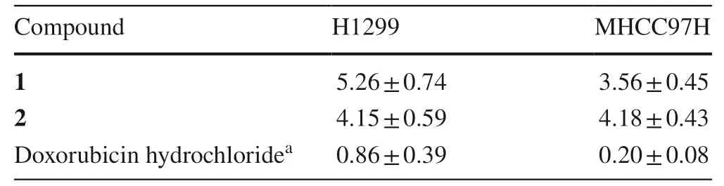
Table 2 Cytotoxicity of saponins 1 and 2 (IC 50 ± SD,μ g/mL)

Table 3 Antimicrobial activity of saponins 1 -4 (IC 50 ± SD, μg/mL)
3.4 Spectroscopic Data of 1
26-O-β- D-glucopyranosyl-22α-hydroxyl-(25R)-Δ 5(6) -furost-3β,26-diol-3-O-α- L-rhamnopyranosyl-(1 → 2)-[β- Dglucopyranosyl-(1 → 4)-α- L-rhamnopyranosyl-(1 → 4)]-β- Dglucopyranoside ( 1): white amorphous powder, [α]46.86 (c0.11, MeOH); IR (νmax ): 3417, 2933, 2851, 1635, 1453, 1382, 1045 cm1; HRESIMSm/z1233.5879 [M + Na] + (calcd for C57H94O27Na, 1233.5875).1 H (pyridine-d5, 600 MHz) and 13 C (pyridine-d5, 150 MHz) NMR spectral data, see Table 1.
3.5 Acid Hydrolysis of 1
The acid hydrolysis of compound 1 was carried out by a previously reported procedure [19].Compound 1 (2.0 mg) was refl uxed at 120 °C for 2 h with 2 M TFA on an oil bath.The aglycone was removed by the extraction with CHCl 3 (5.0 mL) for three times.The reaction residue was fi ltered after neutralizing with 60.0 μL of NaOH (0.3 M).After removing the solvent under reduced pressure, the residue was refl uxed at 75 °C for 1 h with 60.0 μL of PMP (0.5 M in methanol).Moreover, the reaction was quenched with 60.0 μL of HCl (0.3 M) and the reaction mixture was extracted with CHCl3(5.0 mL, three times).Then, the aqueous layer was analyzed over HPLC (18% acetonitrile: 82% sodium phosphate (pH 6.8; 1.5 mL/min).Likewise, the standard monosaccharides D-glucose (1.0 mg) and L-rhamnose (1.0 mg) were derivatized with PMP by the same way as 1, and HPLC analyses were performed under the same conditions as 1.The sugar units in 1 were identifi ed as D-glucose (tR= 14.5 min) and L-rhamnose (tR= 17.0 min) by comparison of the retention times of the corresponding derivatives.
3.6 Cytotoxicity Assay
The cytotoxicity of isolated compounds was determined to use the MTT method with a slight modifi cation [30].Briefl y, two human cancer (MHCC97H and H1299) cell lines were incubated in 96-well plates at a density of 2 × 10 3 cells/well in DMEM medium supplemented with 10% fetal bovine serum at 37with 5% CO2.After overnight incubation, cells were treated with tested compounds at diff erent concentrations (20.00, 10.00, 5.00, 2.50, and 1.25 μg/mL) for 72 h.Subsequently, the culture mediums were exchanged by DMEM medium which contained 10% MTS reagent [3-(4,5-dimethylthiazol-2-yl)-5-(3-carboxymethoxyphenyl)-2-(4-sulfophenyl)-2H-tetrazolium, inner salt] and then cultured for another 4 h.The absorbance was recorded on a microplate reader at 490 nm.
3.7 Antimicrobial Activity Assay
The antimicrobial activity of isolated steroid saponins against 10 strains using the microdilution broth susceptibility assay [31].The strains frozen in the refrigerator at80were activated and inoculated on standard tryptone soy broth agar (TSA) plates at 37for 8 h to observe the bacterial growth.Subsequently, single colonies were selected and inoculated in tryptone soy broth (TSB) plates.After cultivated at 37in shaker (120 rpm) for 8 h, the absorbance of bacterial solution was measured and its concentration was adjusted to 10 5 CFU/mL.Whereafter, an inoculum of 10 5 CFU/mL was made to sterile 96-well plate containing tested compounds at diff erent concentrations (100.00, 50.00, 25.00, 12.50, 6.25 and 3.13 μg/mL) at 37for 8 h.The wells containing only broth served as growth control.The absorbance of bacterial solution was recorded on a microplate reader at 600 nm.
4 Conclusion
In summary, a chemical examination of the roots ofA.cochinchinensisled to the identification of one new furostanol glycoside 26-O-β- D-glucopyranosyl-22α-hydroxyl-(25R)-Δ5(6)-furost-3β,26-diol-3-O-α- Lrhamnopyranosyl-(1 → 2)-[β- D-glucopyranosyl-(1 → 4)-α- Lrhamnopyranosyl-(1 → 4)]-β- D-glucopyranoside ( 1) and three known one ( 2-4).Meanwhile, compounds 1 and 2 exhibited cytotoxic and anti-proliferative eff ects on two human (MHCC97H and H1299) cancer cell lines.At the same time, compounds 14 displayed moderate antimicrobial activity againstC.albicansandB.subtilis, and compound 3 displayed weak antimicrobial activity againstS.aureus.
Supplementary InformationThe online version contains supplementary material available at https:// doi.org/ 10.1007/ s13659- 021- 00321-0.
AcknowledgementsThis study was financially supported by the National Natural Science Foundation of China (Grant Nos.31770388 and U1802281) and the Second Tibetan Plateau Scientifi c Expedition and Research (STEP) program (Grant No.2019QZKK0502).
Declarations
Conflict of interestThe authors declare no competing fi nancial interest.
Open AccessThis article is licensed under a Creative Commons Attribution 4.0 International License, which permits use, sharing, adaptation, distribution and reproduction in any medium or format, as long as you give appropriate credit to the original author(s) and the source, provide a link to the Creative Commons licence, and indicate if changes were made.The images or other third party material in this article are included in the article's Creative Commons licence, unless indicated otherwise in a credit line to the material.If material is not included in the article's Creative Commons licence and your intended use is not permitted by statutory regulation or exceeds the permitted use, you will need to obtain permission directly from the copyright holder.To view a copy of this licence, visithttp:// creat iveco mmons.org/ licen ses/ by/4.0/.
杂志排行

Natural Products and Bioprospecting的其它文章
- Advances in Research on Chemical Constituents and Their Biological Activities of the Genus Actinidia
- Natural Products as Potential Lead Compounds for Drug Discovery Against SARS-CoV-2
- Anticancer Properties and Mechanism of Action of Oblongifolin C, Guttiferone K and Related Polyprenylated Acylphloroglucinols
- Six New 3,5-Dimethylcoumarins from Chelonopsis praecox,Chelonopsis odontochila and Chelonopsis pseudobracteata
- Scutellarin Reduces Cerebral Ischemia Reperfusion Injury Involving in Vascular Endothelium Protection and PKG Signal
- Isoprenylated Flavonoids as Ca v3.1 Low Voltage-Gated Ca2+ Channel Inhibitors from Salvia digitaloides