Bile acid activated receptors:Integrating immune and metabolic regulation in non-alcoholic fatty liver disease☆
2021-10-11MicheleBiagioliStefanoFiorucci
Michele Biagioli,Stefano Fiorucci
Department of Medicine and Surgery,University of Perugia,Perugia,Italy
Keywords:Bile acid Farnesoid X receptor(FXR)G protein-coupled bile acid receptor 1(GPBAR1)Immunity Lipid metabolism Non-alcoholic fatty liver disease(NAFLD)Non-alcoholic steatohepatitis(NASH)
ABSTRACT Bile acids are a family of atypical steroids generated at the interface of liver-intestinal microbiota acting on a ubiquitously expressed family of membrane and nuclear receptors known as bile acid activated receptors.The two best characterized receptors of this family are the nuclear receptor,farnesoid X receptor (FXR) and the G protein-coupled receptor,G protein-coupled bile acid receptor 1 (GPBAR1).FXR and GPBAR1 regulate major aspects of lipid and glucose metabolism,energy balance,autophagy and immunity and have emerged as potential pharmaceutical targets for the treatment of metabolic and inflammatory disorders.Clinical trials in non-alcoholic fatty liver disease(NAFLD),however,have shown that selective FXR agonists cause side effects while their efficacy is partial.Because FXR and GPBAR1 exert additive effects,dual FXR/GPBAR1 ligands have been developed for the treatment of metabolic disorders and are currently advanced to clinical trials.Here,we will review the role of FXR and GPBAR1 agonism in NAFLD and how the two receptors could be exploited to target multiple components of the disease.
1.Introduction
Non-alcoholic fatty liver disease (NAFLD) is a highly prevalent human disorder affecting approximately one quarter of the world population and characterized by an excessive accumulation of lipids in hepatocytes (hepatic steatosis).1NAFLD is categorized histologically into non-alcoholic fatty liver (NAFL) and nonalcoholic steatohepatitis (NASH).Because of the NAFLD terminology is widely established and the term of metabolic-associated fatty liver disease (MAFLD) do not allow a better definition of patients subgroups,in the present review we will maintain the current terminology.In the last two decades,NAFLD has become the leading cause of liver disease on a global scale and it is projected to increase further over the next 10-15 years,becoming the main determinant of liver-related mortality.2To date,there is no approved drug for NAFLD treatment although several candidate drugs have been advanced to Phase II and III.
One growing class of candidate treatments for NAFLD are the ligands of the farnesoid X receptors (FXRs),a member of the bile acid activated receptor (BAR) family.BARs are a family of ubiquitously expressed cell membrane and nuclear receptors.3,4In addition to FXR (NR1H4),that is activated essentially by primary bile acids (Fig.1) chenodeoxycholic acid (CDCA) and colic acid (CA).3,5Secondary bile acids,deoxycholic acid (DCA) and litocholic acid(LCA) function as the physiologic ligands of a cell membrane receptor known as G protein-coupled bile acid receptor 1 (GPBAR1),also known as G-protein-coupled receptor (GPCR) 19,membranetype receptor for bile acids (M-BAR) or Takeda G-protein-coupled receptor 5 (TGR5).3,6

Fig.1.Bile acids biosynthesis.
FXR and GPBAR1 are ubiquitous receptors expressed in the gastrointestinal tract,liver and pancreas,but also in the cardiovascular system,white and brown adipose tissue (WAT and BAT),skeletal muscle and multiple cells of the immune system,especially cells of innate immunity.3The bile acids/FXR and GPBAR1 system integrates signals originated from multiple sources including the intestinal microbiota and host and is central to various entero-liver and entero-endocrine axes that validated targets in the treatment of liver and metabolic disorders.In this review,we will examine preclinical and clinical data that support the advancement of FXR and GPBAR1 ligands in the treatment of NAFLD.
2.Pathogenetic mechanisms of NAFLD
NAFLD is a clinicopathological entity that comprehends various liver diseases,spanning from isolated steatosis (NAFL) to steatohepatitis (NASH),a potentially progressive form of the disease,characterized by hepatocytes ballooning,inflammatory changes and fibrosis.7,8The pathogenesis of NAFLD is multifactorial involving environmental and genetic factors as well as extrahepatic and intrahepatic events.9A major contribution to the pathogenesis of NAFLD is made up by dysregulation of lipid metabolism and the activation of immune system (Fig.2).
2.1.Lipid metabolism
NAFLD arises in individuals with a liver lipid imbalance,leading to hepatocyte lipid accumulation,hepatotoxicity and inflammation/fibrosis.10The liver is central in lipid metabolism and regulates fatty acid (FA) synthesis,transport and redistribution to other organs and their use as substrates for energy production.11All these processes are finely modulated through multiple mechanisms and the alteration or destruction of one or more of these pathways promote the accumulation of lipids in the liver.12The acquisition and disposal of FA by the liver is regulated by four main pathways:uptake of circulating lipids.de novolipogenesis (DNL),fatty acid oxidation (FAO) and the export in the form of very-low-density lipoprotein (VLDL) (Fig.2).
2.1.1.Uptake of circulating lipids
The uptake of circulating FA by the liver is mainly mediated by three FA transporters:the fatty acid transport proteins (FATP),the cluster of differentiation 36(CD36),and caveolins(Fig.2).13Only a marginal contribution is provided by passive diffusion.13,14Of the six FATP isoforms,the liver expresses FATP2 and FATP5.13.FATP2o.FATP5knockout mice have reduced hepatic uptake of FA and develop less steatosis when fed a high-fat diet (HFD).15-17Human data on FATPs are partly conflicting:one study has shown an increased expression of both FATP2 and FATP5 in adolescents with NASH compared to normal controls,18while others have found no difference in FATP5 expression.19
Another mechanism involved in FA uptake by hepatocytes is CD36 (Fig.2).This protein facilitates the import of long-chain FAs by hepatocytes and its expression is regulated by multiple mechanisms including the peroxisome proliferator-activated receptor(PPAR)γ,the pregnane X receptor (PXR) and the liver X receptor(LXR).20The expression of CD36,mRNA and protein,is increased in mice on an HFD,while CD36-/-mice develop less fatty liver disease when feeding an HFD.21,22These animal data have been confirmed in obese NAFLD.Thus,in comparison to control individuals,obese NAFLD subjects show and increased liver expression of CD36 mRNA along with a predominant membrane localization of this protein,suggesting that CD36 translocation from the cytoplasm to the membrane of hepatocytes might be functionally linked to an increased FAs uptake.23,24,25
After their uptake by hepatocytes,hydrophobic FAs do not diffuse freely in the cytosol but are transported by specific fatty acid binding proteins(FABP)of which FABP1,also known as liver FABP,is the predominant isoform in the liver.14FABP1 exerts a protective role against the lipotoxicity exerted by free FAs by facilitating their oxidation or incorporation into triglycerides.26In mice ablation of the gene encoding FABP1 decreases lipid disposal (FA export and oxidation) and worsens the severity of steatohepatitis.27,28In NAFLD individuals,an increase in FABP1 expression has been documented in the early stages of NAFLD likely representing a compensatory mechanism to cope with the increased lipid flow and to decrease hepatic lipotoxicity (Fig.2).As the disease progresses,however,the levels of FABP1 decreases as observed in animal studies.29,30

Fig.2.Pathogenic mechanisms in NAFLD.
2.1.2.DNL
The DNL is a process by which the liver synthesizes FA from acetyl-CoA.The initial step involves the conversion of acetyl-CoA into malonyl-CoA by acetyl-CoA carboxylase (ACC).The newly synthesized FAs are then subjected to numerous successive steps(elongation,desaturation and esterification) before being either stored as triglycerides or exported as VLDL (Fig.2).
Studies using stable isotope tracers suggest that the DNL is abnormally increased in obese individuals with NAFLD in whom approximately 26% of hepatic triglycerides were derived from this pathway.Further on,obese NAFLD subjects seem to be unable to regulate the DNL in the fast to fed transition,suggesting that the inability to repress the DNL is a central feature of NAFLD.31-33The transcriptional regulation of genes involved in the DNL is regulated by two key transcription factors:the sterol regulatory elementbinding protein 1c (SREBP1c),which is activated by insulin and LXRα,and by the carbohydrate regulatory element-binding protein(ChREBP),which is activated by glucose.34-36NAFLD individuals show an up-regulation of the liver expression of SREBP1c and in mouse models of NAFLD,SREBP1c overexpression increases hepatic triglycerides contents,while SREBP1c-/-mice show a downregulation of expression of lipogenic enzymes.37-39The insulin resistance,that typically occurs in NAFLD,prevents the suppression of gluconeogenesis but insulin still retains its ability to induce the DNL by activating SREBP1c.34Furthermore,SREBP1c indirectly contributes to insulin resistance by promoting the liver accumulation of harmful lipid species,such as diacylglycerides,that further deteriorate insulin signaling.
ChREBP,a glucose regulated factor,increases the DNL in response to hyperglycemia and its liver expression is increased by carbohydrates,but not by HFD.ChREBP knockout mice show a strong reduction in hepatic FA content compared to wild-type mice,but are insulin resistant and therefore show a delayed glucose clearance and intolerance to simple carbohydrates due to the inability to dispose fructose through the glycolysis.40An increase in ChREBP expression has been documented in biopsies from NASH individuals,although the ChREBP levels decrease in patients with severe insulin resistance suggesting that ChREBP may segregate hepatic steatosis from insulin resistance.However,insulin sensitivity and glucose tolerance were maintained,likely owing to an increased conversion of saturated FAs (known to cause insulin resistance) to monounsaturated FAs via the stearoyl-CoA desaturase-1 (SCD1).41
ACC1,a key gene in the DNL and its regulatory factor,SREBP1c,is up-regulated along with FA synthesis in both patients and animal models of NAFLD.18,38,39,42-46The liver-specific deletion of ACC1 decreases the hepatic lipids content and the DNL,although mice are not completely protected from hepatic steatosis because a compensatory up-regulation of ACC2 which inhibits the β-oxidation.47Therefore,inhibition of both isoforms could be required to improve hepatic steatosis in NAFLD individuals.48
Insulin resistance is a common feature in MAFLD individuals.Diacylglycerides are considered to be possible mediators of insulin resistance.These lipid species are the precursors of triglycerides and their accumulation in the liver is associated with the induction of protein kinase Cε,a known driver of insulin resistance.49Hepatic over-expression of the diglyceride acyltransferase 2,which catalyzes the conversion of diglycerides to triglycerides,increases the hepatic content of triglycerides without inducing insulin resistance.50Furthermore,silencing of protein kinase Cε protects from diet-induced insulin resistance.51The liver biopsy from NAFLD patients has confirmed that only the cytoplasmic content of diacylglyceride,and not total or membrane-associated diacylglycerides,predicts hepatic insulin resistance suggesting that hepatic diacylglyceride content is a relevant predictor of insulin resistance in NAFLD.52,53In this scenario,the accumulation of triglycerides in the liver can be considered a compensatory mechanism to reduce the levels of the more harmful lipid species.However,hepatic steatosis cannot be considered beneficial because it predisposes to longterm complications such as dyslipidemia and hypertension and correlates with patient survival.54
2.1.3.FA oxidation
The FA oxidation occurs in the mitochondria of hepatocytes and generates ATP (Fig.2).The use FAs as substrates occurs especially when circulating glucose levels are low.55-59This process is controlled by PPARα.The entry of FAs into the mitochondria is mediated by the carnitine palmitoyltransferase 1(CPT1)located in the outer mitochondrial membrane.58In the event that the mitochondria lose the ability to oxidize very-long-chain FAs,they can be metabolized via peroxisomal β-oxidation.Furthermore,FAs can also undergo ω-oxidation in the cytochromes,which however produces a high amount of reactive oxygen species(ROS),oxidative stress,and toxic dicarboxylic acids,potentially promoting inflammation and disease progression.56Activation of PPARα induces the transcription of FAs oxidation-related genes and reduces the hepatic lipid content.55,58-60Accordingly.PPARα knockout mice develop severe hepatic steatosis.61In human liver biopsies,PPARα levels do not differ between NAFLD patients and healthy controls,while PPARα is down-regulated in patients with NASH.62,63A decreased PPARα might also contribute to liver inflammation by increasing the DNA-binding capacity of c-Ju.N-terminal kinase 1(JNK1)and nuclear factor kappa-light-chain enhancer of activated B cells (NF-κB).64
Studies on FAs oxidation in patients with NAFLD/NASH have yielded mixed results.The expression of genes related to mitochondrial β-oxidation and peroxisomes as well as the levels of βhydroxybutyrate,one of the products of β-oxidation,are higher in patients with severe steatosis compared to those with moderate steatosis or healthy individuals.65,66This increase in FAs oxidation can be considered a compensatory mechanism since,although in addition to reducing the hepatic lipid content,the FA oxidation promotes an excessive production of ROS.In patients with NAFLD and in mouse models of NAFLD,the liver content of glutathione,glutathione peroxidase and superoxide dismutase is decreased.64,67Therefore,the induction of FA oxidation as a mechanism to reduce the hepatic lipid content is a double edge sword because it promotes increase in oxidative stress and the progression toward an inflammatory state.
2.1.4.Lipid export from the liver
In addition to FA oxidation,the export of triglycerides is the only other mechanism available to reduce the hepatic lipid content.To be exported,the triglycerides are complexed together with cholesterol,phospholipids and apolipoproteins to form watersoluble VLDL particles.68VLDLs are assembled in the endoplasmic reticulum (ER) by the microsomal triglyceride transfer protein(MTTP) which binds lipids to apolipoprotein B100 (apoB100).Consequently,hepatic steatosis is common in patients who have genetic defects in the apoB100 or MTTP gene.69,70Exposure to moderate levels of FAs increases the production of apoB100 and therefore the export of lipids.On the contrary,prolonged exposure to lipids induces stress on the ER with degradation of apoB100 and impaired secretion(Fig.2).71,72The transcription of the MTTP gene is positively regulated by PPARα.Conversely,both apoB100 and MTTP are negatively regulated by insulin,which reduces hepatic lipid export.73The compensatory mechanism of export of hepatic lipids through VLDL proceeds in line with increasing exposure to lipids until it reaches a plateau:when the hepatic lipid content exceeds 10%,the export capacity of hepatocytes through VLDL decreases dramatically leading to lipids accumulation.68The liver MTTP expression is decreased in NAFLD individuals compared to controls,suggesting that liver lipid export is impaired in these patients.On the other hand,MTTP overexpression in mice reduces the hepatic triglyceride content and improves liver histopathology.
2.2.Immune system
The liver is a metabolic tissue but also an immunological organ.74-77Immune cells,such as liver resident macrophages(Kupffer cells,KCs),blood-derived monocytes and macrophages and lymphocytes represent approximately 15% of total liver cells.78,79The liver immune system might be activated by a variety of mechanisms(Fig.3A).In NAFLD/NASH,the liver immune system becomes activated to cope with hepatocyte injury,since the damaged hepatocytes release a variety of damage-associated molecular patterns (DAMPs) that trigger an immune response.In this setting,the recruitment of inflammatory cells,not only produces the typical lobular inflammation that characterizes NASH,but is a potent driver for disease progression towards fibrosis,cirrhosis and hepatocellular carcinoma.Furthermore,inflammation contributes to the development of extrahepatic complications such as cardiovascular disease and chronic kidney disease.80While there is a consensus that cells of innate immunity are the main driver of immune dysfunction in NAFLD/NASH(Fig.3B),81,82it is increasingly appreciated that lymphocytes might contribute to disease progression (Fig.3C).
2.2.1.Innate immune system
2.2.1.1.Neutrophils.Neutrophils are cells of innate immunity that are recruited at inflammatory sites at the early stages in response to injury.Despite,NAFLD is a slowly progressing inflammation,results from rodent models,have shown that neutrophils are recruited in the liver at early stages in response to hepatocytes injury and that this step is essential for disease progression.Results from mice models have confirmed that mice strains deficient in neutrophils or lacking the expression of neutrophils-derived enzymes,such as elastase and the myeloperoxidase (MPO),are protected against NASH development.83-85In addition to inflammation,neutrophils and MPOs alone directly activate the hepatic stellate cells (HSCs),suggesting a role for neutrophils and/neutrophils-derived factors in fibrosis development at least in models of NASH.86,87Lipocalin 2(LCN2),another neutrophil factor,promotes the expression of CXCR2 and mice lacking LCN2 show less neutrophils and macrophages accumulation and reduced liver damage in NASH models.88The liver expression of chemokines,CXCL1,CXCL2 and interleukin(IL)-8,which are known to promote the neutrophils recruitment,is increased in models of NASH and their inhibition protects/attenuates the severity of liver damage in these models.89,90These data have been partially validated in clinical settings.In NASH patients,the stage of liver inflammation correlates with the elastase/α1-antitrypsin ratio while the circulating levels of elastase and proteinase-3 are increased in obese individuals compared to healthy controls and their levels correlate with the stage of liver fibrosis.91

Fig.3.Immune mechanisms in NAFLD.
2.2.1.2.KCs/infiltrated macrophages.The transition from simple steatosis to steatohepatitis and fibrosis is mediated by the liver resident macrophages,the KCs.92The KCs are“professional antigen presenting cells (APCs)” strategically located in the liver sinusoids and are the first liver cells that enter in contact with immunoreactive materials absorbed in the intestine and perform their function by phagocytosing and processing antigens.Once activated,KCs release various pro-inflammatory mediators including cytokines and chemokines,prostanoids,nitric oxide(NO)and reactive oxygen intermediates.93,94The KCs express several members of the toll-like receptors (TLRs) superfamily and sense both exogenous and endogenous antigens.78There is a consensus that KCs activation is a key step in promoting transition from simple steatosis to steatohepatitis at least in experimental animal models.In the fat liver,hepatocytes injury and death caused by lipotoxicity promotes the release of DAMPs,including mitochondrial DNA (mtDNA) by dead hepatocytes,which directly activate TLR9 in KCs and trigger an inflammatory cascade.95,96In animal models of NAFLD and NASH,the expression of TLR4 is robustly increased.97The high dietary lipids intake that occurs in NAFLD increases the intestinal permeability and the passage of bacterial products from the intestinal lumen to the portal circulation and then to the liver,where they are recognized by the KCs.Animal models have shown that binding of lipopolysaccharide(LPS)to the TLR4 on the surface of KCs promotes the production of IL-6,tumor necrosis factor-alpha(TNF-α)and IL-1β and contributes to the inflammatory process.97-101The role of TLRs in the pathogenesis of NAFLD has been extensively investigated through the use of TLRs deficient mice.TLR4o.TLR9knockout mice are protected from the development of inflammation and fibrosis and similar results have been observed in KCs depleted mice.95,96,100,102
In addition to KCs,other macrophage subsets are recruited in the liver from the bloodstream in response to liver injury.These macrophages can be distinguished from KCs by surface markers:infiltrating macrophages are CD11bhighF4/80lowClec4F-,while KCs are CD11blowF4/80highClec4F+.103In addition,infiltrating macrophages express high levels of C-C motif chemokine receptor 2(CCR2),which allows the recruitment of macrophages to the liver.104The expression of C-C motif chemokine ligand 2 (CCL2),the CCR2 ligand,is strongly up-regulated in the models of hepatic steatosis and this chemokine has proven essential for promoting inflammation and fibrosis.104-106Drugs targeting CCL2/CCR2 axis,such as cenicriviroc,have been investigated for their efficacy in treating liver fibrosis in NASH patients.107-109In addition to CCL2/CCR2,the C-X-C motif chemokine receptor(CXCR)2 and 3 are also required for hepatic recruitment of macrophage in models of NASH and CXCR2 or CXCR3 knockout mice are protected from inflammation and fibrosis caused by an HFD.88,110
2.2.1.3.Dendritic cells(DCs).The DCs are the main APCs in the liver and are critical in initializing an adaptive immune response.In rodent models of NASH,the number of phenotypically activated DCs recruited in the liver microcirculation increases significantly.111There are two major DCs subpopulations in the liver:the classic CD103+DCs that help remove dead cells and limit inflammation and a population of CXCR1+monocyte-derived dendritic cells(MoDCs),that supports inflammation by secreting TNF-α.While removing classic CD103+DCs worsens inflammation,111,112MoDCs promote inflammation and fibrosis and are very abundant in the liver with NASH individuals.113
2.2.1.4.Natural killer (NK) cells.NK cells are the most abundant leukocyte population in the liver and represent 10-20% of total hepatic lymphocytes in mice and 40-50%in humans.114,115NK cells function as effectors of the innate immune system and primarily fighting against viral infections and tumor growth.116In comparison to peripheral NK cells,the liver NK cells display a specific immunophenotype since,in addition to conventional NK cells(NK1.1+CD49b+CD49a-),a population of liver resident NK cells(NK1.1+CD49b-CD49a+) that corresponds to humans NK cells(CD49a+CD56+) has been identified in mouse liver.117-119The number of liver NK increases significantly in rodent models of NASH,although their functional role is poorly defined.In obese individuals,liver NK cells while showing an activated phenotype characterized by high levels of CD69 expression,have a lower cytotoxic and cytokine release capacity than cells isolated from control subjects.120-122NK cells derived tumor necrosis factorrelated apoptosis-inducing ligand (TRAIL) and NKG2D are critical in killing activated HSCs and resolving liver fibrosis.In fact,the exogenous administration of TRAIL improves liver fibrosis.123,124These data indicate that the reduced cytotoxic capacity of NK cells might contribute to liver fibrosis and highlight the need for further investigations to clarify the role of NK cells in NAFLD/NASH.
2.2.2.Adaptive immune system
2.2.2.1.Natural killer T (NKT) cells.NKT cells are a unique immune cell subtype that expresses specific NK cell surface receptors as well as an antigen receptor (TCR) characteristic of conventional T cells.76,125Similar to NK cells,NKT cells are enriched in the liver.NKT cells reside in the sinusoids providing intravascular immune surveillance.126NKT cells can recognize self and foreign lipids and glycolipids antigens presented by the non-classical major histocompatibility complex (MHC) class I-like molecule CD1d.CD1drestricted NKT cells can be further divided into two main subsets:type I (classic or invariant) NKT cells (iNKT) and type II NKT cells(diverse or non-classical NKT).125The two cell subtypes also differ for their specificity in antigen recognition.Several iNKT subtypes have been identified and named as NKT1,NKT2,NKT17,IL17RB,NKTfh,and FOXP3+iNKT,which perform activities and release cytokine patterns that are comparable to their T helper cell (Th)counterparts in T lymphocytes (i.e.,Th1,Th2,and Th17).127,128In addition,an IL-10-secreting,regulatory subtype of NKTcells,named NKT10,has been recently identified.129,130The role and behavior of these cell types vary consistently from one stage to another of NAFLD.Hepatic NKTs are reduced in mouse models of steatosis,131likely because an increased production of IL-12 by macrophages and KCs,and because of a reduced expression of CD1d in hepatocytes.132-134This loss of NKT cells is accompanied by an increased production of pro-inflammatory cytokines and NKT cell transfer t.ob/obmice ameliorates the insulin resistance and steatosis scores.132,135A decrease in the number of NKT cells has been observed in the liver of NAFLD individuals.136Further confirming the role of NKT cells in liver steatosis,administration of probiotics to mice fed an HFD protects against NKT depletion and improves glucose tolerance and liver histopathology.137,138In contrast,NKT cells accumulate in the liver of NASH individuals.139,140Animals studies suggest that NKT cells promote liver fibrosis by producing osteopontin(OPN)which directly activates HSCs.140,141Confirming these data,mice lacking NKT cells are resistant to the development of liver fibrosis in NASH models.140-143Furthermore,NKT cells can directly induce steatosis by promoting the lipids uptake by hepatocytes.This effect is mediated by the release of the LIGHT (also known as tumor necrosis factor superfamily member 14,TNFSF14)which directly promotes the development of NASH.144Taken together,NKT cells are reduced and skewed to a proinflammatory Th1 cytokine profile in the early stage of NAFLD,but in the advanced stage of NAFLD,such as NASH,NKT cells are increased in the liver and contribute to the development of liver fibrosis.
2.2.2.2.T lymphocytes.A typical histopathology feature of NASH is the diffuse lobular infiltration by lymphocytes.145,146In approximately 60% of patients with NASH,B cells and T cells form focal aggregates.Recruitment of CD4+helper T lymphocytes with a Th1 polarization has been observed in both mouse models and patients with NASH.144,147-151Th1 cells are characterized by an increased expression of the characteristic transcription factor T-bet and the production of the interferon-gamma(IFN-γ).132,147Consistent with these findings,IFN-γ knockout mice on a methionine-cholinedeficient diet develop less steatohepatitis and liver fibrosis than wild-type mice.149Histopathology data from both pediatric and adult livers confirm an increase in the percentage of IFN-γ producing CD4+T cells in NASH individuals.150,151In addition to Th1,the Th17 subtype also appears to be involved in the pathogenesis of NAFLD.152,153The possible involvement of this subgroup emerges from the increase of hepatic and circulating Th17 cells observed in patients with NAFLD and/or NASH,together with the increase in Th1 cells.154Interestingly,the progression from NAFLD to NASH is associated with a robust accumulation of Th17 cells in the liver,and this change normalizes one year after bariatric surgery in conjunction with the alleviation of NASH features.In mouse models of NASH,removal of IL-17A,IL-17F or the IL-17 receptor(IL-17R) worsens the severity of steatosis while progression toward steatohepatitis is reduced.155,156In mice feed a methionine-choline deficient diet(that is an imperfect model of NAFLD),the percentage of Th17 cells fluctuates over time with two peaks:one in the initial stages of steatohepatitis and one in the last stages of the disease.157Opposite variations have been observed in another intrahepatic subpopulation of T helper cells,known as Th22,which produce high levels of IL-12.This cell population increases in the liver between the first and second expansion of Th17 cells.157IL-17-/-mice subjected to a methionine-deficient diet show a greater hepatic infiltrate of Th22 cells and develop mild steatohepatitis suggesting a possible antagonist action between Th17 and Th22 lymphocytes in modulating NASH development.153,157Taken together,these data indicate that the hepatic inflow of Th1 and Th17 lymphocytes contributes to the pathogenesis and evolution of NASH,while an opposite role seems to be exerted by Th22 cells.
Progression of NAFLD in both mice and humans is also accompanied by an increase in the number of cytotoxic CD8+T lymphocytes.144,147,158,159These cells are recruited in the liver in response to IFN-α,exacerbating insulin resistance.159Accordingly,β2m-/-mice that lack CD8+T lymphocytes and NKT cells are protected from the development of NASH.Furthermore,selective ablation of CD8+T cells in wild-type mice improves steatohepatitis,suggesting a direct role of these cells in the pathogenesis of NASH.142However,further studies are required to better elucidate the function of these cells.
2.2.2.3.B lymphocytes.B cells are specialized adaptive immune cells producing antibodies.In addition to T cells,B cells are detectable in liver biopsy obtained from patients with NASH.158,160In mouse models of NASH,B cells were activated in parallel with the onset of steatohepatitis and matured to plasma blasts and plasma cells.160While only few studies have investigated the role of B lymphocytes in the pathogenesis of NAFLD,there is evidence that the number of liver-infiltrating B lymphocytes increase in mice fed an HFD along with IL-6 and TNF-α and might contribute to the differentiation of T lymphocytes into Th1 cells.161Furthermore,the B cell-activating factor (BAFF) increases as NAFLD severity progresses and high serum levels of BAFF are considered a risk factor for NAFLD progression toward NASH.160,162
2.2.3.Non-immune cells involved in hepatic immune response
Due to the anatomical position of the liver located between the intestine and the systemic circulation,all the substances absorbed in the intestine necessarily pass through the liver.As a result,the liver has developed the ability to receive,process and store nutrients without developing an inflammatory response but also to respond to exogenous antigens when this is necessary.Therefore,the liver,has the ability to modulate both the local and systemic immune response by interacting with the cells of the immune system.In this view,it is important to remember that both parenchymal and non-parenchymal liver cells participate in immune response.The hepatocytes express some TLRs and the TLR2 and TLR4 are up-regulated in several inflammatory settings,making hepatocytes able to respond to inflammation.163,164Hepatocytes are also able to remove LPS from the circulation.165The liver sinusoidal endothelial cells (LSECs) are the cells that make up the hepatic sinusoids.These cells express a variety of receptors for adhesion molecules,chemokines and TLR1-9 and their exposure to LPS,activates the NF-κB signaling.165,166
Biliary epithelial cells (cholangiocytes) are the cells that form the biliary tree.Murine biliary cells express CD14,MD-2,and TLR2-5 and after exposure to LPS activate the NF-κB pathway and release TNF-α.167Instead,human biliary epithelial cells express TLR1-10.168Several studies have shown an involvement of cholangiocytes in the development of liver fibrosis either in models of liver damage and in patients with primary biliary cholangitis(PBC)and primary sclerosing cholangitis(PSC).There is also preliminary evidence suggesting that cholangiocytes might participate in the development of liver fibrosis in NASH.In fact,the progression of NAFLD associates with an increase in the bile duct size and an upregulation of the senescent markers of these cells.Senescent bile ducts express multiple adhesion molecules,including macrophage chemoattractant protein-1 (MCP-1),which attract immune cells and is also responsible for HSCs activating.169HSCs account for approximately 30% of hepatic non-parenchymal cells.Under physiological conditions,these cells are quiescent and represent the largest content of vitamin A in the body.170In response to liver damage,however,these cells migh.trans-differentiate into HSCs acquiring a myofibroblast-like phenotype.171-173Therefore,activated HSCs switch from resting vitamin A-rich cell to proliferating,fibrogenic,and contractile cells.173,174Activated HSCs promote hepatic fibrosis by releasing extracellular matrix components,such as collagen I and III,and by inhibiting metalloproteinases(MMPs)that degrade the extracellular matrix.The progression of NAFLD in both mouse models and humans is always associated with the proliferation and activation of HSCs.175
2.2.4.Intestinal dysbiosis:the gut-liver axis
The gut and liver communicate via bidirectional links through the biliary tract,the portal vein,the lymphatic system and the systemic circulation.176The intestinal microbiota is the main source of chemical communications between the intestine and the liver and plays a major role in the pathogenesis of NAFLD(Figs.2 and 3).The development of NAFLD associates with intestinal dysbiosis,which is defined as a quantitative and qualitative change in the composition of the intestinal microbiota.In addition to NAFLD,the development of intestinal bacterial dysbiosis is associated with several metabolic disorders including metabolic syndrome,type 2 diabetes mellitus(T2DM)and obesity.Several animal models have provided compelling evidence that intestinal dysbiosis promotes liver inflammation and might participate in the progression of steatosis to NASH.One key finding of these studies has been that exposure to a high caloric intake promotes intestinal inflammation and mucosal barrier dysfunction,allowing the translocation of intestinal bacteria and their product into the mesenteric lymph nodes and the portal and systemic circulation.177This will lead to a portal endotoxemia that triggers the KCs activation via TLR 9 and 4 and production of TNF-α.In addition to cells of innate immunity,intestinal inflammation caused by HFD also redirects the trafficking of several T cell subsets.In mice fed an HFD,the ratios of Th1 to Th2 cells and Th17 to Treg cells in mesenteric lymph nodes are altered and CD4+T lymphocytes from HFD-fed mice tend to migrate to the liver and promote hepatic inflammation.178Furthermore,feeding an HFD increase in the number of α4β7+CD4 T cells,along with the expression of the mucosal addressin cell adhesion molecule 1(MAdCAM-1) in the colonic mucosa and elevated MAdCAM-1 expression correlated with increased mucosa-associate.Proteobacteria.In this model,blocking the heterodimeric integrin receptor α4β7attenuates the development of liver injury and fibrosis.179Confirming these findings,the hepatic expression of MAdCAM-1 has been found elevated in patients with NASH.Intestinal dysbiosis might contribute to liver injury by additional mechanisms,including alterations of the countless chemical messengers generated by the intestinal microbiota in response to food such as shortchain fatty acid(SCFA),bile acids and nutrients intermediates such as choline.A recent study has documented that NAFLD patients had higher fecal acetate and propionate levels and these changes associate with taxonomical differences of fecal bacteria that were dominated by SCFA-producing bacteria.Higher fecal propionate and acetate levels were associated with a reduced number of regulatory T-cells (rTregs) (CD4+CD45RA+CD25++) as well as higher values of Th17/rTreg ratio in peripheral blood of NASH patients.Further on,NASH patients are characterized by higher abundance o.Fusobacteriaan.Fusobacteriaceaecompared to NAFL and healthy subjects.180Others have reported that the intestinal microbiota of NASH patients is enriched in the content o.Parabacteroidesand
Allisonella,while the concentration o.Faecalibacteriuman.Anaerosporobacteris reduced.181In general,however,despite the potential therapeutic relevance of intestinal dysbiosis to NAFLD,human studies have been so far poorly informative and the nature of the dysbiosis in NAFLD patients remains elusive.Although several factors such as obesity have been identified as a potent driver of intestinal microbiota composition,182available studies have identified different patterns that were not reproducible from one study to another.Recently a change in 37 bacterial species includin.Escherichia coli(E.coli) an.Bacteroides vulgatuswas documented in NAFLD individuals and used to construct a model to distinguish mild/moderate NAFLD from advanced fibrosis.183Intestinal dysbiosis characterized by an increase in the gut alcoholproducing bacteria (especiall.E.coli) has been reported in children.182Similarly,inconsistent results have been obtained in NAFLD/NASH subjects treated with probiotics;although counterintuitively,some positive results have been obtained wit.Lactobacillus.Bifidobacteriuman.Streptococciand butyrate-producing bacteria(that are increased in NAFLD/NASH individuals)or by fecal transplantation.184,185
2.2.5.Immune cross-talk between adipose tissue and the liver
An important extrahepatic source of inflammatory mediators in NAFLD is the adipose tissue(Fig.2).While in the liver,immune cells and parenchymal and non-parenchymal cells represent the main source of immunomodulatory molecules,186-190in the adipose tissue the mediators of inflammation are mainly produced by macrophages infiltrating the adipose tissue.191These macrophages show a bias towards the M1 subtype with the production of proinflammatory cytokines that are responsible for low-grade inflammation and insulin resistance,two essential factors in the progression of the NAFLD.192,193Studies in HFD-fed mice showed that genes associated with macrophage recruitment,such as chemokine CCL2,are up-regulated early in adipose tissue relative to the liver,indicating that the inflammation of adipose tissue precedes liver inflammation.194Furthermore,it has been observed that macrophages infiltrating the adipose tissue in mouse models of NASH produce high levels of neutrophil chemotactic proteins,contributing to an increase in the recruitment of neutrophils and macrophages in the liver and therefore to the worsening of the disease.195In line with these data,elevated CD11c expression,one of the main markers of macrophages and DCs,in the adipose tissue of patients with severe obesity correlate with a greater expression of hepatic macrophage and neutrophil activation markers.195Furthermore,macrophages taken from the adipose tissue of obese patients show a greater ability to produce inflammatory mediators compared to those taken from healthy individuals and many studies suggest that the number of macrophages present in the adipose tissue is positively correlated with the severity of liver inflammation and fibrosis.92,196-199
The mediators released by adipose tissue that modulate inflammation are called adipokines:a group of chemically heterogenous mediators produced by the adipocytes and macrophages.200The adipokines involved in the pathogenesis of NAFLD including TNF-α,IL-6,resistin,plasminogen activator inhibitor-1(PAI-1),leptin and adiponectin.TNF-α mediates the increase of apoptosis in many cell types including hepatocytes and upregulates the expression of adhesion molecules by increasing the recruitment of immune cells.201At the metabolic level,TNF-α activates intracellular stress-related kinases,such as inhibitor kβ kinase (IKK)-β,c-Ju.N-terminal kinase (JNK) and p38 mitogenactivated protein kinase (MAPK),which block the insulin signaling making the cells insulin resistant.202-206IL-6 induces factors such as suppressor of cytokine signaling-3 (SOCS-3) that interfere with insulin-mediated suppression of phosphoenol pyruvate carboxy kinase (PEPCK) in hepatocytes.207The elevated expression of PEPCK increases postprandial hepatic gluconeogenesis and promotes a hyperglycemia and compensatory hyperinsulinemia.Hyperinsulinemia induces the desensitization to insulin signaling in many tissues by decreasing the efficiency of systemic glucose disposal.187PAI-1 and leptin are important regulators of HSC activation.These two mediators promote the transition of quiescent to activated HSCs by inducing a myofibroblast-like phenotype leading to hepatic fibrosis.208-210Adiponectin is a protective adipokine produced by small adipocytes that antagonizes the TNF-α and resistin signaling in part by inducing the activation of adenosine monophosphate kinase (AMPK).Activation of AMPK enhances lipid disposal by increasing FAs β-oxidation,while inhibiting the DNL.211Furthermore,adiponectin also decreases the activation of HSCs by reducing the deposition of collagen and prevent liver fibrosis.212
Moreover,the adipose tissue is the main source of free fatty acids (FFA) that are transported to the liver for their metabolism.The excessive accumulation of FFA in hepatocytes,as already mentioned above,is sufficient to promote the production of inflammatory mediators,such as TNF-α and IL-6,which act both in the liver and systemically decreasing insulin sensitivity.186
In summary,NAFLD is a systemic disease involving multiple causative mechanisms.The NAFLD population includes different subsets of patients that have different clinical and therapeutic needs.The development of novel therapeutic approaches designed to target specific patients subsets remains an urgent need,although it is increasingly appreciated that treating NASH will likely require development of combination therapies acting on multiple targets.In the next sections we will examine the potential of bile acid-based therapies in the treatment of NAFLD.
3.Bile acids
Bile acids are a family of atypical steroids derived from cholesterol generated in the liver and intestine by the coordinated action of liver enzymes and intestinal microbiota.213In contrast to cholesterol,bile acids are amphipathic molecules with a hydrophobic side (β face) and a hydrophilic side (α face).This amphipathic structure gives them detergent properties that is essential for solubilizing lipids in the micelles facilitating emulsification and absorption of dietary lipids and fat-soluble vitamins.214,215Bile acids are classified into two main families:primary bile acids,generated in the liver from cholesterol,and secondary bile acids,or degenerated,generated in the intestine from primary bile acids by bacterial enzyme.
3.1.Bile acid synthesis and recycling
CA and CDCA,the primary bile acids,are synthesized by hepatocytes through two pathways known as the neutral (or classical)and the acidic (or alternative) pathway (Fig.1).216In the classical pathway,the first and rate-limiting enzyme in bile acids synthesis is the cholesterol 7α-hydroxylase (CYP7A1) that generates the 7αhydroxycholesterol.In the alternative pathway,the first reaction is catalyzed by the enzyme sterol 27-hydroxylase (CYP27A1) which transforms cholesterol into 27-hydroxy-cholesterol.After this initial step the two pathways converge because CYP27A1 is required for the side chain oxidation in both pathways.The neutral pathway can generate both CA and CDCA while only CDCA is generated using the alternate pathway.The classical pathway generates the large majority of bile acid pool (approximately 90%)while the alternative pathway contributes less than 10%of total bile acid pool.3,217,218In the hepatocytes,the primary bile acids are amidated (i.e.conjugated) with glycine and taurine,giving rise to several bile acid salts glycol-CA and glycol-CDCA(GCA and GCDCA)and tauro-CA and tauro-CDCA (TCA and TCDCA),that are secreted in to the bile ducts and released into the duodenum.In the intestine,several bacterial species operate additional biotransformations of primary bile acids (essentially deconjugation and dehydroxylation) giving rise to the secondary bile acids:DCA and LCA (Fig.1).176,219-221Other bacteria such as th.Bacteroides,Clostridium,Escherichia,Eubacterium(and others)through the C7 βepimerization generates the 3α,7β-dihydroxy-5β-cholanoic acid,UDCA.219Furthermore,the intestinal microbiota generates other bile acid derivates:3-,7- and 12-oxo-bile acid which represent about 20-30%of bile acid metabolites produced by gut microbiota in the colon.Recently,new microbiome-conjugated bile acids with tyrosine,phenylalanine and leucine have also been identified which give rise respectively to tyrosocholic acid,phenylalanocholic acid and leucocholic acid.222
In contrast to humans,in addition to the CA,CDCA,DCA,LCA and UDCA,rodents have specific bile acids,i.e.the α-and β-muricholic acids(MCA),that are primary bile acids,generated in the liver from CDCA,and ω-MCA,a secondary bile acid generated in the intestine by the 7α-dehydroxylation of the α-and β-MCAs,that are not found in humans.3After secretion in the duodenum,the majority of BA are reabsorbed by the intestinal epithelial cells (IECs) and transported back to the liver through the portal vein,completing a cycle called “entero-hepatic circulation”.213,223
3.2.Bile acids pool in health conditions and NAFLD
The bile acid pool size and relative composition are maintained stable by bile acid feedback regulation via multiple regulatory axes,including the gut-to-liver axis.224-226Bile acid pool size comprises the total bile acid content in the liver,serum,intestine and gallbladder,with the liver bile acid pool that most closely represents the newly synthesized and recirculated bile acids.In mice about 95% of this pool is represented by taurine-conjugated bile acids while,in humans,two-thirds of bile acids are glycine-conjugates.227,228In contrast,in the serum,the majority of bile acids are unconjugated:CA,DCA and MCAs in mice and CD,DCA and CDCA in humans.The ileum contains more conjugated than unconjugated bile acids,whereas in the colon and feces the predominant bile acid is the unconjugated DCA (95%) both in mice and humans.228However,the bile acid pool size and composition change in response to various clinical disorders,clinical and preclinical studies have demonstrated that bile acid profiles are altered in patients with NAFLD and rodent models of NAFLD.
3.2.1.Liver bile acid pool in NAFLD
Few studies have examined the composition of bile acid pool in NAFLD individuals.229-231In general,there is an agreement among various studies in reporting that,in comparison to healthy individuals,NASH patients have an increased liver bile acid pool size,but the relative composition of bile acid pool change significantly from one study to another.229,230In contrast,others have reported that overweight and hypercholesterolemia individuals show a decreased liver bile acids pool size because of a reduction in conjugated bile acids.231In aggregate,while these data suggest that hepatic bile acid homeostasis is dysregulated in patients with NAFLD,but the nature of this dysregulation remains undefined.
3.2.2.Fecal and urine bile acids pool in NAFLD
Only one study has investigated the fecal bile acid composition in patients with NAFLD.232This study includes 25 healthy controls,12 patients with steatosis and 17 patients with NASH.The fecal concentrations of primary,secondary,conjugated and unconjugated bile acids in NAFL and NASH patients were significantly higher than in healthy controls,showing a tendency to increase with the severity of liver disease.A correlation analysis revealed that fecal unconjugated primary bile acids positively correlated with steatosis,ballooning,fibrosis,NAS scores,and liver injury(aspartate aminotransferase (AST) and alanine aminotransferase(ALT)levels).The results of this study suggest that in NASH patients,similarly to the liver,the fecal bile acid pool is increased,although it is unclear whether this is due to an impairment of intestine reabsorption or an increase in biliary excretion.Similar data were observed in the urine.A small study,233has shown that the urinary bile acids pool of MAFLD patients is slightly increased in comparison to healthy controls due an increased excretion of DCA,TCA,GCA and GCDCA.
3.2.3.Serum bile acids pool in NAFLD
Several studies have examined the serum bile acids profile in adult individuals with NAFLD.233-238While these studies report a large variability in bile acid pool size and composition,the large majority documented an increased bile acid pool size in NASH individuals compared to healthy individuals.A variable increase of primary,secondary and conjugated bile acids is reported.
In summary,the results of clinical investigations on the bile acid pools in NAFLD patients support the notion that the bile acid pool size is increased in NASH individuals and this tendency is observed constantly in the liver,intestine and blood.The level of fecal and urinary excretion of various bile acid species is increased in NAFLD/NASH patients in comparison with healthy individuals.In contrast,the relative composition of the various bile acid pools in NAFLD individuals changes significantly from one study to another and results of these studies do not allow to draw a firm conclusion on the mechanism that support a dysregulated biosynthesis in NAFLD/NASH,further confirming that NAFLD is an umbrella definition encompassing a spectrum of different disorders.
4.Bile acid receptors
Bile acids are the physiological ligands for GPCR and nuclear receptors collectively known as BARs,mainly expressed in the entero-hepatic system and immune cells.3,218,239The two best characterized BARs are the FXR,deorphanized in 1999,240-242while the GPBAR-1,also known as TGR5,a seven-transmembrane Gprotein coupled receptor was discovered in 2002.240-244FXR is a receptor for primary bile acids(Figs.4 and 5).In human,CDCA is the most potent ligand for FXR,while in mice this role is exerted by CA.Moreover,6α/βMCs and their glycine and taurine derivatives,the main bile acids in mice,act as FXR antagonist.245,246The UDCA derivative,GUDCA is considered an FXR antagonist.3,218,239In contrast,secondary bile acids,LCA and DCA,are the physiological ligands of GPBAR1.3In addition,bile acids can activate other membrane receptors including the sphingosine 1-phosphate receptor (S1PR2),247which binds conjugated bile acids;the muscarinic receptors M2 and M3,activated by DCA and LCA.Additionally,CDCA might act as an antagonist for the formyl peptide receptors(FPR),248and transactivates the vascular endothelial growth factor receptor (VEGFR) in cancer cell lines.The various bile acid species also activate several nuclear receptors:the constitutive androstane receptor (CAR,nuclear receptor subfamily 1 group H member 3(NR1H3)),249and the pregnane X receptor (PXR,NR1H2),250that are activated by CDCA,LCA and DCA,and the vitamin D receptor(VDR,NR1H1),that is activated by LCA and DCA in addition to vitamin D.251Recently,it has also been demonstrated that some oxo derivatives of bile acids bind to the retinoid-related orphan receptor (ROR)ɣt acting as antagonists.252
The two best characterized BARs,FXR and GPBAR1 are ubiquitously expressed in the liver and gastrointestinal tract (Figs.4 and 5),giving rise to a sophisticated network of regulatory mechanisms that include entero-pancreatic and entero-hepatic axes.4,176,253Moreover,both receptors are highly expressed in cells of innate immunity,such as monocytes/macrophages cells,DCs,NK and NKT cells,243,254-260although the RORγt,that bind oxo-bile acids as antagonists,is expressed by the subtype 3 of innate lymphoid cells (ILC3) and by the Th17,a subset of T helper lymphocytes.252

Fig.4.Effects of FXR activation in NAFLD.
4.1.FXR
FXR was identified in 1995 as a putative receptor for farnesol,an intermediate in cholesterol synthesis,by Forma.et al.261and CDCA was identified as the physiological ligand for human FXR in 1999.240-242CDCA activates FXR with an EC50of 6-10 μmol.FXR is a transcription regulatory factor that modulates the expression of target genes both positively and negatively by binding directly on the promoter of these genes or by regulating other transcription factors.FXR binds to specific DNA sequences known as FXR responsive elements(FXR-RE)in the promoter of target genes as a heterodimer in complex with the retinoid X receptor(RXR).262The binding of FXR/RXR heterodimer to FXR-RE always has an upregulating effect on gene expression.In addition,FXR exerts indirect effects by inducing the transcription of other transcription factors such as the small heterodimer partner (SHP),an atypical nuclear receptor that lacks the DNA binding domain,263or hormones such as the fibroblast growth factors (FGFs) 19 and 21 that are released by intestinal and liver epithelial cells in response to the activation of intestinal FXR (iFXR) and liver FXR.264,265Unlike the FXR/RXR heterodimer,SHP and FGF19 can have both positive and negative effects on the expression of target genes (Fig.4).
Several FXR ligands have been developing for clinical use.The first synthetic ligand of FXR,GW4064,was discovered in 2000 but the low plasma bioavailability precluded its clinical development.266-268Subsequently,in 2002,Prof.Fiorucci's laboratory described a derivative of CDCA,the 6-ethyl-CDCA that was later christened as obeticholic acid(OCA),269as a potent semi-synthetic FXR ligand.270In 2016,OCA was granted approval for the treatment of UDCA-resistant PBC patients,and as such,has been the first-inclass of FXR ligands approved for clinical use.In the last decade,several other FXR ligands have been developed and some of them have been advanced in clinical trials.267,271,272

Fig.5.Molecular mechanisms regulated by GPBAR1 in the white and brown adipose tissue(WAT and BAT),muscle,cardiovascular(CV)system and cells of innate immunity.
4.1.1.Functional roles of FXR in NASH:preclinical models
Several studies have shown a beneficial role of FXR in regulating lipid metabolism in rodent models of NAFLD/NASH.Results obtained in mice lacking systemic FXR expression have demonstrated that the absence of the receptor facilitates the development of liver steatosis and a pro-atherogenic lipid profile in response to feeding an HFD.273In an early animal study,it was demonstrated that FXR activation by the isoxazole agent,GW4064,protects against the development of fatty liver disease in mice fed an HFD through a mechanism that involves an SHP-dependent repression of SREBP1c.274The study,elegantly demonstrated that SHP binds to LXR,a positive regulator of SREPB1c transcription,thus inhibiting the recruitment of LXR to the SREPB1c promoter.The downregulation of SREBP1c results in the repression of various lipogenic genes including the fatty acid synthase (FASN) and ACC(Fig.4).Furthermore,other studies have shown that in human hepatocytes activation of FXR induces the expression of PPARα,a positive modulator of FFA β-oxidation.275Unfortunately,these findings have not been confirmed in clinical studies.
Data from preclinical studies also supported the concept that FXR regulates the synthesis of VLDL promoting the VLDL and chylomicrons clearance.276,277Several molecular mechanisms mediated this effect.First,the FXR/RXR heterodimer represses the expression of ApoC-III while inducing ApoC-II and ApoA-V thereby inducing the activity of lipoprotein lipase (LPL),a key enzyme involved in the lipolysis of VLDL and chylomicrons.276,277Second,FXR induces the expression of the very-low-density lipoprotein cholesterol receptor (VLDL-R),an essential component in postprandial lipoprotein metabolism,by inducing LPL activity.These data,however,were not confirmed by human studies.In addition,animal studies suggest that FXR might modulate cholesterol metabolism through the negative regulation of SREBP2,a gene that regulates the DNL of cholesterol.278-281In mice,treatment with FXR agonists down-regulates the expression of SREBP2 by an SHPdependent mechanism(Fig.4).Again,these findings have not been confirmed in clinical trials,since the administration of OCA to NASH individuals worsens the lipoprotein profile.
Some of the beneficial effects exerted by FXR in rodent models of NAFLD are mediated by two FGFs:FGF19/15,which is released by IEC upon iFXR activation,and FGF21 that is released from hepatocytes,in response to liver FXR activation.Both FGF19 and FGF21 act on hepatocytes by binding to a cell membrane receptor complex made up of FGF-R4 and β-klotho.282In mouse model of NASH,administration of FGF15 or FGF21 improves the liver histopathology by attenuating the severity of steatosis,inflammation and fibrosis and promotes an increase in energy expenditure and insulin sensitivity,along with the browning of the WAT,ketogenesis and lipolysis.283-291These entero-liver and liver-liver axes,are considered today the most important target to explain beneficial effects of FXR on glucose metabolism,but their role in clinical settings is poorly defined.Furthermore,while FGF19 analogues have been tested in clinical trials,there is concern over the prooncogenic effects of the FGF19/FGF-R4/β-klotho axis in the liver.292
As mentioned above,several of the beneficial effects exerted by FXR agonists in mouse models of NASH have not been confirmed by clinical trials (Table 1).OCA,the first FXR agonist that has been investigated in patients with liver biopsy-proven NASH,worsened the pro-atherogenic lipid profile by increasing cholesterol and plasma LDL-C levels and decreasing HDL-C levels.269Moreover,the histopathological benefits of OCA on the histopathology benefits on liver steatosis,steato-hepatitis and fibrosis scores in NASH individuals have shown to be inconstant.269

Table 1 FXR agonists in clinical trials and development stage.
At the systemic level,preclinical studies,using pharmacological and genetic approach,have shown that FXR might reduce the formation of atherosclerotic plaques in the aorta,also reducing the expression of inflammatory genes such as IL-6,IL-1β and Cd11b i.ApoE-/-an.Ldlr-/-Fxr-/-/Ldlr-/-double knockout mice.273,293-304However,as already mentioned,the activation of FXR by OCA worsened the plasma lipid profile and therefore the effects of OCA on aortic inflammation,cannot be expected to translate into clinical efficacy.
One of the main target of FXR is the intestine (Fig.4).The intestine plays an essential role in the cholesterol metabolism by regulating its absorption and excretion and,therefore,is an interesting clinical target for the treatment of NASH.iFXR has recently been shown to be one of the factors involved in the regulation of cholesterol excretion (Fig.4).305IECs absorb micelles of bile acids and solubilized cholesterol by phagocytosis.Some of this cholesterol is then excreted back into the intestinal lumen by two transporters,ATP binding cassette subfamily G member (ABCG) 5 and ABCG8,whose expression is reduced in th.Fxr-/-mice,suggesting that iFXR might promote cholesterol excretion.This view has been confirmed by Geg.et al.267through the use of FXR agonist PX20606,since treating mice with this agent increases the fecal cholesterol excretion by an FXR-dependent mechanism.Another mechanism of regulation of cholesterol absorption by iFXR involves an SHP dependent regulation of the Niemann-Pick C1-like 1(NPC1L1) protein.NPC1L1 mediates intracellular cholesterol trafficking from the brush border membrane of the enterocytes to the ER in the proximal ileum.306
Few iFXR restricted agonists have been developed to exploit the entero-liver axis and reduce the side effects linked to generalized activation of FXR.307,308An example is fexaramine,which has been shown effective in several mouse models of obesity and inflammation,although it is not currently advanced in clinical trials.307,308Interestingly,some of the beneficial effects exerted by fexaramine,such as an increased energy expenditure by BAT,browning of WAT and shift in bile acid pool composition are abrogated i.Gpbar1knockout mice,suggesting that this agent might work through a GPBAR1-related mechanism.308
4.1.2.Immune effects of FXR:preclinical models
Recruitment of immune cells is deemed important for the perpetuation of liver damage and is essential for fibrosis development.309,310The fact that bile acids modulate several effector functions in myeloid cells was discovered 30 years ago.254,311-313However,the physio-pathologic relevance of these interactions has remained elusive until the discovery of BARs.313Data indicating that bile acids regulate myeloid cell functions were originally obtained by Kawamat.et al.244for GPBAR1 and by Vavassor.et al.254for FXR.FXR,like other bile acid receptors,is expressed in various cells of innate immunity:monocytes and macrophages,DCs,NK and NKT cells.Conversely,T cells express low levels,if any,FXR.314Results from preclinical models have consistently demonstrated that activation of FXR in cells of innate immunity promotes a tolerogenic phenotype in both the liver and intestine,also decreasing the number of infiltrating leukocytes in models of colitis or acute hepatitis and liver fibrosis.254,257,258,314The anti-inflammatory action exerted by FXR in these cells is mediated by an array of different mechanisms and,in a broad way,can be divided into SHPdependent and SHP-independent (Fig.4).254,315-317Prof.Fiorucci's lab was the first to demonstrate that upon FXR activation,SHP might act as a co-repressor for the production of several cytokines including IL-1β and TNF-α.In macrophages,SHP physicallyinteracts with c-Jun by inhibiting its binding to the promoter of TNF-α and IL-1β.318,319Others have shown that SHP directly binds to CCL2 promoter by preventing the recruitment of NF-κB and repressing the transcription of this chemokine.320Another immunomodulatory mechanism activated by FXR,that is SHPindependent,involves the nuclear co-repressor (NCoR) 1.Activation of FXR stabilizes NCoR1 on the promoter of pro-inflammatory genes preventing NF-κB binding and down-regulating the expression of inducible nitric oxide synthase (iNOS) and IL-1β.255-257In contrast,the activation of TLR-4 causes the release of the NCoR1 complex from the promoter of these genes,promoting their transcription.
In addition to FXR,GPBAR1 is also expressed by monocytes/macrophages.However,since the two receptors have different affinity for various bile acids and different tissue distribution,they contribute to the regulation of macrophages functions in the body in a selective manner.However,FXR and GPBAR1 also share common targets.314One target of FXR and GPBAR1 in macrophages is inflammasome.The liver macrophages express the highest levels of inflammasome components and animal studies have shown that inflammasome activation is required for NASH development.321Inflammasomes are part of the innate immune system and part of the germline-encoded pattern-recognition receptors (PRRs) that are activated in response to harmful stimuli,such as invading pathogens,dead cells,or environmental irritants.76,322,323PRRs recognize the presence of unique microbial components,called pathogen-associated molecular patterns(PAMPs)or DAMPs,which are generated by endogenous stress,and trigger downstream inflammatory pathways to eliminate microbial infection and repair damaged tissues.78The inflammasomes are intracellular multimeric protein complexes that activate caspase-1,leading to the generation of IL-1β and IL-18.An inflammasome is defined by its sensor protein(a PRR),which oligomerizes to form a pro-caspase-1 activating platform in response to DAMP.322There are five members of PRRs that have been confirmed to form inflammasomes:the nucleotide-binding oligomerization domain (NOD),leucine-rich repeat (LRR)-domain receptor (NLR) family members NLRP1,NLRP3,and NLRC4,as well as absent-in-melanoma 2 (AIM2) and pyrin.324The NLRP3 inflammasome is a target for both FXR and GPBAR1 and inhibition of inflammasome might contribute to the anti-inflammatory and immuno-modulatory activities of FXR and GPBAR1 ligands.In a model of systemic inflammation induced by bacterial endotoxin,GPBAR1 ligation by LCA promoted cyclic adenosine monophosphate (cAMP)-protein kinase A (PKA)dependent phosphorylation and inactivation of NLRP3 in macrophages which partially explains the beneficial effect of bile acids in systemic inflammation and diabetes-related inflammation.323In addition to GPBAR1,FXR also inhibits NLRP3 activation in rodent models of cholestasis,and while whole-body Fxr-null mice were more prone to develop a severe shock in response to bacterial endotoxin,FXR-overexpressing mice are resistant to endotoxininduced shock.325In this study,however,DCA and CDCA increased IL-1β mRNA expression and activated NLRP3 inflammasome assembly and FXR expression,but not FXR agonism,suppressed NLRP3 activation by directly interacting with the NLRP3 inflammasome components,raising the question of the translational relevance of this observation.325
4.1.3.FXR agonists in clinical trials
Several FXR agonists are under clinical development for the treatment of NASH (Table 1).267,271The OCA is the first-in-class of FXR steroidal ligands that have reached a clinical stage.It has been approved for clinical use in 2016 as a second line treatment for PBC.326-328OCA,originally described at the University of Perugia as 6-ethyl-CDCA,270and INT-747,318,329-331has recently completed a Phase III study (REGENERATE study) in patients with NASH and liver fibrosis.332The results of this study were that 23.1% of NASH individuals with F2 and F3 fibrosis treated with 25 mg/day of OCA and 11.9% of those treated with the placebo improved the fibrosis status by ≥1,without worsening NASH.However,no change in the NAS score was observed and similarly to results of a Phase II study,the FLINT study,333treatment with OCA worsened the proatherogenic lipid profile:i.e.,increased total cholesterol and LDLcholesterol levels and reduced HDL-cholesterol levels.334Furthermore,OCA caused pruritus,a common side effect associated with the use of OCA,in up to 51% of NASH individuals treated with 25 mg/day,while only 18% of patients treated with placebo experienced this side effect.Additionally,3% of OCA-treated patients developed gallstones or cholecystitis compared to<1%of placebotreated individuals.Because these safety concerns and lack of efficacy,approval of OCA in the treatment of NASH was not granted.335,336Taking into consideration that OCA deteriorates the lipoproteins profile at virtually any dose in NASH patients,the use of atorvastatin has been proposed as a possible remedy to limit the side effects of this FXR agonist.337Additionally,the possibility that OCA might cause liver decompensation in cirrhotic patients has lead to limitations in its use in cirrhotic patients.4,335,336
Another steroidal FXR agonist that is being evaluated in NASH is EDP-305 (Enanta Pharmaceuticals).EDP-305 has completed a Phase IIa study in patients with NASH (NCT04378010).333The results from the phase I studies have shown that EDP-305 activates FX.in vivoand increases the circulating levels of FGF19,while reduces the synthesis of bile acids as measured by assessing the levels of α-hydroxy-4-cholesten-3-one(named C4).At the dose of 20 mg/day,EDP-305 reduced total cholesterol and HDL without increasing the LDL.An increase in the incidence of pruritus compared to placebo was observed in patients treated with EDP-305.
Several second generation non-steroidal FXR agonists are currently under investigation in NASH.267,271Cilofexor (GILEAD),formerly known as GS-9674 or Px-201,is a potent non-steroidal FXR agonist.267A Phase IIb study showed that cilofexor reduced the hepatic steatosis score in a dose-dependent manner with a greater efficacy at doses of 30 and 100 mg/day.338This beneficial effect was accompanied by a trend of reduction of AST values.Among adverse effects,pruritus was reported in 14.3% of patients treated with 100 mg/day cilofexor compared to only 3.6% of placebo.Because some lack of efficacy and a dose-dependent development of pruritus,cilofexor has been also investigated as a part of a combination package with selonsortib and firsocostat,an ACC inhibitor in a Phase II trial on NASH individuals.This trial (ATLAS trial) was designed to investigate the safety,tolerability,and efficacy of monotherapy and dual combination regimens of cilofexor 30 mg,firsocostat 20 mg and selonsertib 18 mg in patients with advanced fibrosis (F3-F4).339,340The study NCT03449446(completed December 2020).Selonsortib was discontinued because lack of efficacy.On the contrary,the cilofexor plus firsocostat combination showed positive results.338
Tropifexor (NOVARTIS),also known as LJN452,267has been tested in a Phase II trial:“Study of safety and efficacy of tropifexor(LJN452) in patients with NASH (FLIGHT-FXR)” (NCT02855164)showed efficacy in reducing hepatic fat content by 5.4%and 10.7%at the dose of 60 μg and 90 μg,respectively,in patients with NASH.A reduction of 8.2% and 11.4% in ALT values was also observed in NASH individuals treated with tropifexor compared to placebo.A more pronounced decrease in the serum C4 levels at week 12 were observed with tropifexor compared to placebo.The interim results of the second part of the trial(part C,NCT02855164)that included 152 patients with NASH,randomized to placebo,140 μg,or 200 μg tropifexor,were also published in abstract form.Secondary endpoints measured at 48 weeks showed a progressive decrease in the hepatic fat fraction of 31-39% using quantitative magnetic resonance imaging (MRI) in tropifexor groups relative to placebo(P<0.001).Similarly,significant decreases in ALT and gammaglutamyl transferase (GGT) were reported in both tropifexor groups compared to placebo.Tropifexor has also been tested in a Phase II trial in PBC patients with inadequate response to UDCA(NCT02516605).Among the adverse effects,pruritus was observed in 14%and 8%of patients treated with 60 μg and 90 μg of tropifexor,respectively,compared with 7%in patients treated with placebo.In addition,tropifexor increased LDL and decreased HDL levels.Tropifexor is currently investigated in combination with cenicriviroc in patients with NASH and fibrosis (Tandem study).
Nidufexor (LMB-763-Novartis),310,341is a non-bile acid FXR agonists based on a tricyclic dihydrochromenopyrazole core,endowed with partial FXR agonistic activit.in vitroand FXRdependent gene modulatio.in vivo.Nidufexor has been advanced to Phase II clinical trials in patients with NASH and diabetic nephropathy.
TERN-101(TERNS Pharmaceuticals Inc.)has recently completed a Phase IIa study(safety,tolerability,pharmacokinetics and efficacy of LMB763 in patients with NASH.ClinicalTrials.gov Identifier:NCT02913105).The primary endpoint of this trial was to evaluate the safety and tolerability of TERN-101 over 12 weeks of treatment plus a 4-week post-treatment follow-up period.Secondary endpoints included percent change from baseline in ALT levels and plasma pharmacokinetics of TERN-101.Exploratory efficacy endpoints included changes in liver fibro-inflammation measured by MRI corrected T1 (cT1),liver fat content by MRI proton density fat fraction (MRI-PDFF),pharmacodynamic parameters,and serum NASH biomarkers.
Additional FXR ligands in clinical trials are:MET409 (Metacrine),that is currently undergoing a Phase II trial in patients with T2DM:study to evaluate MET409 alone or in combination with empagliflozin in patients with TDM2 and NASH (ClinicalTrials.gov Identifier:NCT04702490) and Vonafexor (Enyo Pharmaceuticals)(Table 1).267Other FXR agonists are undergoing preclinical evaluation.267,341
4.2.GPBAR1
GPBAR1,is a seven-transmembrane G-protein coupled receptor,discovered in 2002 by Maruyam.et al.,243mainly activated by secondary bile acids.This receptor is also known as G-protein coupled receptor GPCR19 (hGPCR19) or hBG37,while membrane bile activate receptor(M-BAR)and TGR5 are considered synonyms.DCA,LCA and their taurine and glycine derivatives are considered the physiologic ligands.3The highest expression of the receptors occur,in the ileum and colon (epithelial and endocrine cells and intestinal neurons),in the biliary tree(cholangiocytes),gallbladder,but also in the placenta and spleen.Lower levels are detectable in the adipose tissues,WAT and BAT,lung,heart and lymphatic tissues(Fig.5).In the liver,GPBAR1 is expressed by various nonparenchymal cell types such as the LSECs,KCs and activated HSCs,while no expression has been detected in hepatocytes.342-345In addition,GPBAR1 is abundantly expressed by cells of innate immunity:monocytes,macrophages,DC and NKTcells,although no expression has been identified in T cells.255,258,259,346,347
4.2.1.GPBAR1 in NASH:preclinical models
Despite several studies have highlighted a role of GPBAR1 in regulating energy homeostasis.Gpbar1-/-mice did not gain more weight than their littermates when fed an HFD.348,349In adipocytes and muscle cells,activation of GPBAR1 increases the level of the type 2 iodothyronine deiodinase (D2).D2 is a major thermogenic protein that converts thyroxine (T4) into the active triiodothyronine (T3) in the BAT and muscles.Exposure of BATderived adipocytes and human skeletal muscle cells to bile acids increases D2 activity,oxygen consumption and energy expenditure,suggesting a role for GPBAR1 as an anti-obesogenic receptor(Fig.5).348In the intestine,GPBAR1 is expressed by L cells,a subtype of entero-endocrine cells,that produce GLP-1.Activation of GPBAR1 in these cells induces the secretion of GLP-1 which acts on pancreatic β cells to potentiate glucose-stimulated insulin secretion(Fig.5).350,351The induction of GLP-1 explains how GPBAR1 agonists attenuate liver insulin resistance in metabolic syndrome and T2DM.350,352Consistent with this view,whole body disruption of GPBAR1 slightly increases insulin-resistance in mice fed an HFD.353However,several studies have shown tha.Gpbar1-/-mice subjected to an HFD do not gain more weight than their wild-type counterparts,because these mice are more active than wild-type mice and have an increased basal metabolic rate.353,354Conversely,we and others have shown that GPBAR1 agonism in mice fed an HFD ameliorates vascular function and reduces atherosclerosis and liver fat deposition.349,355,356Preclinical studies with synthetic selective GPBAR1 agonists (INT777 and BAR501) or with dual FXR and GPBAR1 ligand (INT767 and BAR502) have shown that activation of GPBAR1 alleviates liver damage in mice fed with HFD:BAR501,a selective GPBAR1 agonist attenuates hepatic and fat deposition and development of fibrosis and portal hypertension.349,355-359
In addition to liver disease (Fig.5),NAFLD has a robust cardiovascular component that dictates the prognosis of disease more frequently and to a greater extent than the liver component.The cardiovascular component of NAFLD represents a major therapeutic target for the treatment of NASH.Liver and systemic endothelial cells express GPBAR1,342,360and GPBAR1 activation in these cells might account for the long-time known vasodilatory properties of secondary bile acids in the systemic and portal circulation.361,362Keite.et al.342in 2007,were the first to demonstrate that exposure of LSECs to bile acids increases cAMP concentrations and expression of endothelial nitric oxide synthase (eNOS) (Fig.5).342Consistent with these results,exposure of human umbilical vein endothelial cell (HUVEC) to TLCA promotes a Ser1177phosphorylation of eNOS and increases NO production in a GPBAR1-dependent manner.363In its turn,NO attenuates TNF-α-induced adhesion of monocytes to HUVECs and reduces the expression of vascular cell adhesion molecule-1 (VCAM-1) (Fig.5).363,364Reng.et al.361have shown that in addition to eNOS phosphorylation,GPBAR1 agonism modulates the activity of cystathionine-γ-lyase (CSE) an enzyme involved in the generation of hydrogen sulfide (H2S) (Fig.5).H2S,similarly to NO,is a vasodilatory agent in the liver microcirculation and mediates some of the portal pressure-lowering effects of bile acids and OCA.361,362,365,366
As mentioned above,an important component of NASH is the inflammatory state generated by exposure to HFD.This state of subclinical inflammation spans through various tissues including liver,adipose tissues and cardiovascular system.78,309Infiltration of monocytes as well as polarization of macrophages towards a proinflammatory M1 phenotype typically occurs in the liver of mice fed with HFD and is thought to promote the progression from NAFLD to NASH.92,367Several reports have highlighted the potential of GPBAR1 agonism in modulating immune responses in models of NAFLD.3,79,310GPBAR1 regulates myeloid cells functions,244and the effects of the receptor on these cells overlap those of other nuclear receptors,including LXR and FXR.314In general,both FXR and GPBAR1,once activated with natural or synthetic ligands,exert counter-regulatory effects on monocytes and macrophages,i.e.,attenuates the macrophage responses to pro-inflammatory stimuli,suggesting that the two receptors could be part of the regulatory network involved in maintaining a telegenic phenotype in enterohepatic and vascular tissues.6,254,257,258,342,344,355,368Consistent with this view,mice deficient for FXR or GPBAR1 are more prone than their wild-type counterparts to develop inflammation with age and react to challenges with pro-inflammatory agents with an exaggerated generation of inflammatory mediators.273,353GPBAR1 exerts anti-inflammatory activity through multiple mechanisms.Perin.et al.368in 2014 have shown that GPBAR1(TGR5)activation induces a PKA/protein kinase B (AKT) dependent phosphorylation of mTOR and inhibits the generation of chemokines and their receptors in LPS-activated macrophages(Fig.5).In addition,we have shown that GPBAR1 agonism increases the recruitment of pCREB to CRE sequences expressed in the promoter of IL-10,increasing the production of this anti-inflammatory cytokine (Fig.5).258,369Importantly.IL-10knockout mice do not respond to GPBAR1 activation,indicating that part of the anti-inflammatory activity exerted by GPBAR1 is mediated by this cytokine.In addition to IL-10,GPBAR1 regulates the expression/function of several chemokines,including CCL2 and BAR501,6b-Ethyl-3a,7b-dihydroxy-5b-cholan-24-ol,a GPBAR1 ligand,effectively reverses the CCL2/CCR2 activation at the sinusoidal cell/macrophages interface via a forkhead box protein O1 (FOXO1)-dependent inhibitory pathway (Fig.5B).260Additionally,similarly to FXR,GPBAR1 negatively regulates NLP3 assembly,thereby reducing the production of pro-inflammatory mediators.323,324,370The pharmacological effects exerted by BAR501 on these inflammatory mediators are likely involved in the beneficial results obtained with this agent in preclinical models of NASH.Thus,despite the fact that GPBAR1 is not expressed in hepatocytes,BAR501 reverts both the hepatic and vascular damage induced by an HFD in .Gpbar1-dependent manner.356Furthermore,Gpbar1-/-mice show an increase in systolic blood pressure,while there is no difference in diastolic blood pressure and heart rate compared to wild-type mice confirming a role for GPBAR1 in regulating the vascular tone.Further studies are needed to fully understand the mechanisms by which the activation of GPBAR1 has a protective role in metabolic and immune liver diseases,but the data obtained so far identify this receptor as an interesting therapeutic target.
4.3.GPBAR1 agonists in clinical trials
Several natural triterpenoids,including oleanolic acid,betulinic acid and ursolic acid,activate GPBAR.in vitroan.in vivo,suggesting a role for this receptor in the beneficial effects exerted by these diet components and opening interesting opportunities for developing GPBAR1-based nutraceuticals.371,372Synthetic GPBAR1 ligands,are INT777 (Intercept Pharmaceuticals) and BAR501 (BAR Pharmaceuticals) and SB-756050 (GlaxoSmithKline).271,358,373Results from a Phase IIa trial are available only for SB-756050(Clinical trial:NCT00733577).374In this placebo-controlled study carried out in T2DM individuals,treatment with SB-756050,a non-steroidal GPBAR1 ligand,375exerted no effects on glucose,insulin and GLP-1 plasma levels.

Fig.6.Molecular targets of dual GPBAR1 and FXR agonists in NAFLD/NASH.
4.4.Dual GPBAR1 and FXR agonists
The phenotype of doubl.Gpbar1an.Fxrknockout mice has been described recently.376These mice show a profound dysregulation of bile acid homeostasis and are more prone to develop liver fibrosis when challenged with hepatotoxic agents.There are dual GPBAR1/FXR agonists that have been tested in models of NAFLD:INT767 and BAR502.INT767 is an FXR preferential agonist,while BAR502 is a slightly preferential for GPBAR1.271Preclinical studies have shown that these agents exert beneficial effects in models of NASH (Fig.6).357,377Based on preclinical data,a Phase I trial assessing safety and pharmacokinetic of BAR502 in healthy volunteers has been announced.The use of dual ligands might have several benefits,including a broader activity in comparison to single agents,spanning over multiple targets that could be desirable in a complex disease.Additionally,the use of less potent FXR agonists might reduce the weight of some FXR-related side effects that have manifested in clinical trials (Fig.6).Results of clinical study are therefore awaited.
5.Conclusions
Here,we have revised how a dysregulated immune system contributes to the development of NAFLD.While NAFLD is a multisystemic disease,a dysregulated immune response contributes to lipotoxicity caused by the lipid overflow in liver and adipose tissues.A dysregulated immune system integrates pathogenic signals from multiple sources explaining the role of intestinal dysbiosis in promoting progression of liver disease from simple steatosis to steatohepatitis and fibrosis and might represent an additional target in the treatment of NAFLD.While several FXR agonists have entered clinical trials for the treatment of NAFLD,there are no clinical data on the effects of these agents on immune mechanisms,although reduction of liver fibrosis involves immune-mediated pathways.In contrast to FXR,GPBAR1 is expressed by multiple cells of innate immunity,and ligands for this receptor exert beneficial effects in models of inflammation,378suggesting a potential role for this receptor in modulating the immune response ignited by a fat-enriched diet.Furthermore,while the effects of the two receptors are generally overlapping,there is some evidence that GPBAR1 and FXR act as antagonists on some targets including GLP-1 release and autophagy.Thus,while FXR represses the liver expression of autophagic genes in response to feeding,GPBAR1 exerts an opposite effect and promotes liver autophagy in response to feeding or in mice fed with HFD.336,379In addition,GPBAR1 exerts beneficial cardiovascular effects that might be exploited in the treatment of cardiovascular component of NAFLD.Dual GPBAR1/FXR ligands are currently developed to target these additional mechanisms in NAFLD,highlighting the potential of bile acids based therapies in complex disorders.
Author’s contributions
Both authors contributed equally.
Declaration of competing interest
Prof.Fiorucci,a co-founder of Intercept Pharmaceuticals,is listed as an inventor in patents mentioned in this manuscript,specifically INT767,Intercept Pharmaceuticals,and BAR501 and BAR502,BAR Pharmaceuticals.Prof.Fiorucci has received grants from BAR Pharmaceuticals.
杂志排行
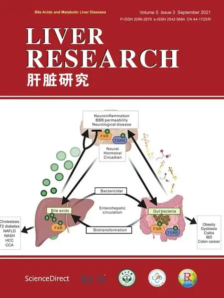
Liver Research的其它文章
- Bile acid metabolism and bile acid receptor signaling in metabolic diseases and therapy☆
- Gut microbiome in liver pathophysiology and cholestatic liver disease☆
- Mitigation of cholestasis-associated hepatic and renal injury by edaravone treatment:Evaluation of its effects on oxidative stress and mitochondrial function☆
- Metformin alleviates cholestasis-associated nephropathy through regulating oxidative stress and mitochondrial function☆
- Bile acids and metabolic surgery☆
- Farnesoid X receptor and fibroblast growth factor 15/19 as pharmacological targets☆