Bile acid receptors and signaling crosstalk in the liver,gut and brain☆
2021-10-11JessicaFerrellJohnChiang
Jessica M.Ferrell,John Y.L.Chiang
Integrative Medical Sciences,Northeast Ohio Medical University,Rootstown,OH,USA
Keywords:Bile acid metabolism Farnesoid X receptor (FXR)Gut-brain axis Microbiome Neurodegenerative disease Takeda G protein-coupled receptor (TGR5)
ABSTRACT Bile acids are physiological detergents derived from cholesterol that aid in digestion and nutrient absorption,and they play roles in glucose,lipid,and energy metabolism and in gut microbiome and metabolic homeostasis.Bile acids mediate crosstalk between the liver and gut through bactericidal modulation of the gut microbiome,while gut microbes influence the composition of the circulating bile acid pool.Recent research indicates bile acids may also be important mediators of neurological disease by acting as peripheral signaling molecules that activate bile acid receptors in the blood-brain barrier and in the brain itself.This review highlights the role of bile acids in maintaining liver and gut microbe homeostasis,as well as their function as mediators of cellular signaling in the liver-gut-brain axis.
1.Bile acid synthesis in the liver-gut axis
1.1.Primary bile acid synthesis in the liver
Bile acids are physiological detergents that solubilize dietary fats,vitamins,and xenobiotics so they can be absorbed across the intestinal epithelium.They also serve as metabolic regulators that mediate glucose and lipid homeostasis in the liver and small intestine via receptor binding,and they interact with gut microbes to prevent bacterial overgrowth.Bile acids are synthesized from cholesterol in the liver by either the classical or the alternative pathway and are the major source of cholesterol catabolism and removal in humans.The liver exclusively contains all the enzymes required for the classical bile acid synthesis pathway while other tissues,including macrophages,adrenal glands and the brain,have the alternative bile acid synthesis enzymes to metabolize cholesterol to oxysterols and bile acid intermediates that can be shunted to the liver for further synthesis of primary bile acids.1
The classical bile acid synthesis pathway is initiated in the endoplasmic reticulum of hepatocytes by the rate-limiting enzyme cholesterol 7alpha-hydroxylase (CYP7A1) to form 7α-hydroxycholesterol,which is then converted to 7α-hydroxy-4-cholesten-3-one (C4) by hydroxysteroid dehydrogenase.C4 is the metabolic precursor for the two major primary bile acids in humans,cholic acid(CA)and chenodeoxycholic acid(CDCA),and serum C4 is used as a biomarker for bile acid synthesis.2Sterol 12α-hydroxylase(CYP8B1) catalyzes 12α-hydroxylation of C4 to produce CA;without this step CDCA is formed.Thus,CYP8B1 determines the ratio of CA to CDCA and the hydrophobicity of the circulating bile acid pool.Steroid side-chain oxidation is catalyzed by mitochondrial sterol 27-hydroxylase (CYP27A1) and cleavage of the steroid side-chain by beta (β)-oxidation in peroxisomes forms cholyl-CoA and chenodeoxycholyl-CoA.Finally,conjugation to the amino acids taurine(T)or glycine(G)by bile acid-CoA:amino aci.N-acyl transferase forms T/GCA and T/GCDCA,which reduces hydrophobicity and increases the solubility of these bile acids.In mice,the murine-specific enzyme sterol 6-β-hydroxylase(Cyp2c70)converts CDCA to α-muricholic acid(α-MCA),which can then be epimerized to β-MCA by 7-α/β-epimerase.3
The alternative synthesis pathway begins with the hydroxylation of cholesterol by CYP27A1,which is expressed in the liver,macrophages,adrenal glands,and some regions of the brain,forming 27-hydroxycholesterol.Oxysterol 7α-hydroxylase(CYP7B1) then catalyzes hydroxylation at the 7α position to form 7α-27-dihydroxycholesterol,which can be converted to CDCA and CA in the liver.In macrophages,both CYP27A1 and CYP7B1 are involved in the production of oxysterol intermediates that can be shunted to the liver for bile acid synthesis.In the brain,cholesterol is hydroxylated by sterol 24-hydroxylase (CYP46A1) to form 24-hydroxycholesterol,which can then be transported across the blood-brain barrier (BBB) and taken up by the liver where it is converted to 7α-24-dihydroxycholesterol by 24-hydroxyc holesterol 7α-hydroxylase (CYP39A1).4,5Fig.1 shows the key regulatory enzymes involved in bile acid synthesis in the liver-gut axis in humans and mice.Detailed bile acid biosynthesis pathways can be found in a recent review.6

Fig.1.Bile acid metabolism and feedback regulation.
Conjugated bile acids are transported from hepatocytes across the canalicular membrane into bile via bile salt export pump(BSEP).A small amount may be reabsorbed by cholangiocytes,transported back to hepatocytes,and re-secreted into bile(forming the cholehepatic shunt).Bile formation in the biliary system also involves the transport of other organic molecules.Phospholipids are transported from hepatocytes into bile by multidrug resistant protein 3,while cholesterol is transported by adenosine triphosphate (ATP)-binding cassette subfamily G member 5/8 protein.Bilirubin and glutathione are transported via multidrug resistanceassociated protein 2,and cholangiocytes also secrete bicarbonate,water,and ions into bile.Bile acids,phospholipids,and cholesterol form mixed micelles and the bile is stored in the gallbladder until meal intake.7
1.2.Gut microbiota and bile acid biotransformation
Upon release from the gallbladder postprandially,T/GCA and T/GCDCA are deconjugated in the small intestine by bacterial bile salt hydrolase (BSH).BSH is widely expressed throughout major bacterial phyla,and expression is high in gener.Lactobacillus.Bifidobacterium,Enterococcus,Bacteroides,andClostridium.8Deconjugation of bile acids by bacterial BSH may enhance bile acid tolerance and survival of bacteria in the gut.BSH overexpression in mice reduced weight,serum cholesterol and liver triglycerides by regulating transcription of genes involved in lipid and cholesterol metabolism and circadian rhythms in the liver and small intestine.9In the colon,CA and CDCA are dehydroxylated by bacterial 7α-dehydroxylase,forming the secondary bile acids deoxycholic acid(DCA)and lithocholic acid(LCA),respectively.This multiple-step 7α-dehydroxylase activity may be limited to anaerobic species of the phylum Firmicutes,and has been identified i.Clostridium(clusters I,XI,XIVa).Eubacterium,an.Ruminococcus.10,11.Clostridiumcluster XIVa plays a central role in bile acid composition,metabolism and metabolic diseases.The bile acidinducible (Bai) operon in severa.Clostridiumclusters have been sequenced and th.Baigenes encoding bile acid 7α-dehydroxylase(BaiE) and 7β-dehydroxylase (BaiI) have been identified.12In humans,a small amount of CDCA is dehydrogenated and epimerized to the highly soluble ursodeoxycholic acid (UDCA) by 7αand 7β-hydroxysteroid dehydrogenases (7α/β-HSDH) in severa.Clostridiumclusters,Bacteroides fragilisan.Escherichia coli.13Thus,bile acids regulate gut bacteria growth and composition,which reciprocally regulates the circulating bile acid pool size and composition to affect host metabolism and physiology (Fig.1).In addition,physiological concentrations of intestinal bile acids protect agains.Clostridium difficileinfection in gnotobiotic mice and 7α-dehydroxylation of bile acids b.C.scindensis associated with resistance t.C.difficilecolonization and pathogenesis.14
Bile acids and gut bacteria exist in a bi-directional relationship.As discussed above,primary bile acids are transformed to secondary bile acids through the actions of gut bacteria that express BSH and 7α-dehydroxylase.Conversely,bile acids act as bactericidal agents that prevent bacterial overgrowth in the intestine and colon through the degradation of the bacterial membrane and damage to bacterial DNA.A recent study demonstrated that taurocholic acid(TCA) and DCA feeding in mice resulted in greater toxicity to bacterial communities and greater impairment of bacterial metabolism compared to LCA,possibly due to efficient detoxification of LCA.15Interestingly,another study demonstrated that hydrophilic β-MCA displayed similar bactericidal activity as the more hydrophobic and toxic DCA and CDCA,indicating a potential fundamental difference in controlling gut bacterial growth between humans and rodents.16Bile acids conjugated with quaternary aluminum or choline and other amino acids are currently being studied as antimicrobials and may represent a novel therapeutic approach to combat antibiotic resistance.17
1.3.Enterohepatic circulation of bile acids
Postprandially,bile is released into the duodenum to aid in fat and nutrient absorption.Most bile acids are reabsorbed in the terminal ileum by apical sodium bile acid transporter (ASBT).In enterocytes,bile acids bind to ileal bile acid-binding protein and are secreted from the sinusoidal membrane into portal circulation by organic solute transporter α and β(OSTα/β).These bile acids return to the liver and re-enter hepatocytes via sodium taurocholate cotransport peptide (NTCP),completing the cycle of enterohepatic circulation.18The enterohepatic circulation of bile acids is an important physiological process for intestinal absorption of nutrients and drugs which are then transported to the liver for metabolism and distribution to other tissues.The enterohepatic circulation of bile acids is highly efficient and provides feedback regulation of bile acid synthesis in the liver to maintain metabolic homeostasis (Fig.1).
1.4.Dysbiosis
Diet,drugs,antibiotics,alcohol,circadian disruption,and hormones can alter gut microbial populations to have negative effects on host metabolism,termed dysbiosis.Dysbiosis is a clinical feature of many conditions and can be characterized by a loss of beneficial gut bacteria,overgrowth of pathogenic bacteria,and/or a loss of biological diversity in gut bacterial populations.Cirrhotic patients with low levels of serum and fecal bile acids were shown to have bacterial dysbiosis,19indicating that reduced bile acid function may contribute to bacterial overgrowth in these patients.This study also demonstrated that advanced cirrhosis was associated with decreased conversion of primary bile acids to secondary bile acids,with increased Enterobacteriaceae and reduced Blautia(which has 7α-dehydroxylase activity).Conversely,patients with fatty liver disease and non-alcoholic steatohepatitis (NASH) had increased fecal primary bile acids that correlated to intestinal dysbiosis.20Dysbiosis is also present in patients with obesity and Type 2 diabetes,often manifesting as an increased ratio o.Firmicutest.Bacteroidetes,21and high fat and high sugar diets may increase inflammation via alteration of the gut microbiome.22,23
2.Bile acid-activated receptors
The synthesis,absorption,and secretion of bile acids are tightly regulated to ensure a constant bile acid pool and to prevent cellular toxicity.Bile acid pool size and composition may be altered by diet,dysbiosis,or liver-related disease.Several receptors are activated by bile acids and bile acid metabolites,including the nuclear receptors farnesoid X receptor(FXR),pregnane X receptor(PXR),vitamin D3receptor(VDR)and constitutive androstane receptor(CAR),and the G protein-coupled receptors Takeda G protein receptor 5 (TGR5),and sphingosine 1-phosphate receptor 2(S1PR2).24These receptors mediate bile acid,glucose,and lipid homeostasis as well as xenobiotic detoxification and inflammatory processes.The metabolic functions of these bile acid receptors are briefly described.
2.1.FXR
FXR is widely distributed in the liver and gut and acts as a metabolic feedback sensor for bile acid synthesis by binding bile acids (CDCA >DCA >LCA >CA).25Upon bile acid binding,FXR induces transcription of the nuclear receptor repressor small heterodimer partner(SHP),which inhibits the transcription o.CYP7A1an.CYP8B1in hepatocytes.In enterocytes,bile acids activate FXR to induce fibroblast growth factor 19 (FGF19;mouse homolog Fgf15)secretion into the portal circulation;FGF19/Fgf15 then binds to the membrane fibroblast growth factor receptor 4 (FGFR4)/β-Klotho complex in hepatocytes (Fig.1).FGF19/Fgf15 triggers c-Jun and extracellular signal regulated kinase 1/2 (ERK1/2) signaling of the mitogen-activated protein kinase pathway to inhibit CYP7A1.26,27FXR also prevents bile acid toxicity by regulating the secretion and reabsorption of bile acids in hepatocytes;activation of FXR induces canicular BSEP and inhibits sinusoidal NTCP,resulting in an overall net reduced concentration of bile acids within hepatocytes.Lastly,FXR signaling represents a critical component of the livergut axis responsible for maintaining bile acid homeostasis and preventing bile acid-induced liver injury.
In addition to mediating bile acid feedback,FXR regulates lipid and cholesterol transport and lipoprotein metabolism as evidenced i.Fxr-/-mice.These animals have increased serum high-density lipoprotein,low-density lipoprotein,and very low-density lipoprotein cholesterol and increased serum triglycerides,in addition to increased bile acid synthesis.28Age.Fxr-/-mice develop spontaneous hepatocellular carcinoma (HCC) while FXR agonism suppresses HCC and reduces xenograft growth.29,30The functional binding sites of FXR are highly tissue-specific,with only 11%of sites shared between the liver and intestine.31Consequently,liver- and intestine-specific studies were designed to further evaluate the functions of FXR.Liver-specific deletion of FXR in mice fed a cholesterol-containing diet resulted in increased lipid accumulation which was not present in intestine-specifi.Fxr-/-mice.32In contrast,liver-specifi.Fxr/Shpdouble knockout mice exhibited an antisteatotic phenotype with improved glucose metabolism,possibly due to reduced hepatic triglyceride synthesis or increased autophagy.33Selective activation of intestinal FXR protected mice from cholestatic liver disease,in part through the suppression o.Cyp7a1by Fgf15 and reduction of the bile acid pool.34
FXR is an attractive therapeutic target for the treatment of cholestatic liver diseases,including primary biliary cholangitis(PBC)and primary sclerosing cholangitis,and for the prevention of metabolic-associated fatty liver disease.Global FXR agonism reduces lipogenesis via the suppression of sterol regulatory elementbinding protein 1c (SREBP-1c) and CCAAT/enhancer-binding protein expression,35which likely occurs through both SHP-dependent and SHP-independent pathways.36FXR agonism also improves insulin resistance and reduces hepatic expression of gluconeogenic genes in mice.37FXR interacts with the pro-inflammatory nuclear factor κ B (NF-κB) or stabilizes its co-repressor NCoR to suppress pro-inflammatory cytokine production.38,39The potent FXR agonist obeticholic acid,a semi-synthetic derivative of CDCA,is the secondline therapy for PBC and in clinical trials for NASH,as are the nonbile acid FXR agonists cilofexor and tropifexor.40,41Interestingly,the intestine-restricted FXR agonist fexaramine reduced weight gain and serum glucose,insulin,leptin,and cholesterol levels while also promoting white adipose browning in mice,42indicating that both liver and gut FXR may play an important role in regulating bile acid homeostasis and preventing liver disease.
FXR antagonism may also affect peripheral metabolic health.Short-term administration of UDCA to morbidly obese patients resulted in reduction of hepatic and serum cholesterol,which was attributed to increased bile acid synthesis due to presumed FXR antagonism.43Mice fed UDCA for 8 weeks had reduced intestinal FXR,SHP and FGF15 protein levels,coupled with accelerated turnover of bile acid circulation.44Finally,glycoursodeoxycholic acid (GUDCA) was identified as an FXR antagonist in a seminal study of human patients with metformin-treated Type 2 diabetes.45These patients had increased GUDCA,reduce.B.fragilis,and inhibition of FXR signaling.When obese mice were treated wit.B.fragilis,the beneficial effects of metformin were reversed,while GUDCA treatment improved insulin resistance in an FXRdependent manner.45Overall,this study suggests that metformin may act through the inhibition of intestinal FXR to improve glycemia and other metabolic defects.
2.2.TGR5
TGR5 is a membrane G protein-coupled bile acid receptor expressed in cholangiocytes,stellate cells,sinusoidal endothelial cells and Kupffer cells of the liver.It is also expressed in gallbladder,skeletal muscle,brown adipose tissue,and neurons and astrocytes.Activation of TGR5 by bile acids(LCA ≥DCA >CDCA >CA)induces an intracellular accumulation of cyclic adenosine monophosphate(cAMP) that leads to tissue-specific cell signaling cascades.46A recent study identified UDCA as a TGR5 agonist that could exert beneficial effects in a mouse model of high-fat diet-induced NASH.47Likewise,dual agonism of FXR and TGR5 stimulated adipose tissue browning and reversed high fat diet-induced hepatic steatosis and fibrosis in mice.48In the pancreas,TGR5 mediates crosstalk between α and β cells to stimulate insulin sensitivity,49and in the gut it stimulates glucagon-like peptide-1 (GLP-1)release from enteroendocrine L cells.50TGR5 may mediate energy expenditure in brown adipose tissue through the activation of type 2 iodothyronine deiodinase,which converts thyroxine to tri-iodothyronine,51and TGR5 also stimulates gallbladder filling.52Lastly,TGR5 reduces inflammation through inhibition of NF-κB.53
Tgr5-/-mice have reduced gallbladder volume and a small bile acid pool size compared to wild-type controls,with reduced expression o.Cyp7a1an.Cyp7b1.54,55The mechanism by which TGR5 may regulate these enzymes in hepatocytes is unclear.Tgr5gene expression is regulated in part through FXR by an FXRresponsive element in th.Tgr5promoter,indicating that FXR and TGR5 may coordinately crosstalk to regulate metabolism in the liver and gut.56Indeed,many metabolic phenotypes,including gallbladder filling,liver regeneration,adipose tissue browning and protection against metabolic insults are promoted by either FXR or TGR5 specific ligands.
Several studies implicate TGR5 activation by bile acids in the improvement of metabolic symptoms following metabolic bariatric surgery (MBS),often occurring prior to weight loss.MBS,such as vertical sleeve gastrectomy (VSG) and Roux-en-Y gastric bypass,increases serum bile acids and FGF19 which correlate to increased serum GLP-1.Both FXR and TGR5 have been shown to be involved in improving metabolism and insulin sensitivity after MBS.57-59Diet-induced obese mice that received VSG exhibited body weight-independent improvements in glucose tolerance,hepatic inflammation,insulin signaling,and islet morphology,and these effects were blunted i.Tgr5-/-mice.59VSG did not improve glycemic control i.Tgr5-/-mice,suggesting a critical role for TGR5 in promoting insulin resistance.Lastly,this study demonstrated a TGR5-dependent reduction i.Cyp8b1expression after VSG,reflected by an increase in 12α-OH bile acids i.Tgr5-/-mice that received VSG.59VSG performed in mice and humans resulted in a significant increase in cholic acid-7-sulfate (CA7S) in the gut and feces,which increase.Tgr5expression to improve insulin sensitivity and glucose intolerance.60Interestingly,these investigators determined that VSG decreased gut bacteria of clas.Clostridiaas well as LCA production,but increased the expression of ileum bile acid uptake transporter ASBT and the ileum bile acid efflux transporter OSTα.It is not clear how VSG increased the expression of these ileum bile acid transporters,though bile acids are known to reduce ASBT expression and induce OSTα expression via FXR.These investigators suggested that increasing bile acid efflux to portal blood might increase portal vein LCA,which activates hepatic VDR to induce sulfur transferase 2A1 and CA7S production.60These studies suggest that VSG may improve metabolism through gut bacteria,and CA7S could be utilized as a non-invasive method for reducing weight and treating diabetes.
2.3.PXR,CAR and VDR
PXR and CAR are mainly expressed in the liver and gut and play an important role in drug metabolism,conjugation and transport.61Activation of either PXR or CAR induces heterodimerization with retinoid X receptor and the subsequent activation of gene transcription.PXR downstream targets include the CYP3A family of xenobiotic processing enzymes,while CAR induces CYP2B enzymes.62LCA is a potent PXR ligand that stimulates all three phases of drug metabolism and detoxification.63It has been reported that guggulsterone,an FXR antagonist,activates PXR to inhibi.CYP7A1gene transcription.64Rifampicin promotes PXR binding to the huma.CYP7A1gene promoter to block transcription factor-mediate.CYP7A1gene transcription,65while loss of PXR sensitizes mice to lithogenic diet-induced cholesterol gallstone formation.66PXR also protects against LCA- and drug-induced hepatoxicity by inducing sulfotransferases,which conjugate sulfur to LCA and xenobiotics to promote excretion.67Bile acids do not bind CAR,but activation of CAR by an inverse agonist induced sulfotransferase to reduce LCA toxicity.68Conversely,deficiency of both PXR and CAR increased LCA toxicity and lipid accumulation in mouse liver.69
In recent years,the roles of these nuclear receptors expanded to include metabolic regulation.PXR and CAR play a role in energy metabolism through regulation of fatty acid,lipid and glucose metabolism.70,71CAR and PXR were shown to downregulate SREBP-1c to reduce hepatic lipid accumulation,which was dependent upon insulin induced gene-1 protein.72Treating obese mice with a CAR agonist reduced hepatic steatosis and improved insulin sensitivity,whil.Car-/-mice developed insulin intolerance.73A recent comprehensive metabolomic study found that CAR agonism in mice significantly reduced hepatic gluconeogenesis and paradoxically,increased hepatic levels of genes involved i.de novofatty acid synthesis,but these changes were altered over different times.74Activation of PXR induced hypercholesterolemia and atherosclerosis i.apoE-deficient mice whil.Pxr-/-mice were protected against diet-induced obesity via induction of FGF15.75,76Conversely,the novel PXR agonist ginkgolide B reduced the expression of lipogenic genes in mice,which were also protected from diet-induced steatosis.77
Another nuclear receptor,VDR,is activated by 1,25-dihydroxyvitamin D3 and plays a critical role in calcium homeostasis,bone formation and anti-inflammation.LCA is a potent VDR ligand and activates VDR in the intestine to induce CYP3A4,a predominant drug metabolizing cytochrome P450.78VDR is not expressed in mouse hepatocytes,but LCA activation of VDR in hepatic stellate cells inhibited hepatic fibrogenesis,79and in human hepatocytes LCA activated nuclear VDR to inhibit CYP7A1.80,81VDR is strongly expressed in cholangiocytes and plays an antiinflammatory role in cholangiopathy.82.Vdr-/-mice are dysbiotic with reduce.Lactobacillus and increase.Bacteroidesan.Clostridium,83while intestine-specifi.Vdr-/-mice have a significantly carcinogenic fecal bacterial profile.84
2.4.S1PR2
S1PR2 is a G protein-coupled receptor widely expressed in hepatocytes,cholangiocytes and gut epithelial cells that binds sphinogosine-1-phosphate as well as bile acids.In the liver,conjugated bile acids activate S1PR2 to mediate glucose and lipid homeostasis through ERK 1/2 and protein kinase B (AKT)signaling pathways.85.S1pr2-/-mice rapidly developed liver steatosis when fed either a normal or high fat diet which was accompanied by reduced mRNA expression of key genes involved in lipid metabolism.86Interestingly,S1PR2 was found to be the dominant S1PR expressed in cholangiocytes.87This study also found S1PR2 was upregulated by bile duct ligation(BDL)in mice,an.S1pr2-/-mice were protected against BDL-induced cholestatic injury.Similarly,blockade of S1PR2 signaling by the antagonist JTE-013 significantly reduced inflammasome activation caused by BDL in mice,88and macrophage-specific knockdown of S1PR2 attenuated liver fibrosis and inflammation caused by BDL.89.S1pr2-/-mice also have exacerbated intestinal barrier damage,90though another study demonstrated that DCA can upregulate S1PR2 and lead to more severe colitis in mice.91
3.Bile acids in the liver-gut-brain axis
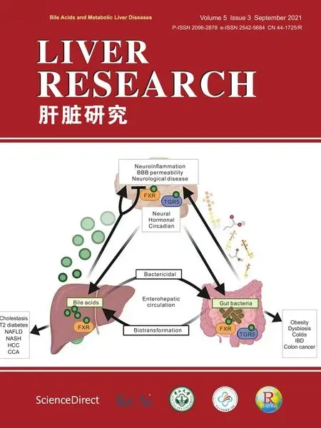
The liver-gut-brain axis is crucial for the maintenance of metabolic homeostasis and is based on neural and hormonal communication signals between the central nervous system(CNS),the peripheral nervous system (PNS) and the liver,intestine,and gut microbiota (Fig.2).Sensory signals from the periphery,including gastric distension and nutrient activation of gut receptors,trigger hormone release that feeds back to the gut,PNS and CNS to signal food intake,nutrient absorption,and satiety.The CNS exerts its own neurohormonal and circadian control over meal size,gut motility,hepatic glucose use,and immune responses.Meanwhile,the gut microbiome mediates signaling through the excretion of metabolites such as short-chain fatty acids and the inflammatory endotoxin lipopolysaccharide (LPS),while bile acids regulate host metabolism and functions of distant organs.92
3.1.Bile acids and their receptors in the brain
Several studies demonstrated the presence of bile acids in the cerebrospinal fluid (CSF) of rodent models and humans,but it is unclear whether these bile acids were synthesized in neural cells or if they originate from the systemic circulation.93The full suite of enzymes required for the classic synthesis pathway exists only in the liver but neurons produce 24-hydroxycholesterol,a cerebral sterol synthesized via CYP46A1,that can be transported to the liver for further bile acid synthesis.Bile acid transporters (including BSEP,NTCP,and ASBT)and bile acid receptors have been identified in the brain,recently reviewed by Merten.et al.94Thus,systemic circulating bile acids may cross the BBB,the tightly-regulated network of brain microvascular endothelial cells that utilize tight junction proteins and transport proteins to securely protect neurons of the CNS from the contents of the circulatory system.A recent study in rats confirmed that CA,DCA,and CDCA detected in the brain correlated to concentrations in the serum,and that bile acids may passively diffuse from the periphery.95C27and C24bile acid intermediates have been detected in human CSF,96and later studies confirmed the CYP27A1 metabolite 7α,25-dihydroxy-3-oxo-cholest-4-en-26-oic acid in CSF and its significant reduction in patients with Alzheimer's disease (AD).97,98
Bile acids may influence the function of the brain and CNS through activation of bile acid receptors and through modulation of the gut microbiome.The receptor for FGF15/19,FGFR4,is expressed in the rodent brain and suppressed in rats fed a high-fat diet.Several studies indicate that central administration of FGF19 improves glucose tolerance and insulin sensitivity in rodents;99,100however,the transport of FGF19 across the blood-brain barrier may be inefficient.101.Fgf15 mRNA expression was identified in mouse brain and significantly upregulated in response to a high-fat diet.102
Both FXR and TGR5 have been detected in the brains of mice and humans (Fig.3).103-105TGR5 may have several distinct functions within the CNS and PNS;it is expressed in both neurons and astrocytes,it reduces neuroinflammation and microglial activation,106and its activity was induced by neurosteroids and suppressed by ammonia.107It was first thought that bile acids may activate TGR5 on sensory nerves to mediate itch and analgesia during cholestatic liver disease.108,109However,a bile acid receptor for cholestatic itching has recently been identified in human dorsal root ganglia and is co-expressed with the itch receptor histamine receptor H1(HRH1).110,111This study also showed that a TGR5-specific agonist failed to reduce itching in human subjects.111
The role of FXR in neuronal function is far less understood.It was recently demonstrated that FXR may be differentially distributed in neurons,with cultured neurons expressing FXR mainly in the nucleus an.in vivopreparations expressing FXR in the cytoplasm.104Another study confirmed FXR localization to the neuronal cytoplasm under basal conditions,which was shifted to the nucleus after treatment with bile acids,112indicating that bile acids may influence the activity of neuronal FXR similarly to what occurs in the periphery.Interestingly.Fxr-/-mice exhibited reduced depressive-and anxiety-like behaviors,which was associated with altered neurotransmitter expression including increased γ-aminobutyric acid in the hippocampus and increased norepinephrine and 5-hydroxyindole acetic acid (a metabolite of serotonin) in the cerebellum.113In mice exposed to stress-induced depression,hippocampa.Fxrgene expression was significantly upregulated,while disrupting FXR signaling reduced stress-induced depressionlike behaviors in these mice.114

Fig.2.Bile acids in the liver-gut-brain axis.
The function of S1PR2 within the CNS is also unclear.S1PR2 may play a role in axonal inhibition and synaptic plasticity by acting as a receptor for the negative outgrowth protein Nogo-A.115.S1pr2-/-mice exhibit spontaneous seizures coupled with increased gliosis and dysfunction in spatial memory,116and interestingly,S1PR2 protein expression was downregulated in epileptic rats and patients with temporal lobe epilepsy.117With respect to bile acids,TCA treatment in a mouse model of hepatic encephalopathy (HE)activated S1PR2 signaling to increase inflammatory gene expression,while pharmacological blockade of S1PR2 reduced neuroinflammation.118In a mouse model of ischemic stroke,blocking S1PR2 protected the BBB and prevented injury-induced reductions in tight junction proteins.119The roles bile acids may play in exerting protective effects via TGR5 and potentially detrimental effects via FXR and S1PR2 are still unknown.
CAR and PXR have been identified in rodent and human brain tissue,particularly in the cortex and the endothelial cells that make up the BBB in the cortex.Car-/-mice have memory impairment and anxiety-like behavior,and peripheral administration of the neurotoxin kainic acid induced convulsions in these mice,but not in wild-type mice,indicatin.Car-/-mice may have increased BBB permeability.120PXR activation by rifampicin or hyperfori.in vitroresulted in upregulation of the ATP-binding cassette (ABC) drug transporters brain multidrug resistance protein an.P-glycoprotein(PG),121whil.in vivodexamethasone-mediated activation of PXR increased PG in rats.Activation of both PXR and CAR induced the expression of PG in human cerebral endothelial cells,122while inhibition of CAR prevented drug-induced increases in PG in rat brain microvessels.123VDR is widely expressed in the brain and this receptor has been localized to the neuronal plasma membrane and astrocytes.124,125In rat brain capillaries,activation of VDR upregulated PG,indicating that VDR may be involved in mediating substrate and drug efflux from the brain across the BBB.126Modulation of the nuclear receptors that regulate the permeability of the BBB,like PXR,CAR and VDR,maybe a novel approach to prevent the drug resistance that is often endemic to the management of chronic neuropathological conditions.127

Fig.3.Function of bile acid receptor in the brain.
3.2.Circadian regulation of the liver-gut-brain axis
Circadian rhythms exist in nearly all organisms and ensure that internal physiology is temporally synchronized to the external environment,important for both homeostasis and adaptation.Circadian rhythms are endogenously generated by the suprachiasmatic nucleus of the hypothalamus through a functional transcriptional translational feedback loop comprised of core clock genes and proteins.The forward limb of the clock is driven by circadian locomotor output cycles kaput (CLOCK) and brain-andmuscle ARNT-like 1 (BMAL1),while period (PER1/2) and cryptochrome (CRY1/2) modulat.Clock/Bmalgene transcription.A secondary regulatory loop is maintained by REV-ERBα and retinoidrelated orphan receptor alpha (RORα),and altogether these proteins oscillate with a period of approximately 24 h.Rhythms are mainly entrained by photic signals(daily environmental light/dark cues) but are also influenced by non-photic signals like social interaction,food,and locomotor activity.These input signals can influence the timing of the clock,which then produces rhythmic biochemical,hormonal,and behavioral physiological outputs.In humans,circadian misalignment can occur with shift work,jet lag,or chronic sleep loss,and can desynchronize internal physiology from the external environment to contribute to the pathology of metabolic syndrome and liver disease,128,129cancer,130,131cardiovascular diseases,132,133neurodegenerative diseases,134,135and mood disorders.136,137
Within the liver,bile acid homeostasis is regulated by circadian rhythms and circadian disruption alters the normal rhythm o.Cyp7a1gene expression in mice.138Likewise,hepatic Krüppel-like factor 15 regulates Fgf15.Cyp7a1gene expression,and bile acid synthesis in a circadian fashion.139In mice,chronic circadian disruption led to global deregulation of lipid and bile acid metabolic pathways,and interestingly.Fxr-/-mice were more susceptible to HCC induced by chronic circadian disruption whil.Car-/-mice were protected.140
Disruptions to circadian clock function contributes to disease progression,as evidenced extensively i.Clockknockout mouse models.ClockΔ19 mutant mice are obese and hyperphagic,141and bot.ClockΔ19 mutant mice an.Bmal1-/-mice exhibit reduced insulin secretory responses.142Interestingly,pancreatic-specific ablation o.Bmal1was sufficient to induce hyperglycemia and hypoinsulinemia in mice,indicating individual tissue clocks may be critical to maintaining metabolic homeostasis independent of rhythmic behavior.142Similarly.Cry1/2double knockout mice and liver-specifi.Bmal1o.Rev-Erbα knockout mice were obese and hyperphagic when give.ad libitumaccess to a high-fat diet.When fed an isocaloric portion of high fat-diet restricted only to the active(dark) phase,these mice were lean with reduced liver and serum lipids compared t.ad libitum-fed mice.143.Per1/2double knockout mice were shown to have elevated serum and hepatic bile acids coupled with a complete loss of the circadian expression o.Cyp7a1an.Cyp8b1.144Studies such as these illustrate the bidirectional relationships between clock-generated rhythms that influence hepatic metabolism and how timing of peripheral metabolic signals may override the effects of clock defects.
Circadian rhythms also alter the gut microbiota,and gut bacteria may influence rhythms.Unconjugated bile acids,generated by gut microbes,altered the expression of circadian gene.in vitroand in mice,while equivalent tauro-conjugated bile acids had little effect.145While one study reported that sleep restriction in rats and human did not significantly alter the composition of the gut microbiome,146another demonstrated that a single sleep-cycle shift of 2-4 h produced changes in bot.Bacteroidesand subsequent downstream functional pathways in purine and short-chain fatty acid metabolism in humans.147Four weeks of circadian disruption in mice resulted in decreased intestinal barrier function that may be attributed to microbial changes and increased expression of genes responsible for expression and transport of LPS.148Another recent study demonstrated that microbial diversity was significantly and positively correlated with sleep efficiency and total sleep time,and was negatively correlated with sleep fragmentation in humans.149
4.Bile acids in neurological diseases
Bile acid physiology and signaling have been implicated in the treatment and etiology of many neurological conditions.Bile acids,particularly UDCA,have been studied for the treatment of AD,Huntington's disease,Parkinson's disease(PD),amyotrophic lateral sclerosis,multiple sclerosis,and retinal disease,among others.93,150The mechanisms of action are still unknown but may involve protection against apoptosis and modulation of neurotransmitter activity.TUDCA was first shown to ameliorate amyloid-β-induced apoptosis via a phosphatidylinositol 3-kinase (PI3K) dependent pathway.151Later studies demonstrated TUDCA inhibited apoptosis via p53 and the inhibition of PI3K reduced the protection against apoptosis by TUDCA.152PC-12 neuronal cells treated with nanospheres containing UDCA were protected against glutamateinduced toxicity via downregulation of cleaved caspase-3 protein,153and in rats exposed to subarachnoid hemorrhage TUDCA reduced BAX,cleaved caspase-3,and brain water content through a TGR5-sirtuin 3 signaling pathway.154Still,relatively little is known about the therapeutic potential for T/UDCA,other bile acids,and bile acid receptors in the treatment of neurological diseases.
4.1.BBB permeability
Bile acids can disrupt the integrity of the BBB,and disruption of the BBB is a hallmark feature of many neurological diseases that can lead to impaired glucose uptake and reduced metabolite clearance from neurons,and may potentially expose neurons to cytotoxic molecules.155Mice that had undergone BDL had significantly increased expression of matrix metalloproteinase-9 and tissue inhibitors of metalloproteinase in brain tissue coupled with loss of tight junction proteins,156while another study demonstrated that BBB tight junctions were disrupted and permeability was increased in rats that underwent BDL or bile acid injections.157Conversely,utilizing a.in vitromodel of the BBB,it was shown that G/UDCA reduced apoptosis induced by unconjugated bilirubin (UCB),and UDCA administration reduced cytokine release and prevented UCBinduced barrier permeability,158while activation of TGR5 reduced BBB breakdown in a model of ischemic stroke in rats.159The role of specific bile acids play in the protection of BBB structure and function versus their roles in mediating pathology are still unclear.
4.2.HE
HE is a neurological condition that comprises a spectrum of disorders ranging from personality changes,lethargy and disrupted sleep to cerebral edema,coma,and death,and can result from either acute or chronic liver failure.160A potential pathological mechanism involves excessive ammonia,which is synthesized through the deamination of glutamine in enterocytes and by urease-producing gut microbes.Ammonia may accumulate due to failing liver function and result in pro-inflammatory cytokine release,microglial activation and astrocyte swelling,and dysfunction in glutamatergic and dopaminergic signaling leading to further inability to clear ammonia from the brain.161However,first-line treatments that reduce ammonia may not sufficiently treat symptoms of HE,162and blood ammonia levels are not a reliable diagnostic marker for HE.163,164Rather,systemic inflammation,oxidative stress,and permeabilization of the BBB also contribute to the pathogenesis of HE.162
Bile acids may also be involved in the development of HE.The enterohepatic circulation of bile acids is often impaired during liver injury,due to release of bile acids from damaged hepatocytes,reduced reuptake of bile acids from the portal circulation,or both.This results in significantly increased serum levels of bile acids,which may serve as a stratification biomarker for cirrhosis and liver failure.165In a mouse model of acute liver failure and HE,elevated TCA was detected in cortical tissue,while treatment with the bile acid binding resin cholestyramine reduced bile acids,cerebral edema,neurological decline,and pro-inflammatory cytokine expression in these mice.112,118A recent study in patients with cirrhosis determined serum TCA positively correlated with disease severity,and that increased TCA may promote disease progression by activating hepatic stellate cells.166Concomitantly,elevated TCA,GCA,and GUDCA were confirmed in the CSF of patients with HE.167Cortical activity of Cyp46a1 was significantly reduced and resulted in FXR-dependent cholesterol accumulation in the brains of mice with liver failure,and preventing cholesterol accumulation improved neurological deficits in these mice.168Peripheral circulating bile acids may contribute to the pathogenesis of HE,and may also serve as a serum biomarker for the disease.
4.3.Alzheimer's disease (AD)
AD is a neurodegenerative disease characterized by a progressive decline in cognitive ability.AD is thought to be caused by accumulation of neuron-damaging lesions in the form of plaques,consisting of amyloid-β aggregates(Aβ),and neurofibrillary tangles consisting of hyperphosphorylated tau protein.169,170Central and peripheral changes in glucose and insulin metabolism,increased inflammation,and mitochondrial dysfunction may also play key roles in AD pathology.171,172Recently,liver disease and bile acids have come to the forefront of AD research,173and several studies identified altered bile acid composition and physiology in patients with AD.Of particular interest are two studies which demonstrated significantly increased secondary bile acids in the serum of AD patients,indicating a potential role for the gut microbiome in the development or pathology of AD.174,175Marksteine.et al.174identified LCA and its glycine- and tauro-conjugates as significantly upregulated in AD patients,with a 3.2-fold increase in plasma LCA detected during the progression from mild cognitive impairment to AD.Similarly,MahmoudianDehkord.et al.175found significant reduction of primary bile acids in the serum of patients with AD and a significant increase in serum DCA.The ratio of DCA to CA in serum was significantly associated with AD and worsened cognition,indicating bacterial dehydroxylation may be an important contributor to the altered bile acids profiles in patients with AD.
TUDCA is the most common bile acid studied in the context of AD.Long-term intraperitoneal(i.p.)injection of TUDCA reduced Aβ accumulation in the brains of amyloid precursor protein/presenilin 1 (APP/PS1) AD mice,both before and after the onset of amyloid pathology.176,177TUDCA also reduced Aβ-induced synaptic toxicity and cognitive impairment in these mice.178,179In a separate study,i.p.injection of TUDCA in streptozotocin-induced AD mice for 10 days improved both central and peripheral glucose homeostasis,and reduced neuroinflammation and improved memory.180Treating tissue derived from AD patients with TUDCA restored mitochondria membrane potential and rescued mitochondrial morphology.181Importantly,TUDCA combined with sodium phenylbutyrate (AMX0035) is in a phase II clinical trial for mild cognitive impairment and early dementia due to AD.
Limited data are available regarding the roles of bile acid receptors in AD pathology.TGR5 agonism conveyed neuroprotection against both Aβ-induced and LPS-induced AD symptoms.Mice receiving a single intracerebroventricular injection of the TGR5 agonist INT-777 were resistant to apoptosis and dendritic spine loss,182as well as microglial activation and behavioral deficits.183Conversely,FXR was shown to be upregulated in Aβ-induced neuronal apoptosis.Overexpression of FXR resulted in increased Aβ-induced neuronal apoptosis,whil.Fxrknockdown decreased Aβ-induced apoptosis in mouse hippocampal neurons.184Interestingly,VDR gene expression was reduced in APP/PS1 mouse hippocampus and cortex,185and VDR was shown to associate with APP and amyloid processing protein in neurons.125Coupled with localization to the neuronal membrane,this may imply the existence of non-genomic functions for VDR in the AD brain.These limited studies provide some initial insight into the potential physiological roles for bile acid signaling in the pathology and treatment of AD.
4.4.Parkinson's disease (PD)
PD results from the impairment or loss of dopaminergic neurons in the substantia nigra,with subsequent formation of Lewy bodies in the brain (composed of aggregates of the protein α-synuclein)and disordered locomotor movement.Mechanistically,neuroinflammation and mitochondrial dysfunction are thought to contribute to the pathology of PD.Patients with PD may also have dysbiosis resulting in abnormal bile acid metabolism.186Plasma metabolomic screening identified reduced TUDCA in serum of human PD patients and in a mouse model of PD,187,188while another study found patients with PD had increase serum CA,DCA,and LCA without changes in serum TUDCA.189
Conversely,bile acids may represent a novel therapeutic option for the treatment of PD.In a mouse model of PD,oral gavage of UDCA improved locomotor deficits and protected against the loss of dopaminergic neurons.in vitroUDCA prevented apoptosis and mitochondrial dysfunction.190Likewise,UDCA prevented apoptosis and inflammation and improved dopamine content and locomotor performance in a rat model of PD.191In a mouse model of chemically-induced PD.i.p.TUDCA attenuated both microglial activation and striatal loss of dopamine.192A small study in five patients with PD showed that UDCA was well-tolerated and might improve mitochondrial function in the form of increased ATP concentration and synthesis,193and UDCA is currently in a phase II clinical trial for the treatment of PD.194
4.5.Dysbiosis and neurological diseases
Gut dysbiosis is linked to systemic inflammation and may contribute to the pathogenesis of neurological conditions like HE,AD,PD,and mood disorders.Neurotransmitters like dopamine and serotonin can affect microbial populations and bacterial virulence in the gut.Conversely,metabolites produced by gut bacteria,including SCFA,can influence brain physiology and behavior.Patients with HE or liver failure may have increased bacterial translocation due to intestinal permeability and oxidative damage to the intestinal mucosa,resulting in increased circulation of inflammatory cytokines.195Recently,it was shown that a mouse model of familial AD had a significant enrichment o.Prevotella,a genus present within the human microbiome that may degrade intestinal mucin.196This study also demonstrated that antibiotic treatment in these mice reduced Aβ pathology and ameliorated cognitive impairment.Similarly,APP mice were shown to have age-related changes i.Bacteroidetesan.Firmicutes,and changes in bacterial families correlated to increased Aβ content.197Germ-free APP mice had reduced Aβ content,while colonization of these mice with conventional APP mice increased Aβ pathology.Patients with AD had reduce.Bifidobacterium,with increased representation o.Firmicutesand decrease.Bacteroidetes,198similar to patients with obesity and metabolic syndrome.21
Several studies have identified alterations in the gut microbiome of patients with PD.PD progression and worsened symptoms were associated with lower counts o.Bifidobacteriuman.Bacteroides,199while another study demonstrated decrease.Roseburiaan.Faecalbacteriumwith increase.Lactobacillaean.Akkermansia.200In contrast to AD,reduce.Prevotellawas associated with PD and may represent a prodromal marker for the disease.201,202
Metabolites and SCFA generated by the gut microbiome may be associated with progression of neurological disease,though the exact pathways of action are unclear.Colonization of germ-free mice with butyrate-,acetate-,or propionate-producing bacterial strains reduced BBB permeability,possibly through the upregulation of cell adhesion molecules.203A.in vitrostudy demonstrated that valeric acid,butyric acid,and propionic acid supplementation inhibited Aβ aggregation,204while APP/PS1 mice given acetate demonstrated improved cognitive performance.205Conversely,a study in human patients with dementia found a positive association between serum LPS,acetate,and valerate and brain amyloid load,while serum butyrate was negatively associated with amyloid load.206One recent study found that fecal SCFA were reduced in patients with PD compared to healthy controls,207while another found increased serum acetate associated with PD after adjusting for covariates.208In a mouse model of PD,butyrate reduced αsynuclein,suppressed microglial activation,and inhibited inflammation in the brain.209The use of metabolites like SCFA for therapeutics or biomarkers of neurological disease progression warrant further investigation.
Fecal microbiota transplant (FMT) studies have provided some insight into the mechanism by which gut microbes and their metabolites contribute to disease pathology.FMT reduced hospitalizations and improved cognition in patients with HE.210Interestingly,this was accompanied by an increased ratio of secondary to primary bile acids.Mice that received FMT from patients with cirrhosis had increased neuroinflammation which was attenuated with FMT from cirrhotic patients that had received FMT from a healthy donor.211Several studies indicate that depressiveand anxiety-like behaviors are reduced after FMT from healthy donors in rodents.212,213FMT from a mouse model of PD induced motor impairment in normal mice,while PD mice that received FMT from healthy mice had improved locomotor activity and increased striatal dopamine.214FMT improved motor and nonmotor symptoms in patients with PD,215though another study demonstrated this may be a temporary effect lasting only a few months.216FMT may develop into efficient and novel treatments for a variety of neurological conditions,though issues like safety and route of administration still require further research.
5.Conclusions and future perspectives
Physiological crosstalk between the brain,liver and gut is crucial for homeostatic maintenance of metabolism,and strong evidence demonstrates that disruption of this axis contributes to the liver,gut,and neurological disease pathology.Neurological conditions like HE,AD,PD,and mood disorders likely have peripheral contributors that exacerbate disease progression.Bile acids are being studied as potential signaling molecules that can mediate crosstalk signaling between the brain and periphery,as they play major roles in the regulation of liver and gut homeostasis.UDCA is in clinical trials for the treatment of AD and PD,and changes in bile acid composition may serve as biomarkers of neurological diseases.Research efforts should be focused on integrative approaches to better understand the interactions of these physiological systems;concurrently,targeting bile acids,bile acid receptors,and the gut microbiome may lead to novel therapeutics beyond the liver and gut.
Authors’ contributions
J.M.Ferrell developed the concept and wrote the manuscript;J.Y.L.Chiang developed the concept and edited the manuscript.
Declaration of competing interest
The authors declare that they have no conflict of interest.
Acknowledgements
This work was supported by the USA National Institutes of Health AA015951 to J.M.Ferrell,and DK44442 and DK58379 to J.Y.L.Chiang.
杂志排行
Liver Research的其它文章
- Bile acid metabolism and bile acid receptor signaling in metabolic diseases and therapy☆
- Gut microbiome in liver pathophysiology and cholestatic liver disease☆
- Mitigation of cholestasis-associated hepatic and renal injury by edaravone treatment:Evaluation of its effects on oxidative stress and mitochondrial function☆
- Metformin alleviates cholestasis-associated nephropathy through regulating oxidative stress and mitochondrial function☆
- Bile acids and metabolic surgery☆
- Farnesoid X receptor and fibroblast growth factor 15/19 as pharmacological targets☆