剪切波弹性成像联合超声造影评价颈动脉斑块稳定性的临床研究
2021-05-08粘迪
粘迪

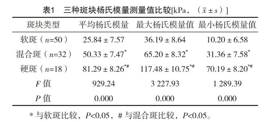
【摘要】 目的:探討剪切波弹性成像(shear wave elastography,SWE)结合超声造影(CEUS)评价颈动脉斑块稳定性的临床价值。方法:选取2017年1月-2018年12月本院收治的患有动脉斑块的患者75例作为研究对象,均对其行CEUS及SWE检查。观察斑块回声类型、厚度、所致狭窄率。比较不同类型斑块的剪切波传播速度(SWV)及杨氏模量。结果:75例患者中常规超声共检出100处颈动脉板块,根据斑块回声图像的特点将斑块分为低回声斑块(软斑)50处,斑块厚度1.5~4.9 mm,斑块所致狭窄率14%~67%;混合回声斑块(混合斑)32处,斑块厚度1.5~5.1 mm,斑块所致狭窄率17%~86%;强回声斑块(硬斑)18处,斑块厚度1.5~4.8 mm,斑块所致狭窄率15%~67%。100处颈动脉板块中,混合回声斑块中有2处斑块、强回声斑块中有6处斑块测不出SWV。三种回声的斑块的SWV比较,差异均有统计学意义(P<0.05),其中强回声斑块SWV值高于低回声斑块(P<0.05);混合回声斑块SWV高于低回声斑块(P<0.05);强回声斑块SWV高于混合回声斑块(P<0.05)。三种斑块平均杨氏模量、最大杨氏模量值及最小杨氏模量值比较,差异均有统计学意义(P<0.05)。软斑的平均杨氏模量、最大杨氏模量值及最小杨氏模量值均低于混合斑和硬斑,其中混合斑均低于硬斑(P<0.05)。结论:SWE结合CEUS能够更好地显示颈动脉斑块内的结构特征,有助于颈动脉斑块稳定性的评估。
【关键词】 剪切波弹性成像 超声造影 颈动脉斑块 稳定性
Evaluation of Carotid Plaque Stability by Shear Wave Elastography combined with Contrast-enhanced Ultrasound/NIAN Di. //Medical Innovation of China, 2021, 18(05): 117-121
[Abstract] Objective: To investigate the clinical value of shear wave elastography (SWE) combined with contrast-enhanced ultrasound (CEUS) in evaluating the stability of carotid artery plaques. Method: A total of 75 patients with arterial plaque in our hospital from January 2017 to December 2018 were selected as the research objects. They were examined by CEUS and SWE. The echo type, thickness and stenosis rate of plaque were observed. The shear wave propagation velocity (SWV) and Youngs modulus of different types of patches were compared. Result: In 75 patients, a total of 100 carotid artery plaques were detected by conventional ultrasound. According to the characteristics of plaque echo images, 50 plaques were classified into low-echo plaques (soft plaques). The plaque thickness was 1.5-4.9 mm, and the stenosis rate caused by plaque was 14%-67%. 32 plaques of mixed echo plaque (mixed plaque), the plaque thickness of 1.5-5.1 mm, plaque induced stenosis rate 17%-86%. 18 plaques of strong echo plaque (hard plaque), the plaque thickness was 1.5-4.8 mm. The stenosis rate caused by plaque was 15%-67%. In 100 carotid artery plaques, SWV were not detected in 2 of the mixed echo plaques and 6 of the strong echo plaques. There were significant differences in SWV among the three type echo plaques (P<0.05), and the SWV of the strong echo plaques were higher than those of the low-echo plaques (P<0.05). The SWV of mixed echo plaques were higher than those of low echo plaques (P<0.05). SWV of strong echo plaques were higher than those of mixed echo plaques (P<0.05). There were significant differences in the average Youngs modulus, maximum Youngs modulus and minimum Youngs modulus among the three type plaques (P<0.05). The average Youngs modulus, maximum Youngs modulus and minimum Youngs modulus of soft plaques were lower than those of mixed plaques and hard plaques, and mixed plaques were lower than those of hard plaques (P<0.05). Conclusion: SWE combined with CEUS can better display the structural features of carotid plaque and contribute to the evaluation of carotid plaque stability.
[Key words] Shear wave elastography Contrast-enhanced ultrasound Carotid plaque Stability
First-authors address: Fushun Central Hospital, Fushun 113006, China
doi:10.3969/j.issn.1674-4985.2021.05.030
腦梗死是造成中老年人群永久性残疾的重要因素。动脉硬化与脑卒中密切相关,研究发现血管狭窄不是导致脑卒中的唯一原因,因此评价动脉斑块的稳定性意义重大。剪切波弹性成像(shear wave elastography,SWE)技术作为一种无创的能够反应组织硬度的功能性成像方法[1-2]。目前剪切波弹性成像的研究已经涉及肝脏、甲状腺、乳腺、胃肠道、肌骨等方方面面,对于弥漫性病变的分级、局灶性病变的鉴别诊断、疾病的监测及随访等都有重要的意义[3]。本研究探讨SWE结合超声造影评价颈动脉斑块稳定性的临床价值,现报道如下。
1 资料与方法
1.1 一般资料 选取2017年1月-2018年12月于本院超声科检查患有动脉斑块的患者75例作为研究对象,纳入标准:经超声检测确认存在颈动脉粥样硬化斑块,符合《血管和浅表器官超声检查指南》对颈动脉斑块定义标准[4]。……