改进3DU-NET在CT影像分割中的应用研究
2021-05-07李林静侯军浩吴建峰杨小军
李林静 侯军浩 吴建峰 杨小军
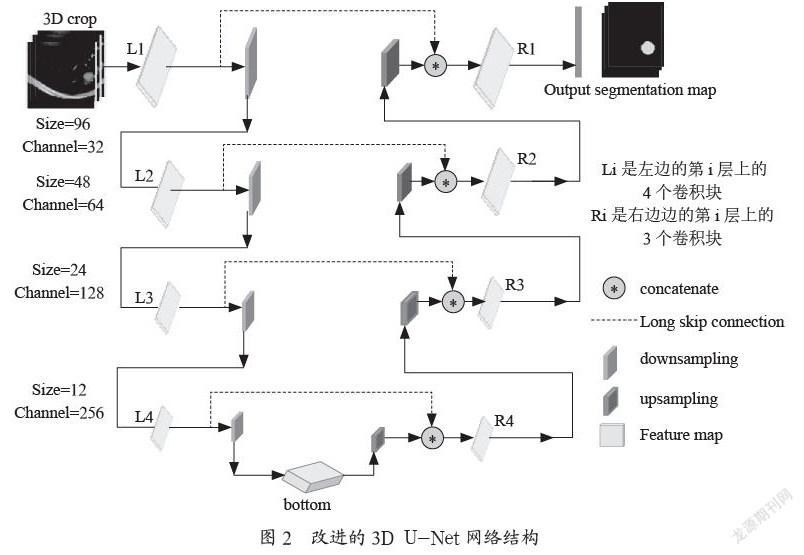
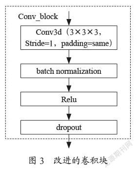
摘 要:对3D U-NET网络结构进行改进,提出一种CT影像中结节的自动分割方法。该项目在3D U-Net的基础上对其进行改进,改进的内容是卷积块操作采用3×3×3,Stride=1,padding=same的卷積,每个卷积后面相继增加Batch Normalization、Relu 和Dropout操作,池化被卷积操作代替,同时加入long skip connection长链接,实现浅层、低水平、粗粒度特征传递下去而不消失,提升网络对形状在10 mm以下但亮度高结节的轮廓表示能力,同时扩大了感受野、加速了网络的收敛。实现对CT影像的自动、准确描述。
关键词:3D U-NET;CT影像;长链接;感受野;浅层;低水平;粗粒度特征
中图分类号:TP391.4 文献标识码:A文章编号:2096-4706(2021)21-0105-04
Abstract: The structure of 3D U-NET network is improved, and an automatic segmentation method of nodules in CT images is proposed. The project improves it on the basis of 3D U-NET. The improved content is that the convolution block operation adopts the convolution of 3×3×3, stripe=1 and padding=same. After each convolution, Batch Normalization, Relu and Dropout operations are added successively. Pooling is replaced by convolution operations. At the same time, long skip connection long links are added to realize the transmission of shallow layer, low level and coarse grained characteristics without disappearing, so as to improve the ability of the network to express the contour of nodules of shape less than 10 mm with high brightness, at the same time, it expands the receptive field and accelerates the convergence of the network. And then realize the automatic and accurate description of CT images.
Keywords: 3D U-NET; CT image; long link; receptive field; shallow layer; low level; coarse grained characteristic
0 引 言
随着CT在肺癌早期筛查应用的普及,筛查数量倍增。通常情况下,一名患者的CT影像在80幅~250幅之间,影像科医生在医用竖屏显示器上调阅影像图片,逐幅观察。对于肺结节,它的直径约3 mm到30 mm的球状阴影,有实性结节、肺内混合磨玻璃密度结节和肺内纯磨玻璃密度结节,实性结节肺内亮度较高,容易观察,肺磨玻璃结节表现为密度轻微增加,呈云雾状,小于3 cm的局限性病变,其结节的密度不足以遮挡支气管及血管的结构,形状多变,容易和周围的血管、气管组织混淆,医生识别难度加大,加上长时间高强度的阅片,医生难免会产生视觉疲劳,不可避免地导致小结节的漏诊和误诊。如何在大量图像资料中准确检测出结节并早期定性,已成为临床迫切需求。
1 项目介绍
随着医疗大数据库的建立及计算机硬件水平的发展,深度学习算法在医疗诊断领域优势明显,为实现医学影像的自动智能诊断提供了新的契机。……