全景超声成像技术在浅表软组织及小器官诊断中的诊断效果
2021-01-28黄士元
黄士元
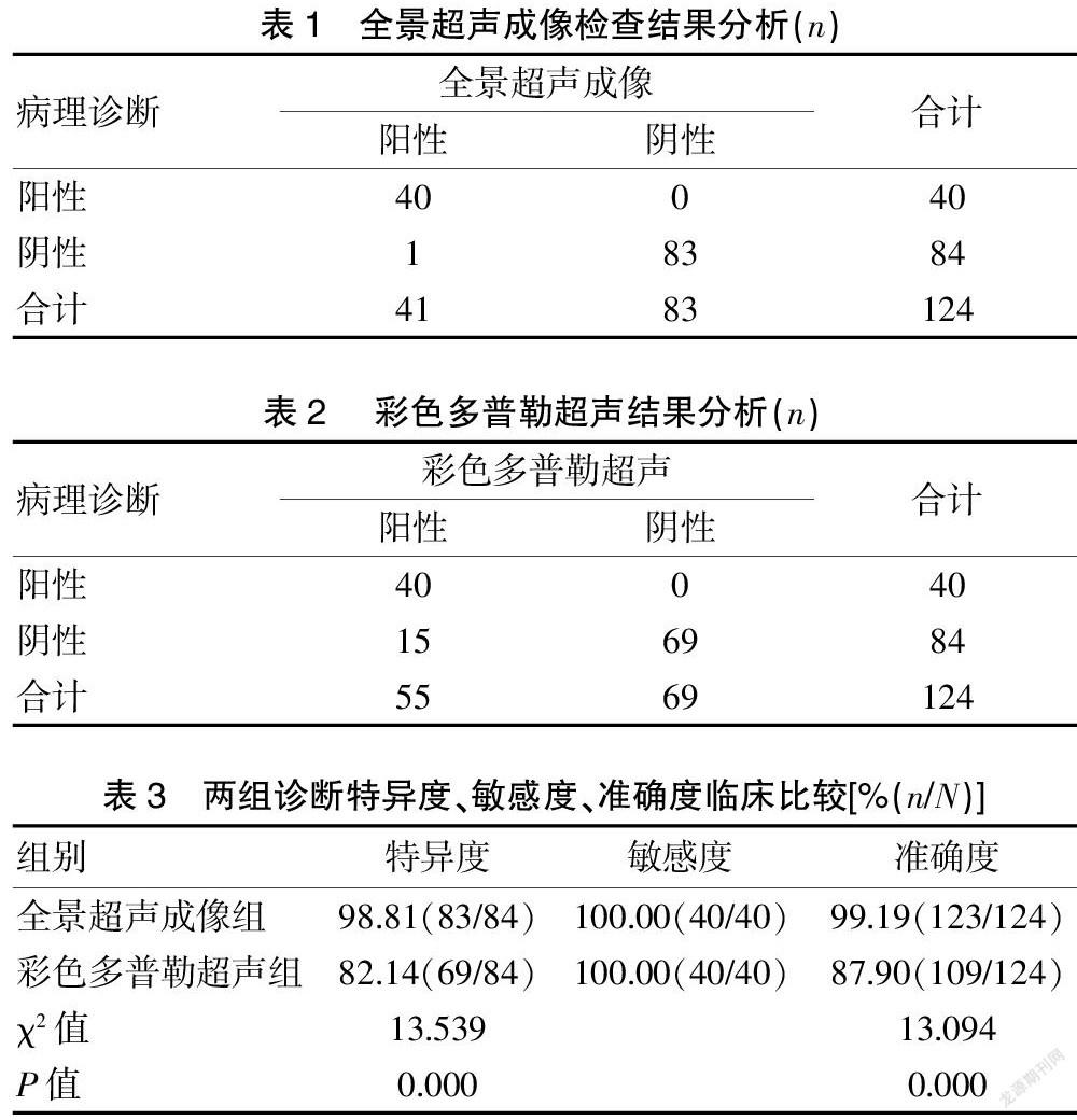
[摘要] 目的 探討对浅表软组织与小器官采用全景超声成像技术进行疾病诊断的临床效果。 方法 选取2018年1月1日至2020年12月31日收治的124例浅表软组织与小器官肿块患者进行研究。临床对其均展开全景超声成像检查、彩色多普勒超声检查;将病理诊断作为参考标准,比较全景超声成像检查、彩色多普勒超声检查对浅表软组织与小器官疾病的诊断价值。 结果 所有患者经病理检查后发现,恶性肿瘤40例,良性肿瘤84例。全景超声成像检查发现,恶性患者40例,良性患者83例,诊断准确率99.19%(123/124);彩色多普勒超声检查发现,恶性患者40例,良性患者69例,诊断准确率87.90%(109/124);两组诊断准确率比较,差异有统计学意义(P<0.05)。全景超声成像检查发现,诊断特异度、敏感度分别为98.81%(83/84)、100.00%(40/40);彩色多普勒超声检查发现,诊断特异度、敏感度分别为82.14%(69/84)、100.00%(40/40);两组诊断特异度比较,差异有统计学意义(P<0.05),诊断敏感度比较,差异无统计学意义(P>0.05)。结论 全景超声成像检查方式科学运用,诊断效果显著,可以充分鉴别良恶性肿块,可早期确诊浅表软组织与小器官肿块,能够为疾病治疗方案的早期研究与应用奠定基础。
[关键词] 浅表软组织;小器官;全景超声成像技术;病理诊断
[中图分类号] R445.1 [文献标识码] B [文章编号] 1673-9701(2021)34-0113-03
[Abstract] Objective To explore the clinical effect of using panoramic ultrasound imaging technology for disease diagnosis of superficial soft tissues and small organs. Methods A total of 124 patients with superficial soft tissue and small organ masses admitted from January 1, 2018 to December 31, 2020 were selected for disease diagnosis research. Panoramic ultrasound imaging and color Doppler ultrasound were performed clinically; under the condition of pathological diagnosis as a reference, the clinical value of panoramic ultrasound imaging and color Doppler ultrasound for superficial soft tissues and small organs was compared. Results After pathological examination, all patients found 40 cases of malignant tumors and 84 cases of benign tumors. Panoramic ultrasound imaging found 40 malignant patients and 83 benign patients, with a diagnostic accuracy rate of 99.19% (123/124); color Doppler ultrasonography found 40 malignant patients and 69 benign patients, with a diagnostic accuracy rate of 87.90% (109/124); The comparison of the diagnostic accuracy between the two groups was statistically significant(P<0.05). Panoramic ultrasound imaging revealed that the diagnostic specificity and sensitivity were 98.81% (83/84) and 100.00% (40/40), respectively; color Doppler ultrasonography revealed that the diagnostic specificity and sensitivity were 82.14% (69). /84), 100.00% (40/40), the difference in diagnostic specificity between the two groups was statistically significant (P<0.05); the difference in diagnostic sensitivity between the two groups was not statistically significant(P>0.05). Conclusion The panoramic ultrasound imaging method is scientifically used, and the diagnosis results are observed, and the results are significant. It can fully differentiate between benign and malignant masses. It can ensure the early diagnosis of patients with superficial soft tissue and small organ masses, and the early research and application of its treatment options, lay the foundation.
[Key words] Superficial soft tissues; Small organs; Panoramic ultrasound imaging; Pathological diagnosis
浅表软组织以及小器官肿块均呈现出广泛起源组织,神经外胚以及间叶组织脂肪、血管、肌肉以及纤维等组织均可表现[1]。其病理组织学表现以及组成组织细胞对肿物声像图会产生影响,但因为肿块病理分类复杂,难以进行准确判定[2-3]。以往在实施超声诊断期间,因为扫描角度以及探头宽度有所限制,针对肢体、躯干、盆腔、腹部以及血管等系列器官无法进行充分显示,出现误诊[4]。在此种情形下,运用全景超声成像检查,可以充分显示肢体各部位软组织,明确声学性质、病变形态、侵犯层次以及范围,提高诊断价值[5-6]。……