经会阴盆底超声在产后女性前盆腔功能评估中的应用价值
2020-06-24余彩茶焦岩汪晓茜林媛媛水旭娟
余彩茶 焦岩 汪晓茜 林媛媛 水旭娟
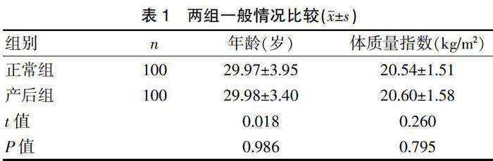
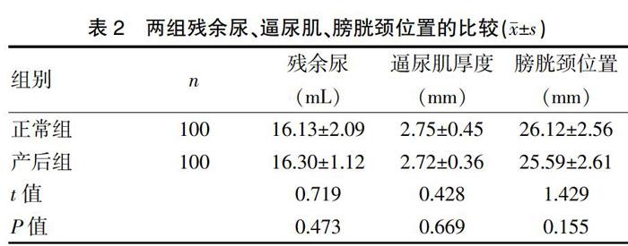
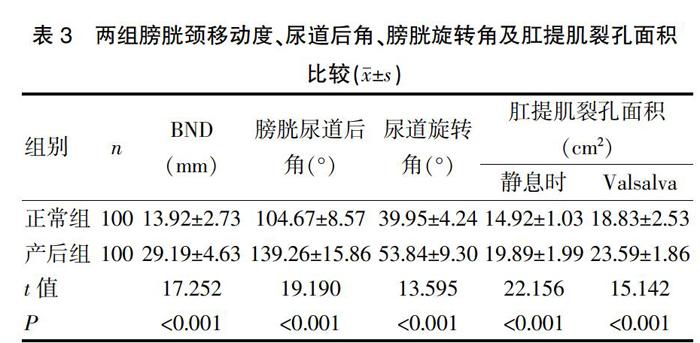
[摘要] 目的 探討经会阴盆底超声在产后女性前盆腔功能评估中的应用价值。 方法 随机选取2017年1月~2018年12月在我院就诊的未孕未育育龄期正常妇女100名,设为正常组,选取初产妇产后42 d妇女100名,设为产后组,分别获取两组女性静息状态及最大Valsalva状态下前盆腔的超声图像及各项盆底相关超声指标,比较两组间的差异。 结果 所有入选患者均能较好的完成Valsalva动作并取得两种不同状态下二维、三维超声图像及各种指标。正常组膀胱残余尿为(16.13±2.09)mL、静息状态下逼尿肌厚度为(2.75±0.45)mm、膀胱颈位置为(26.12±2.56)mm,产后组膀胱残余尿为(16.30±1.12)mL、静息状态下逼尿肌厚度为(2.72±0.36)mm、膀胱颈位置为(25.59±2.61)mm,两组比较差异无统计学意义(P>0.05);静息状态下盆隔裂孔面积正常组为(14.92±1.03)cm2,产后组为(19.89±1.99)cm2,两组差异有统计学意义(P<0.05);最大Valsalva动作下正常组膀胱颈移动度为(13.92±2.73)mm、膀胱尿道后角(104.67±8.57)°、尿道旋转角(39.95±4.24)°、尿道漏斗形成率为5.00%、盆隔裂孔面积(18.83±2.53)cm2,产后组膀胱颈移动度为(29.19±4.63)mm、膀胱尿道后角(139.26±15.86)°、尿道旋转角(53.84±9.30)°、尿道漏斗形成率为25.00%、盆隔裂孔面积(23.59±1.86)cm2,两组差异有统计学意义(P<0.05)。 结论 经会阴二维、三维超声联合使用能定量评估前盆腔结构和功能状态,为产后早期发现FPFD提供临床依据,并为临床诊治提供更加全面翔实的信息。
[关键词] 产后;女性;前盆腔;超声;盆底功能障碍
[中图分类号] R445.1;R713.4 [文献标识码] B [文章编号] 1673-9701(2020)11-0115-04
Application value of transperineal ultrasound of pelvic floor in the evaluation of prepelvic function in postpartum women
YU Caicha1 JIAO Yan1 WANG Xiaoqian2 LIN Yuanyuan1 SHUI Xujuan1
1.Department of Ultrasound in Obstetrics and Gynecology,Wenzhou People's Hospital in Zhejiang Province,Wenzhou 325000,China;2.Department of Obstetrics and Gynecology,Wenzhou People's Hospital in Zhejiang Province,Wenzhou 325000,China
[Abstract] Objective To investigate the application value of transperineal ultrasound of pelvic floor in the evaluation of prepelvic function in postpartum women. Methods 100 normal women of non-pregnant and non-child-bearing age who were admitted in our hospital from January 2017 to December 2018 were randomly selected as the normal group,while 100 primiparae(42 days after delivery) were selected as the postpartum group. Ultrasonic images of the anterior pelvic cavity and various pelvic floor-related ultrasonic indexes were obtained respectively under resting state and maximum Vasalval state of the two groups,and the differences between the two groups were compared. Results All enrolled patients were able to complete Vasaval motion and obtain 2D and 3D ultrasonic images and various indexes under two different states. The residual urine volume in the bladder was (16.13±2.09) mL,detrusor thickness in resting state was(2.75±0.45) mm,and bladder neck position was(26.12±2.56) mm in the normal group,while those in the postpartum group were (16.30±1.12) mL,(2.72±0.36) mm, and (25.59±2.61) mm correspondingly,the difference was no statistically significant between the two groups (P>0.05); in resting state,the area of pelvic septal hiatus was(14.92±1.03) cm2 in the normal group and (19.89±1.99) cm2 in the postpartum group,the difference was statistically significant(P<0.05); under the maximum Vasaval motion, bladder neck mobility was(13.92±2.73) mm,posterior urethral angle was (104.67±8.57)°,urethral rotation angle was(39.95±4.24)°,urethral funnel formation rate was 5.00%,and area of pelvic septal hiatus was (18.83±2.53) cm2 in the normal group,while those in the postpartum group were(29.19±4.63) mm,(139.26±15.86)°, (53.84±9.30)°,25.00%,and (23.59±1.86) cm2 correspondingly,the difference in results was statistically significant between the two groups(P<0.05). Conclusion Combined use of transperineal 2D and 3D ultrasound can quantitatively evaluate the structure and functional state of the anterior pelvic cavity,provide clinical basis for the early postpartum detection of FPFD,and provide more comprehensive and accurate information for clinical diagnosis and treatment.
[Key words] Postpartum; Female; Anterior pelvic cavity; Ultrasound; Pelvic floor dysfunction
近年来,女性盆底功能障碍性疾病(female pelvic floor dysfunction,FPFD)已成为当前影响我国女性身心健康的最常见慢性病之一,其致病原因主要是盆底支持结构的损伤、缺陷及其功能障碍,不仅发病机制复杂,且与盆腔器官脱垂、尿失禁等多种症状并存[1],已严重影响女性的生活和健康。研究表明[2],孕期和分娩时由于腹压增加、胶原减少等生理变化均可导致盆底结构发生改变,进而引起盆底肌力降低、组织松弛,严重时出现FPFD,且首次妊娠与分娩风险更高[3]。超声对盆底的检查目前被认为是一种新型且有效的方法。本研究旨在探讨经会阴二维、三维超声联合应用在观察正常盆底超声结构的同时,分析妊娠与分娩对前盆腔结构的影响,现报道如下。
