Clinical Study on CT-guided Modified Akupotomye in the Treatment of Lumbar Nerve Posterior Branch Compression
2020-02-20QIAOJinlin乔晋琳LIJinniu李金牛LINJingfu林井副JIGuangcheng汲广成XIANGDongdong向东东SHENHongxing沈红星
QIAO Jin-lin (乔晋琳), LI Jin-niu (李金牛), LIN Jing-fu (林井副), JI Guang-cheng (汲广成),XIANG Dong-dong (向东东), SHEN Hong-xing (沈红星)
1. Department of Rehabilitation Medicine, the Sixth Medical Center of the General Hospital of the PLA, Beijing 100048, China
2. Department of Chinese Medicine, Zhongguancun Hospital of Beijing, Beijing 100190, China
3. Imaging Center, the Sixth Medical Center of General Hospital of the PLA, Beijing 100048, China
4. Affiliated Hospital of Changchun University of Chinese Medicine, Changchun 130021, China
ABSTRACTObjective:To observe the clinical effect of modified akupotomye closed lysis under CT guidance on compression of posterior lumbar nerve branch. Methods:Patients were diagnosed by HRCT 3-D reconstruction combined with clinical symptoms and signs. After HRCT three-dimensional reconstruction combined with clinical symptoms and signs, the patients were confirmed as posterior lumbar nerve compression. After CT accurate surface positioning, CT-guided modified akupotomye was used for closed lysis of the posterior lumbar nerve branch. Oswestry Dysfunction Index Questionnaire (ODI) was used for quantitative scoring, 7 days before and after treatment and 6 months after treatment. Results:In 62 cases,20 cases were cured, with 25 cases markedly effective, 11 cases effective, and 36 cases ineffective. The total effective rate was 90.3%. ODI score: Self-paired t test 7 days before after treatment, P < 0.01; Before treatment and 6 months after treatment, self-paired t test (P < 0.01); Self-paired t-test was performed 7 days after treatment and 6 months after treatment (P > 0.05). Conclusion:With CT precise positioning, the modified akupotomye can be used to do closed lysis, to relieve the adhesion and compression, so that the low back pain can be relieved, with good clinical. The akupotomye closed lysis, combined with modern imaging technology has not only achieved good clinical effect, but also can improve the accuracy, safety and scientificity of akupotomye treatment.
KEYWORDS Compression of posterior branch of lumbar nerve; Akupotomye; CT Three-dimensional reconstruction
INTRODUCTION
Posterior lumbar nerve branch entrapment syndrome, also known as posterior lumbar nerve branch fibrous tube syndrome, is a kind of nonspecific low back pain (LBP). About 30% of LBP is caused by the compression of the posterior branch of the lumbar nerve. It is a common and frequentlyoccurring disease in the clinic, but it has often been misdiagnosed for a long time. It is often confused with low back pain such as lumbar protrusion and lumbar muscle strain. Clinical manifestations[1]: Compression of the posterior branch of lumbar nerve and posterior inner branch, is more common in middle-aged and elderly people, with a history of sprained or chronic strain of lumbar spine; lower lumbar pain or soreness can be involved in the buttocks. There is more pain in the early morning and little less after mild activity.And then the pain worsens after exertion. Signs:Physiological bending of the waist becomes straight.The movement is limited, especially the limitation of forward bending is obvious. There is deep tenderness in the affected lumbar spine of patients, but tenderness in the involved pain area is not obvious.Straight leg elevation tests were mostly negative.X-ray films often have degenerative changes such as lumbar hyperplasia and calcification of ligaments etc. Compression of the posterior external branch of the lumbar nerve is more common in young adults and in those with a thin body shape. It has a history of lumbar trauma with different degrees. It shows chronic low back pain and hip and femoral pain with one side as the main pain. The morning pain is obvious or sitting and standing in a single posture is difficult to last. The pain is relieved after a little activity, and it is obviously worsened after exertion. It can involve the hip and the back. But cough has no effect on this. Signs: Waist movement was slightly restricted, with obvious local tenderness or soreness centered on the L3 transverse process,and painful nodules can be touched. Numbness or tactile hypersensitivity are often found in the lower outer waist and lateral hips. The lower limb tendon reflex and straight leg elevation tests were mostly normal. X-ray examination often indicates that the L3 transverse process is longer or left-right asymmetry.
With the appearance of spiral CT, threedimensional reconstruction technology is possible.Today, we have been able to image the intraspinal nerve and the external nerve at any angle and level, and achieve the "same layer display" of the longitudinal direction of the nerve through reconstruction technology, which makes it possible to image the pressed part of the posterior branch of the lumbar nerve. Meanwhile, since July 2007,we have taken that lead to establish accurate position under CT, guide the akupotomye to close release the posterior branch of the lumbar nerve under compression, close release adhesion and compression, and thereby relieve the low back pain.A definite clinical effect has been achieved and is reported as follows:
DATA AND METHODS
General Data
A total of 34 patients with lumbar nerve posterior branch compression were diagnosed by HRCT three-dimensional reconstruction results combined with clinical symptoms and signs in our outpatient department. There were 16 males and 18 females,aged from 27-76 years (mean 54.7 ± 10.5 years),with a disease history of 3 months to 20 years.
Diagnostic Criteria:
(1) History: History of trauma or fatigue;
(2) Symptoms and signs: Low back pain (the propterty is referred pain); Lower lumbar finger pain (refers to the pain area outside the lumbar intervertebral foramen, but also in the posterior branch nerve distribution area, that is, the radiation pain area), but no tenderness or tenderness is not obvious, no percussion pain; Pain of rest; Restricted activities; Straight leg lifting and reinforcement test (-);
(3) In that outlet correspond to the posterior branch of the lumbar nerve, the blocking treatment is effective;
(4) Lumbar spine X-ray, CT, MRI, excluding other diseases;
(5) Three-dimensional CT reconstruction of lumbar nerve shows that the posterior branch of lumbar nerve is compressed outside the vertebral canal.
Exclusion Criteria:
(1) Lumbar disc herniation with compression of the posterior branch of the lumbar nerve;
(2) Third lumbar transverse process syndrome with compression of the posterior branch of the lumbar nerve
(3) Lumbar muscle strain with compression of posterior lumbar nerve.
Treatment
CT-guided close release lysis with akupotomye:① Placement of positioning needles: In the corresponding lumbar segment of the posterior branch of the lumbar nerve under compression,12 positioning needles arranged in parallel and equidistantly parallel to the posterior midline were placed and fixed with adhesive tape; ② Fixed position: CT scanning was performed on the corresponding lumbar segment with a slice thickness of 2.5 mm. According to the image obtained, the position of the corresponding bone fiber tube of the posterior branch of the lumbar nerve under compression was determined: The upper edge of the transverse process of the lower vertebral body and the lateral side of the superior articular process;The parameter and distance between the bone fiber tube and the body surface was measured by CT measurement, and the location of the bone fiber tube on the body surface was determined by CT localization on the corresponding body surface localization needle. ③Insertion of puncture needle:Routine disinfection of skin, application of sterile hole towel, vertical penetration of intracardiac puncture needle from skin surface to corresponding depth at positioning point, injection of 0.5% lidocaine 1.5 ml at each treatment point during puncture; At this time,the patient had local swelling, but no downward radiation; CT scanning is performed again to adjust the puncture angle and depth so that the needle tip reaches the upper edge of the transverse process of the lower vertebral body and the lateral side of the superior articular process. ④Insert akupotomye:Withdraw the intracardiac puncture needle and enter the akupotomye along the original puncture path.Most of the patients have local weight and swelling sensation, and adjust the needle feeding direction and depth slightly. If there is radiation sensation to the lower limb, slightly adjust the direction of needle insertion; Scan by CT again, adjust the angle and depth of the akupotomye, make the needle tip to the upper edge of the lower vertebral body transverse process, the lateral side of the upper articular process; ⑤ Akupotomye release : The improved akupotomye clings closely to the upper edge of the root of the corresponding transverse process and the lateral edge of the upper articular process, and releases the 2-3 knife in a small range; The needle hole was pressed with sterile gauze for 2 min, and the bandage was applied to the needle hole after a week. ⑥ Medical advice: 24 hours in bed rest,3 days to avoid fatigue, forbid eating spicy food,re-examination after the outpatient for a week; ⑦The modified akupotomye closed lysis under the guidance of CT was usually treated once, and 2 cases were treated with the second treatment, 2 weeks after the first treatment.
Evaluation of Efficacy
Therapeutic effect evaluation criteria: Clinical cure: After treatment, self-conscious pain symptoms disappear. Waist and leg activity function returns to normal, and can participate in general work;Obvious effect: The symptom of self-conscious pain basically disappears, and the function of waist and leg movement is slightly restricted, or walking has slight pain or no pain; Effective: The symptom of conscious pain is obviously improved,but the function of waist and leg movement still has obstacle, and walking fashion feeling has some pain or lameness; Ineffective: No obvious improvement or repeated onset of symptoms.
ODI scores Oswestry Dysfunction Index Questionnaire (ODI) was adopted[2], 7 days before and after treatment, and 6 months after treatment,and scored according to ODI, ODI = actual cumulative scores÷45×100%, 0% is normal. The closer to 100%, the more severe the dysfunction is[3]. The data obtained was expressed as mean ±standard deviation
Statistical Methods
SPSS11.0 statistical analysis software was used, the paired t test was performed before and after ODI score. There was significant difference in P < 0.05 and very significant difference in P < 0.01.
RESULTS
The Situation after Three-dimensional Reconstruction of CT
After three-dimensional reconstruction of CT, the compression of the posterior branch of the lumbar nerve in the follow-up pathway was clearly displayed. Statistics of the compression of the posterior branch of the lumbar nerve in 34 patients showed that the compression of the posterior branch of the L4 and L5 nerves was more common, which were in 51 and 81 sides, respectively. The left and right sides were compared respectively. They were in 65 and 78 sides, and the right side was slightly more than that in the left (see Table 1).
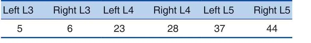
Table 1. CT Three-Dimensional Reconstruction Shows the Compression Distribution of the Posterior Branch of the Lumbar Nerve (Example)
Clinical Evaluation
After 7 days, the re-examination rate was 100%in 65 patients. Up to 6 months after treatment, the follow-up was successful in 51 cases. The cure rate of the patients was shown in Table 2.
ODI scores 7 days before and after treatment and 6 months after treatment were shown in Table 3.

Table 2. Effective Rate of 62 Patients
Table 3. ODI (%) t, 7 Days Before and after Treatment, and 6 Months after Treatment

Table 3. ODI (%) t, 7 Days Before and after Treatment, and 6 Months after Treatment
Before and 7 days after treatment, △P < 0.01 (t1 = 14.51, t2 = 13.69); 6 months before and after treatment, △△P < 0.01 (t3 = 12.78);Self-paired t-test was performed 7 days after treatment and 6 months after treatment, #P > 0.05 (t4 = 1.35).
Group Before treatment 7 days after treatment 6 months after treatment All patients (n = 62) 47.3±11.8 27.9±12.6△t1 -6 months follow-up (n = 51) 46.6±10.9 25.6±11.8△t2 24.8±10.7△△t3#t4
DISCUSSION
Injury to the posterior branch of the lumbar nerve is the main factor inducing lower back pain. Defining the specific site of nerve injury in patients with lower back pain caused by posterior branch injury of the lumbar nerve is the key to successful treatment[3]. Since the second half of 2006, we have performed three-dimensional CT reconstruction on suspected cases of posterior lumbar nerve compression[4]. It was found that the lumbar nerve root (intraspinal canal) or posterior lumbar nerve branch (extraspinal canal) in most of the patients had different degree of compression,which showed that the nerve was thickened, or the thickness was uneven, the tissue around the nerve was hyperplastic and adhesion, and the density was uneven. Local hyperosteogeny resulted in pathological changes such as nerve translocation and osteo-fibrous canal stenosis. According to preliminary statistics, the coincidence rate between imaging diagnosis and clinical diagnosis is as high as 91.4%. HRCT reconstruction of the same layer display technology can clearly show the anatomy of the posterior branch of the lumbar nerve and related diseases, providing a credible basis for the diagnosis of the disease[5]. The reliability of this new diagnostic technique is relatively high, which provides a new way and method for clinical diagnosis and treatment.There are many clinical reports on the treatment of nerve entrapment with
Akupotomye therapy through imaging localization[6,7], and the use of ultrasound guidance for radiofrequency thermocoagulation of the posterior branch of the lumbar nerve[8,9]. However,the accurate diagnosis of lumbar nerve entrapment is the first of modern imaging techniques combined with HRCT three-dimensional reconstruction of the same layer display image technology.
The posterior branch of lumbar nerve and the posterior medial branch and the posterior lateral branch of lumbar nerve pass through the bone fiber holes formed by transverse processes,articular processes, and ligaments, and bone fiber ducts between the lumbar mastoid and accessory processes, respectively[10]. WANG, Bin et al.[11], by dissecting, confirmed that there were at least two bone fiber ducts in the posterolateral branch of the lumbar nerve, one on the back of the transverse process of the lower vertebral body, and the other on the bone fiber ducts of the superior gluteal nerve into the buttock. In normal condition, these holes,tube or fissures have that function of protecting blood vessels and nerve passing there through,but because the pores are small, the surrounding structures are tough and lack elasticity[12]. Various acute and chronic injuries cause soft tissue damage around the vertebrae, resulting in changes in tissue structure and physical and chemical properties,which cause pain in waist by soft tissues on the side or segment of the spinous process to stimulate or compress the posterior branch of the spinal nerve[13].
JIN Yu-xiao and others believe that the pathogenesis of this disease is due to the stagnation of qi and blood in channels, resulting in qi and blood dysfunction, and causing pain. Akupotomye loosening can stimulate channel qi, loosen adhesion, and clear the meridians to achieve the treatment goals,and clinical research results show that the effect of akupotomye loosening treatment is significant,which can effectively relieve clinical symptoms of the patients and improve the degree of pain in patients,with the advantages of accurate positioning, less trauma, and good curative effect[14]. The akupotomy loosens the surrounding soft tissues of the compressed nerve, reduces tension and pressure,releases the compressed part of the nerve, relieves the compression[15], improves the local blood supply,promotes the absorption of inflammatory factors, and restores the nerve function.[16]
Before treatment, self-paired t test 7 days after treatment, and 6 months after treatment, the score of ODI of 62 patients with self-matched t test and self-matched t-test (P < 0.01) decreased significantly after closed release lysis, and the difference was statistically significant. There was no significant difference in self-paired t test between 7 days after treatment and 6 months after treatment, (P > 0.05),indicating that there was a good long-term curative effect after ackupotomy lysis, which was difficult to be achieved by closed therapy alone.
The authors believe that the relaxation and decompression of the posterolateral lumbar nerve branch can be achieved by releasing the transverse ligament of the lumbar spine. Because of the lower edge of the transverse process of the lumbar spine,there are branches from the anterior artery and vein of the transverse process[17], it is necessary to close to the upper edge of the transverse process of the lumbar spine when releasing the ligament between the transverse processes, so as to avoid damage to the blood vessels. The posterior medial branch of the lumbar nerve is relatively thin, and follows the fibrous hole at the root of the transverse process after bifurcation, and enters the fibrous canal between the mastoid and the accessory process along the outer edge of the superior articular process of the lower vertebral body[18]. Therefore, the needle body is perpendicular to the skin, the incision line is in line with the longitudinal axis of the human body, quickly pass through the skin, and slowly explore the needle.
Akupotomye passes through the skin,thoracolumbar fascia, erector spinae, and the lumbar vertebrae in order and it can close to the bone edge shovel cut[19], in order to loosen the accessory process, the mastoid bone between the fibrous tube,achieves the goal of the lumbar nerve posterior medial branch to reduce tension decompression. In the case of the compression of the posterior medial branch of the L5 spinal nerve, the lateral bone margin of the superior articular process close to S1 is scooped[20]to release the soft tissue of the lateral edge of the superior articular process of S1.
In that method, an improved akupotomye is adopt because the nerve and blood vessels at the outlet of the back branch of the lumbar nerve are rich, and a rectangular flat blade with a tip of the Hanzhang I type 3 akupotomye is ground to form an arc-shaped blunt blade with the tip of the akupotomye, In that invention, the improve akupotomye can play the effect of blunt separation when peeling the back branch of the compress lumbar nerve and avoid stabbing the nearby nerves and blood vessels. There was no hematomas and postoperative nerve injury in 62 patients during akupotomye closed lysis, which indicated that this method avoided iatrogenic injury while releasing the posterior branch of the lumbar nerve under compression, and it was a safe and effective method. In that operation, the low-dose local infiltration anesthesia with lidocaine can reduce the pain of the patient. The operator can communicate with the patient in time, and the operator can adjust the direction and depth of the akupotomye in time if the patient has a downward radiation feeling during the akupotomye release process.
At the same time of summing up this therapy,the author analyzed the condition of 3 cases of invalid patients. 2 of the 3 patients had a history of intervertebral disc orthopaedic surgery and were all fitted with internal fixators, one of which underwent 2 orthopaedic operations and one minimally invasive radiofrequency surgery. In that author's opinion, because the original mechanical structure of the lumbar spine can be chan by biting away the structure of the lumbar spine and place the internal fixation during the orthopaedic operation, and the extensive tissue adhesion after the operation may aggravate the compression of the posterior branch of the lumbar nerve. Needle-knife lysis can only be performed in a limited range of small-scale release,and it is difficult to improve the wide range of tissue adhesion and mechanical structure changes.
CT three-dimensional reconstruction can accurately diagnose the pathological condition of the posterior branch of the spinal nerve and establish a reasonable akupotomye treatment plan. In that treatment process, the angle and depth of the akupotomye can be adjusted according to the CT scan image thereof, and the akupotomye can be guided for accurate positioning and release. The combination of closed akupotomye lysis and modern science and technology has not only achieved good clinical effect, but also improved the accuracy, safety and scientificity of akupotomye therapy.
杂志排行

World Journal of Integrated Traditional and Western Medicine的其它文章
- World Integrated Medicine Master TANG You-zhi—Doctor of Ophthalmological Surgery for Chairman Mao
- Multicenter Randomized Double-Blind Controlled Clinical Study of Huoxue Tongluo Recipe (活血通络方) External Washing in the Treatment of Chemotherapy-Induced Peripheral Neuropathy
- Study on the Biological Basis of Qi, Blood, and Vessel in Immune Thrombocytopenia Patients With Syndrome of Qi Failing to Govern Blood Based on the Theory of Qi and Blood
- Effects of Shenmai Injection (参麦注射液) Combined with Meglumine Adenosine Cyclophosphate Injection on Cardiac Function and Peripheral Serum Levels of TNF-α,TGF-β1 and IFN-γ in Patients with Viral Myocarditis
- Two Definitions of Life Will Highlight on Physiological Understanding of Six Meridians