1,3-二(4-咪唑基)苯构筑的两个金属-有机框架化合物:合成、晶体结构和荧光性质
2020-02-11刘志强武峻峰
刘志强 武峻峰 陈 俊 吴 夏 王 彦
(1 安庆师范大学化学化工学院,光电磁功能材料安徽省重点实验室,光电磁功能配合物和纳米配合物安徽省重点实验室,安庆 246011)
(2 南京大学化学化工学院,配位化学国家重点实验室,南京 210023)
0 Introduction
Benefiting from large surface areas, adjustable pore size, readily modifiable functional groups and enough active sites, metal-organic frameworks (MOFs)are widely used in the fields of gas adsorption and separation, drug delivery, sensing and detection,catalysis and so on[1-7].It is well-known that the potential applications of MOFs largely depend on their structural characteristics. Due to MOFs syntheses affected by many factors including organic ligands,solvent systems, pH, metal ions and so on, so it still has a large challenge the syntheses of target MOFs.Of all these conditions, the organic ligand is of vital aspect because physical properties of ligands directly affect the frameworks and properties of MOFs[8-12]. Our previous studies have demonstrated that organic compounds with 1H-imidazol-4-yl groups are versatile ligands for the construction of MOFs with definite structures and properties[13-16].
Herein we focused our attention on reactions of 1,3-di(1H-imidazol-4-yl)benzene (L) with Cd(Ⅱ)or Ni(Ⅱ)salts. In this work, two MOFs were successfully synthesized, namely [Cd(L)(SO4)]n(1) and {[Ni(L)(SO4)]·H2O}n(2). The MOFs were characterized by X-ray crystallography, IR spectroscopy, elemental and thermal analyses. In addition, photoluminescence property of the MOF 1 was investigated.
1 Experimental
1.1 Materials and methods
All commercially available chemicals and solvents are of reagent grade and were used as received without further purification. The ligand L was synthesized according to the procedure reported in the literature[17]. FT-IR spectra were recorded in a range of 400~4 000 cm-1on a Bruker Vector 22 FT-IR spectrometer using KBr pellets. Thermogravimetric analysis(TGA) was performed on a Mettler-Toledo (TGA/DSC1) thermal analyzer under nitrogen with a heating rate of 10 ℃·min-1. Powder X-ray diffraction (PXRD)patterns were obtained on a Bruker D8 Advance diffractometer using Cu Kα (λ=0.154 18 nm) in 2θ range of 5°~50° at 293 K, in which the X-ray tube was operated at 40 kV and 40 mA. Photoluminescence spectra were measured on an Aminco Bowman Series 2 spectrofluorometer with a xenon arc lamp as the light source. In the measurements of emission and excitation spectra, the pass width is 5 nm.
1.2 Synthesis of complexes 1 and 2
1.2.1 Synthesis of complex 1
3CdSO4·8H2O (17.6 mg, 0.05 mmol) and L (10.5 mg, 0.05 mmol) were dissolved in H2O (10 mL) was sealed in a Teflon-lined stainless steel container and heated at 90 ℃for 3 days. The resulting colorless block crystals were collected in 68% yield based on L. Elemental analysis Calcd. for C12H10N4O4SCd(%): C 34.42, H 2.41, N 13.38; Found(%): C 34.47, H 2.53,N 13.29. Selected IR peaks (cm-1, Fig.S1): 3 423 (s),3 108 (s), 2 880 (s), 1 620 (w), 1 586 (w), 1 497 (m),1 366 (w), 1 126 (s), 853 (s), 794 (s), 603 (s).
1.2.2 Synthesis of complex 2
The synthesis was similar with that of 1 except that NiSO4·6H2O (13.1 mg, 0.05 mmol) was used instead of 3CdSO4·8H2O. The resulting green block crystals were collected in 82% yield based on L.Elemental analysis Calcd. for C24H22N8O5SNi(%): C 48.59, H 3.74, N 18.89; Found (%): C 48.53, H 3.83,N 18.82. Selected IR peaks (cm-1, Fig.S1): 3 427 (s),2 855 (s), 1 623 (m), 1 581 (m), 1 449 (m), 1 245 (s),1 120 (s), 1 092 (s), 983 (s), 830 (m), 747 (w).
1.3 Crystallographic data collection and refinement
Crystallographic data for 1 and 2 were collected on a Bruker Smart Apex Ⅱ CCD with graphite monochromated Mo Kα radiation source (λ=0.710 73 nm). The diffraction data as well as the intensity corrections for the Lorentz and polarization effects,was carried out using the SAINT program[18]. Semiempirical absorption correction was applied using SADABS program[19]. The structures of 1 and 2 were determined by direct methods and refined anisotropically on F2by the full-matrix least-squares technique with SHELXTL-2016 program package[20-21]. The hydrogen atoms except for those of water molecules were generated geometrically and refined isotropically using the riding model. For 2, the SQUEEZE subroutine of the PLATON software suite was used to remove the scattering from the highly disordered solvent molecules[22]. The final formulas were calculated from the SQUEEZE results, TGA and elemental analysis.Selected bond lengths and angles are listed in Table S1 (Supporting information). The hydrogen bonding data are provided in Table S2.
CCDC: 1920722, 1; 1920723, 2.
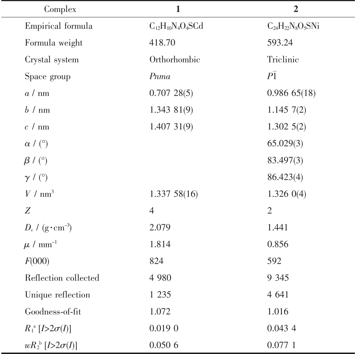
Table 1 Crystal data and structure refinements for complexes 1~2
2 Result and discussion
2.1 Crystal structures of 1 and 2
2.1.1 Crystal structure of 1
Single crystal X-ray structural analysis reveals that complex 1 crystallizes in the triclinic Pnma space group. As exhibited in Fig.1a, the Cd (Ⅱ) is sixcoordinated by four oxygen atoms (O1, O2, O3, O4)from the bridging sulfate anion and two N atoms (N2,N2ii)from distinct L.The asymmetric unit of 1 contains one molecule of [Cd(L)(SO4)]. The Cd-O bond distances are from 0.236 2(2) to 0.253 7(3) nm, while the Cd-N ones are 0.218 39(19) and 0.218 40(19) nm.The coordination angles around Cd (Ⅱ)in 1 are from 57.09(7)° to 163.59(9)° (Table S1). Each L links two Cd(Ⅱ)to form an infinite one-dimensional (1D) chain,and the Cd-L 1D chains are further connected by sulfate anion to generate a 2D network (Fig.1b), which is further extended into a three-dimensional (3D)supramolecular architecture through O-H …O hydrogen bonding interactions (Fig.1c and Table S2).
2.1.2 Crystal structure of 2
When NiSO4·6H2O instead of 3CdSO4·8H2O was used under the same reaction conditions for preparation of 1, complex 2 was obtained. The results of single crystal X-ray structural analysis showed that 2 crystallizes in triclinic space group P1. As shown in Fig.2a, Ni1 is six-coordinated by four N atoms (N2,N4i, N5, N8ii) from four different L ligands, two O atoms (O1, O2) from sulfate anion. The Ni-N bond distances are from 0.204 0(2) to 0.209 6(2) nm, while the Ni-O ones are 0.218 56(18) and 0.219 42(18) nm.The coordination angles around Ni (Ⅱ) in 1 are from 65.25(6)° to 172.49(9)° (Table S1). Each L links two Ni (Ⅱ)to generate a 2D network (Fig.1b), which is further extended into a three-dimensional (3D) supramolecular architecture through O-H …O hydrogen bonding interactions (Fig.1c and Table S2).
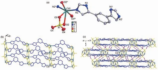
Fig.1 (a) Coordination environment of Cd(Ⅱ)ion in 1 with the ellipsoids drawn at 30% probability level;(b) 2D structure of 1; (c) 3D structure of 1 with hydrogen bonds indicated by dashed lines

Fig.2 (a) Coordination environment of Ni(Ⅱ)ion in 2 with the ellipsoids drawn at 30% probability level;(b) 2D structure of 2; (c) 3D structure of 2 with hydrogen bonds indicated by dashed lines
2.2 Powder X-ray diffraction and thermogravimetric analyses
The purity for the as-synthesized samples was ensured by PXRD measurements and the results are provided in Fig.S2. Each as-synthesized sample gives consistent PXRD pattern with the corresponding simulated one, indicating the pure phase of 1 and 2.
The thermal stability of 1 and 2 was examined by TGA in the N2atmosphere from 30 to 650 ℃, and the TG curves are given in Fig.3. The result was that MOF 1 did not show obvious weight loss before the decomposition of the framework at about 450 ℃,which is in agreement with the result of the crystal structure analysis. For MOF 2, the first weight loss of 3.1% from 30 to 130℃is in accordance with the loss of free H2O molecules (Calcd. 3.03% ), and the framework maintained before about 430 ℃.
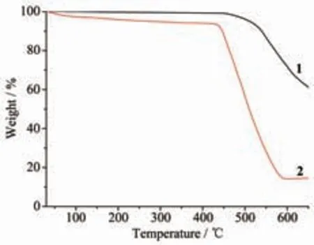
Fig.3 TGA curves of 1 and 2
2.3 Photoluminescence property
It is known that the frameworks with d10metal centers and π-conjugated organic ligands may have photoluminescence. Luminescence in MOFs generally arises from the building components: conjugated organic ligands and/or metal ions or clusters. Organic linkers with aromatic moieties or extended π systems are commonly used in the construction of porous MOFs due to their rigid molecular backbone. MOFs with d10metal centers show luminescent properties and is likely to be candidates for luminescent materials[23-29].Accordingly, the solid-state luminescent emission spectra of L and 1 were collected at room temperature.As shown in Fig.4, the free ligand L exhibited an emission maxima at 381 nm (λex=335 nm), and MOF 1 gave emission maxima at 363 nm (λex=330 nm).Compared with the emission of free L, the blue-shifts of the emissions of 1 are considered to be caused by the coordination of the ligand to the metal centers.
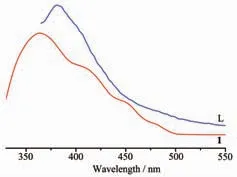
Fig.4 Emission spectra of 1 as well as free L ligand in solid state at room temperature
3 Conclusions
In summary, two MOFs based on 1,3-di(1Himidazol-4-yl)benzene(L)and corresponding metal salts were synthesized and characterized. The structures of them are 2D network, which are further linked together by hydrogen bonds to give eventual 3D architectures.Furthermore, the thermal stability and photoluminescence property of the MOFs were investigated.
Supporting information is available at http://www.wjhxxb.cn