心脏脚踝指数对冠状动脉粥样硬化的预测诊断价值
2019-09-25欧邦豪李博方胜先
欧邦豪 李博 方胜先

[摘要]目的 探討心脏脚踝指数(CAVI)对冠状动脉粥样硬化的预测诊断价值。方法 选取2015年2月~2017年2月龙岗中心医院心内科及合作医院心内科收治的疑似冠心病拟行冠状动脉造影(CAG)的108例患者作为研究对象,根据冠脉造影检查结果及Gensini评分,分为冠脉正常组(0分,34例)、1~80分组(36例)、81~160分组(38例)。均于造影前空腹采血,测定CAVI、颈总动脉最大内膜中层厚度(Max IMT)及平均内膜中层厚度(Mean IMT)指标。结果 三组患者CAVI、Max IMT及Mean IMT值水平随Gensini评分升高而递增,差异有统计学意义(P<0.05);CAVI、Max IMT及Mean IMT评分与Gensini评分均成正相关(r=0.57、0.29、0.46,P<0.01、0.05、0.01),且CAVI与Gensini评分的相关性最强;比较CAVI、Max IMT及Mean IMT对预测粥样硬化的ROC曲线下面积(AUC),CAVI显著大于Max IMT和Mean IMT;与Max IMT及Mean IMT相比,CAVI假阳性率、阴性似然比最低,阴性预测率最高。结论 CAVI水平随动脉粥样硬化程度加重而逐渐升高,与Max IMT、Mean IMT相比,CAVI与冠状动脉粥样硬化病变显著相关,判断动脉粥样硬化更为敏感准确。
[关键词]心脏脚踝指数;冠状动脉粥样硬化;最大颈动脉内中膜厚度
[中图分类号] R541.4 [文献标识码] A [文章编号] 1674-4721(2019)7(a)-0032-03
[Abstract] Objective To explore the value of cardiac ankle index in predicting and diagnosing coronary atherosclerosis. Methods A total of 108 patients suspected with coronary artery disease (CAG) in Longgang Central Hospital cardiology department and cooperative hospitals from February 2015 to February 2017 were selected as the subjects of study. According to the results of coronary angiography and Gensini score, they were divided into normal coronary group (0 points, 34 cases), 1-80 points group (36 cases) and 81-160 points group (38 cases). The cardio-ankle vascular index (CAVI), the maximum intima-media thickness (Max IMT) of common carotid artery (CCA) and mean intima-media thickness (Mean IMT) were measured before angiography. Results The levels of CAVI, Max IMT and Mean IMT increased with the increase of Gensini score in the three groups, and the differences were statistically significant (P<0.05). The scores of CAVI, Max IMT and Mean IMT were positively correlated with Gensini score (r=0.57, 0.29, 0.46, P<0.01, 0.05, 0.01), and the correlation between CAVI and Gensini score was the strongest. CAVI, Max IMT and Mean IMT were compared to predict ROC curve of atherosclerosis. Under-line area (AUC), CAVI was significantly larger than Max IMT and Max IMT. Compared with Max IMT and Mean IMT, CAVI had the lowest false positive rate, the lowest negative likelihood ratio and the highest negative predictive rate. Conclusion The level of CAVI gradually increases with the severity of atherosclerosis. Compared with Max IMT and Mean IMT, CAVI is significantly correlated with coronary atherosclerosis lesions and is more sensitive and accurate in judging atherosclerosis.
[Key words] Cardiac foot stamp index; Coronary atherosclerosis; Maximum carotid intima-media thickness
冠状动脉粥样硬化是动脉硬化中最为常见的类型,血脂异常及血管壁成分改变都与冠状动脉粥样硬化密切关联。高血压指南将用于无创血管硬化的检查指标颈动脉内膜中层厚度等作为判断靶器官损伤的主要指标。心踝血管指数(CAVI)可反映主动脉、股动脉及脚趾动脉的粥样硬化程度,且具有操作简便,对机体损伤小等优势,国外研究显示,通过冠脉介入手术(PCI)或心电图检测有缺血性改变的患者CAVI值高于无动脉硬化的患者[1-2]。本研究以龙岗中心医院心内科及合作医院疑诊冠心病拟行冠脉造影的108例患者作为研究对象,旨在探讨CAVI对冠状动脉粥样硬化的预测诊断价值,现报道如下。
1资料与方法
1.1一般资料
选取2015年2月~2017年2月龙岗中心医院心内科及合作医院心内科收治的疑似冠心病拟行冠状动脉造影(CAG)的108例患者作为研究对象,其中男69例,女39例;平均年龄(61.51±6.05)岁。纳入标准:患者均拟行冠脉造影,且年龄40~70岁。排除标准:患有急性冠脉综合征、严重肝肾功能不全者,踝肱指数(ABI)<0.9、左心射血分数(LVEF)<50%、心房颤动(扑动)、Ⅱ~Ⅲ度房室传导阻滞、先天性心脏病及恶性肿瘤患者。根据冠脉造影检查结果及Gensini评分[3],分为冠脉正常组(0分)、1~80分组、81~160分组。正常组34例,其中男20例,女14例;平均年龄(61.41±8.14)岁。1~80分组36例,其中男21例,女15例;平均年龄(60.14±7.12)岁。81~160分组38例,其中男28例,女10例;平均年龄(61.95±9.14)岁。三组患者的一般资料比较,差异无统计学意义(P>0.05),具有可比性。本研究经医院医学伦理委员会批准通过。
1.2方法
测定三组患者的CAVI、颈总动脉最大内膜中层厚度(Max IMT)及平均内膜中层厚度(Mean IMT)。采用大连欧姆龙电子仪器公司的BP-203RFEIII多功能动脉硬化检测仪检测患者的CAVI值,测定10个连续波动,包括一个完整的呼吸周期。采用siemenssequoia512型彩色多普勒超声仪,测量正常部位舒张末期颈动脉内膜中层厚度(IMT)。分别测量两侧颈前后壁总动脉IMT,根据测得的4个IMT,得到Max IMT值及Mean IMT值。每例患者均测量3次取平均值。
1.3观察指标及评价标准
阳性预测值是指筛检试验检出的全部阳性例数中,真阳性所占的比例;阳性似然比=敏感性/(1-特异性);阴性似然比=假阴性率/真阴性率。
1.4统计学方法
采用SPSS 13.0统计学软件对数据进行分析,计量资料以均数±标准差(x±s)表示,采用t检验;计数资料以率(%)表示,采用χ2检验;样本均数比较采用单因素方差分析,两两比较采用SNK-q检验。CAVI、Max IMT及Mean IMT与 Gensini积分相关性分析使用偏相关系数,比较使用ROC曲线。CAVI与心血管危险因素的关系采用单变量及多元逐步回归分析,以P<0.05为差异有统计学意义。
2结果
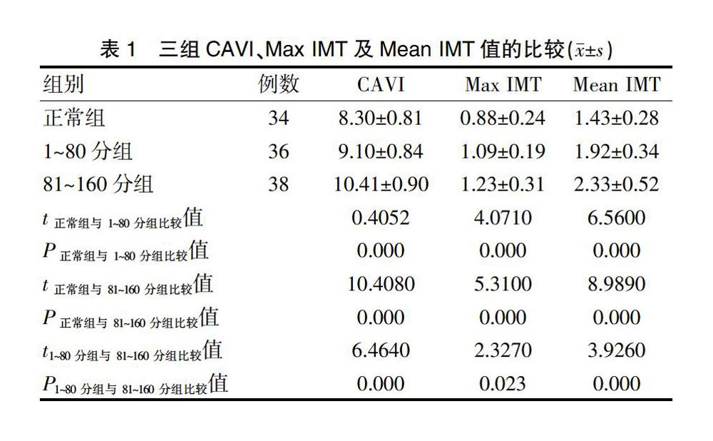
2.1三组CAVI、Max IMT及Mean IMT值的比较
2.2 CAVI、Max IMT及Mean IMT评分与Gensini评分的相关性
2.3 CAVL、Max IMT及Mean IMT预测冠状动脉粥样硬化的ROC曲线
ROC曲线显示,CAVI曲线下面积(AUC)显著大于Max IMT和Mean IMT,Mean IMT的AUC最小(图1)。CAVI预测冠状动脉粥样硬化cut-off值、敏感性及特异性分别为8.78、85.15%、68.44%;MaxIMT预测冠状动脉粥样硬化cut-off值、敏感性及特异性分别为1.31、72.64%、75.15%;Mean IMT预测冠状动脉粥样硬化cut-off值、敏感性及特异性分别为1.05、66.48%、74.68%。
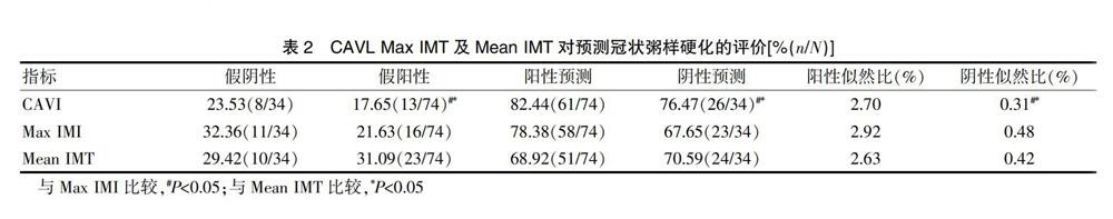
2.4 CAVL、Max IMT及Mean IMT预测冠状粥样硬化的价值
3讨论
冠状动脉粥样硬化常伴发冠状动脉痉挛[4-6],加剧原有管腔狭窄程度,导致供血的中断,引起心绞痛、心肌梗死等病变,也是导致心源性猝死的原因之一[7-8]。CAVI基于脉搏波传导速度相同的原理,并根据僵硬系数β得出CAVI值,僵硬系数β的介入避免传统脉搏波传导速度测量易受血压波动影响的缺陷,具有较高的重复性[9-12]。相关研究显示,冠状动脉硬化多在主动脉粥样硬化之后发生,而CAVI主要反映主动脉的粥样硬化改变,故而CAVI预测冠脉粥样硬化更具有前瞻性[13-15]。
本研究中三组患者CAVI、Max IMT及Mean IMT值水平随Gensini评分升高而遞增,差异有统计学意义(P<0.05),提示CAVI、Max IMT、Mean IMT数值随Gensini积分的增加而增高。CAVI、Max IMT及Mean IMT评分与Gensini评分均成正相关(r=0.57、0.29、0.46,P<0.01、0.05、0.01),且CAVI与Gensini评分的相关性最强,提示CAVI、Max IMT、Mean IMT均有助于冠状动脉粥样硬化的诊断。比较CAVI、Max IMT及Mean IMT预测粥样硬化的AUC,结果显示,CAVI显著大于Max IMT和Mean IMT;与Max IMT及Mean IMT相比,CAVI假阳性率最低,阴性预测率最高,进一步提示CAVI与冠状动脉粥样硬化病变的显著相关,判断动脉粥样硬化更为敏感准确。
综上所述,CAVI、Max IMT、Mean IMT水平均随动脉粥样硬化程度加重而逐渐升高,均可应用于冠状动脉粥样硬化患者的早期预测诊断,且与Max IMT、Mean IMT相比,CAVI与冠状动脉粥样硬化病变的显著相关,判断动脉粥样硬化更为敏感准确。
[参考文献]
[1]Koji Y,Tomiyama H,Ichihashi H,et al.Comparison of anklebrachial pressure index and pulse wave velocity as markers of the presence of coronary artery disease in subjects with a high risk of atherosclerotic cardiovascular disease[J].Am J Cardiol,2012,94(7):721-868.
[2]McLeod AL,Uren NG,Wilkinson IB,et al.Noninvasive measures of pulse wave velocity correlate with coronary arterial plaque load in humans[J].J Hypertens,2012,22(2):363 281.
[3]Censini CC.A more meaningful scoring system for determining the severity of coronary heart disease[J].Am J Cardiol,1983,51(3):606.
[4]Takuro K,Masaaki M,Kiyo U,et al.Clinical significance and reproducibility of new arterial distensibility index[J].Circ J,2014,71(1):89-94.
[5]Asmar R,Benetos A,Topouchian J,et al.Assessment of arterial distensibility by automatic pulsewave elocity rement:validation and clinical application studies[J].Hypertension,2011,26:485-490.
[6]Lehmann ED.Clinical value of aortic pulse-wave velocity measurement[J].Lancet,2007,354(9178):528-529.
[7]陈红伟,石宗华.颈动脉粥样硬化、狭窄与急性冠状动脉综合征的相关性研究[J].中国现代药物应用,2014,12(7):44-45.
[8]Matsushima Y,Kawano H,Koide Y,et al.Relationship of carotid intima media thickness,pulse wave velocity,and ankle brachial index to the severity of coronary artery atherosclerosis[J].Clin Cardiol,2013,27(11):6292341.
[9]李博,方勝先,朱平先,等.心脏脚踩指数对冠状动脉粥样硬化的预测价值分析[J].南昌大学学报(医学版),2016, 14(13):1454-1457.
[10]潘展群,吴清华.心脏脚躁指数对冠状动脉粥样硬化的诊断价值[J].山东医药,2012,52(21):64-66.
[11]程云鹏,张娜,祝艳秋,等.心踝血管指数对高血压及合并糖尿病患者血管功能的评价[J].大连医科大学学报,2015,8(3):242-247.
[12]黄剑,项丽,朱静,等.冠状动脉粥样硬化性心脏病患者颈动脉粥样硬化彩色多普勒超声特征及与冠状动脉病变关系研究[J].创伤与急危重病医学,2018,11(2):224-227.
[13]Akira T,Hiroshi O,Takatoshi W,et al.Cardio-ankle vascular index is superior to brachial-ankle pulse wave velocity as an index of arterial tiffness[J].Hypertens Res,2013,31(7):1347-1355.
[14]开金津.急性冠状动脉综合征同颈动脉粥样硬化狭窄的相关性探讨[J].河北医学,2015(2):249-251.
[15]孙涛,秦淮,程宇彤,等.急性冠状动脉综合征颈动脉粥样斑块稳定性及相关因素分析[J].中华实用诊断与治疗杂志,2014,28(2):137-139.
(收稿日期:2019-02-22 本文编辑:闫 佩)