Allogenic tooth transplantation using 3D printing:A case report and review of the literature
2019-03-14HuDiXuRichardMironXiaoXinZhangYuFengZhang
Hu-Di Xu,Richard J Miron,Xiao-Xin Zhang,Yu-Feng Zhang
Abstract
Key words: Transplantation;Allografts;Dental implants;3D printing;Case report
INTRODUCTION
The history of allogenic tooth transplantation can be traced back to the 16thcentury,particularly during the last decades of the 20thcentury.While individual organs are often transplanted from one individual to the next,in the dental field,transplantation of teeth is much less frequently performed.Several innovations from basic and clinical translational research has improved patient matching tests and clinical techniques[1,2].Allogenic tooth transplantation refers to tooth grafting between two individuals of the same species whereas autogenic tooth transplantation indicates tooth grafting from one site to another in the same individual[3].Atkinson reported in the 1970s that an autograft can be orthotopic (when transplanted to the natural location) or heterotopic(when transplanted to a completely different site).Generally,allografts are rejected by immune cells,which may cause the most common histopathological symptoms including chronic inflammatory infiltration of the grafted tissue[4,5].However,a few cases of allogenic tooth transplantation have been reported[6].Little progress has been made owing to the high rate of potential immunological rejections and the increased use of titanium dental implants[7].Nevertheless,allografts can still function normally and often symptomless for many years[8].
There are many successful cases of allo-transplantation.Ole Schwartz reported 73 allotransplanting cases which were carried out by three surgeons in humans from 1956 to 1980[8].The mean functional (without symptoms) time of the grafted teeth was 6.8 years (maximum 28.5 years,which is also the maximum observation period)[4,6,9].This study illustrates the potential long-term survival rates of allograft tooth transplants with rates influenced by a series of factors.
In previous cases,since differences in root shape and length exist,surgeons have had to reposition the donor tooth back to its original socket and remodel the recipient site with a round implant bur[10].The remodeling of the recipient site adds additional time to the surgical procedure with the possibility of surgically removing more bone than needed,which could totally be improved by the utilization of 3D printing.Today,3D printing has been used widely in many fields of organ engineering[11-17].A typical 3D printing process involves data collection,model analysis,structure design,and final manufacture.Specifically,data initially can be collected by a variety of systems including computed tomography (CT),digital scanning,magnetic resonance imaging,and other image modality systems[18].Computer-aided design (CAD)software was followed by a precisely conducted 3D printing process controlled by a computer-aided manufacturing printer.The advantages of 3D printing include accurate control of material distribution,fast speed,scalability,and cost-effectiveness,which render this technology highly suitable and relevant in many areas of medicine including dentistry[19,20].One of the greatest advantages of 3D printing in tooth transplantation is that osteotomy drills can be customized to the three-dimensional geometry of the tooth root of the individual donor[21].Such drills allow an optimal integration of transplanted tooth without unnecessary compression of tissues that may occur with standard burs,minimize unnecessary bone loss,and improve the implant stability[22,23].Furthermore,allogenic tooth transplantation could be an alternative solution for financially compromised patients that by-pass the need for additional costs including an implant,abutment,and final crown.This case report is the first published case combining 3D printing technology for tooth transplantation,which aims to inspire future research endeavors into this largely unstudied field.
CASE PRESENTATION
Chief complaints
A 47-year-old Chinese patient presented to the dental clinic at Wuhan University with a missing 1stmolar for 2 mo.Her daughter (the donor) was a 21-year-old dental student with an impacted wisdom tooth.
History of present illness
The first left maxillary molar of the patient had been extracted in a private dental clinic 2 mo prior to the tooth transplantation.The root canal treatment for the 1stmolar has failed,and tooth extraction was conducted after the roots fractured.The donor recently extracted two right third molars 2 wk prior to the donation (upper and lower).
History of past illness
No other health conditions are reported.
Medication history
The patient is allergic to penicillin.
Personal and family history
The mother has hypertension.
Physical examination upon admission
In the initial physical examination,the patient had a blood pressure of 128/84 mmHg with a pulse rate of 79 beats per minute (bpm).And the donor had a blood pressure of 92/62 mmHg with a pulse rate of 82 bpm.The extraction wound was well healed,and the anteroposterior gap of the defect was acceptable.The mandibular and maxillary distance had not been compromised,and the buccolingual width was about 7 mm.No obvious inclination of adjacent teeth was observed for the patient.The patient’s oral hygiene was acceptable,with little microbial plaque accumulation or associated trauma (Figure 1).
Laboratory examinations
The patient has an O blood type.The donor presents with an A blood type.Routine blood count and coagulation profile for both the patient and donor were within normal limits.
Imaging examinations
Cone beam computed tomography (CBCT) of the donor revealed a tapered root shape different in morphology from the recipient’s residual 1st maxillary molar root (3 roots),which eliminated the potential for an immediate transplantation.The lower 3rdmandibular molar (the donor tooth) was scanned,and the data of the morphology were collected and analyzed before an individualized implant drill was designed to match the contour of the donor tooth roots,with expectation that the shape of the recipient site could be identical to that of the original socket.A direct metal laser sintering 3D printing system was introduced to manufacture the drill (Figure 2).
FINAL DIAGNOSIS
Maxillary dentition defect.
TREATMENT
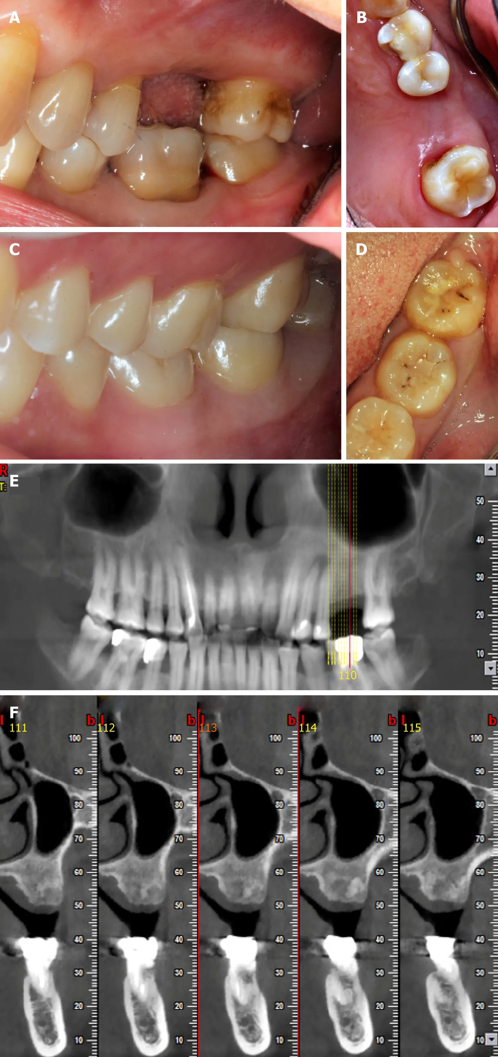
Figure1 Preoperative photos and computed tomography scan of the donor and the recipient.
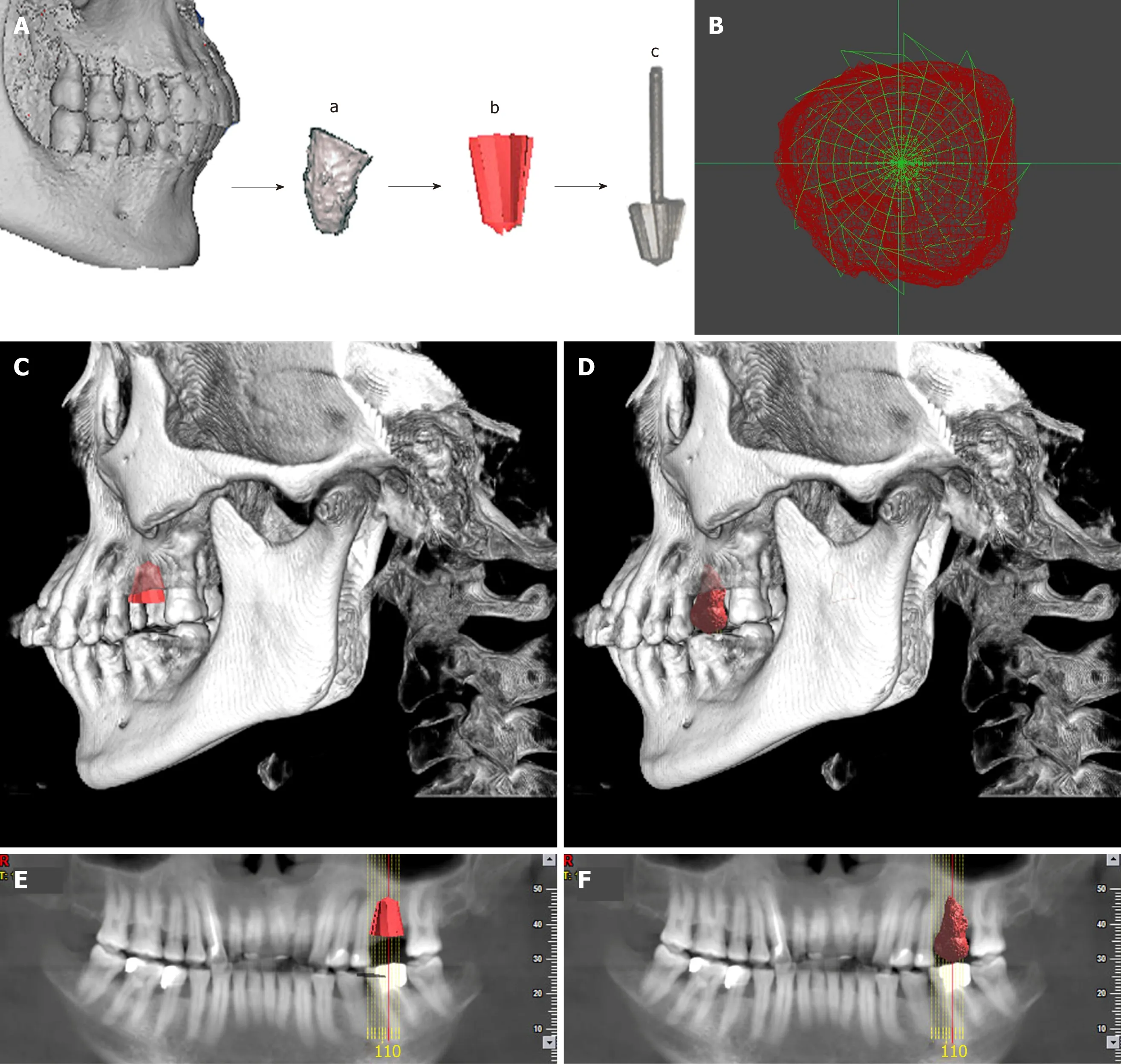
Figure2 Individual drill compared with donor’s tooth.
The individual drill was produced by Wuhan Tianyu Intelligent Manufacturing Company using direct metal laser sintering as follows:(1) The 3d image of the donor’s tooth was extracted from the CBCT;(2) The data of the donor’s CBCT were imported into a STEP format CAD file using CAD software;(3) A CAD model was built using a measuring instrument to gather information regarding the shape and size of the tooth;(4) The shape of the drill was designed according to the donor’s tooth,and the taper and the base of the drill were in accordance with the tooth’s taper and the cervix,and then transferred into a CAD model;(5) The model was then sliced into discrete horizontal layers;and (6) A high-powered laser beam was focused onto a bed of powdered metal to fuse the model layer by layer.Each layer was printed followed by a subsequent layer of powder and printing to produce the next slice of the framework and fused with the first until all layers have been completely built.The laser was utilized to fuse the metal powder into metal solid particles and control their trajectory accurately.
In order to mitigate a potential immunological rejection,the tooth which was carefully extracted with minimal trauma was treated with solutions of 10 g/L cephalosporin,75 g/L clindamycin hydrochloride,and 50 g/L aspirin in sequence.Then the pilot hole was expanded with progressively wider drills,and the preparation was completed using the 3D printed individual drill.The autogenous bone was collected during the entire drill preparation technique and utilized to fill the compartment.After suture,the tooth was splinted with an acid-etch resin composite splint (Figure 3).The recipient was treated with antibiotics and a compound gargle solution of chlorhexidine gluconate was utilized for 2 wk.The follow-up radiographs and photos were taken after surgery (Figure 4).
OUTCOME AND FOLLOW-UP
Inflammation in the periodontal area was observed during healing 1 wk after transplantation.Resorption initiated after 2 wk,which was further increased after 4 and 8 wk.Replacement resorption on the external surface showed increasing appearance time,initiating at 4 wk.The periodontal ligament was no longer visible by X-ray after 4 wk.No pain or swelling was present;however,the height of the peri-implant papillae began to show some recession.The outcome was less than optimal or reported previously and further observations are needed.Other tooth treating methods such as sintering will be studied and carried out in the future.
DISCUSSION
The history of tooth transplantation
Allogenic tooth transplantation has been utilized for five decades with publications occurring in the literature at irregular intervals[3].While studies regarding the immunological aspects of tooth transplantation were not published until 1963[24],more recently,a greater number of studies concerning these aspects have been performed,demonstrating that teeth can be antigenic and elicit an immune response from the host patient[4].Today,an overgrowing research development is placed on whole tooth regeneration,yet research in this field remains wishful.Advances in molecular biology,experimental embryology,developmental biology,and bioengineering may one day overcome present limitations[25].Nevertheless,tooth transplantation has received very little research in comparison owing largely to the developing field of dental implants.In general,the number of tooth transplantation cases have been decreasing and the utilization of new and modern bioengineering techniques and findings from immunological research have been seldom applied to transplantation research in the dental field.This makes this case report provide an insight into the potential development of future avenues of research and discovery in the field of tooth transplantation.
Complications
Root resorption is reported to be the most relevant complication in tooth transplantation and plays an important role in the long-term prognosis of allotransplanted teeth.However,it is also very common in the first few weeks following surgery.It has been suggested that the selection of autogenic tooth versus allogenic tooth is prominent to reduce the apoptosis of odontoblast-lineage cells,but this study also showed that the immunological rejection may have nothing to do with periodontal tissues[26,27].
Root resorption usually happens as replacement resorption and inflammatory resorption[28].In previous cases,a surgeon had to remodel the recipient site to the according donor root sizes,which would definitely increase the surgical time required to transplant the tooth,and therefore worsen the prognosis as well as potentially lead to greater bone loss without the precise bur size created from the donor tooth[10].The replacement resorption is reported to be a response to the operative trauma,which can be lightened by the use of 3D printed burs.Alloimmune rejection also plays an important role during root resorption.Inflammatory resorption can be affected by the endodontic treatment and interestingly the age of recipient[8].
There is also a potential for loss of marginal periodontal attachment which can be caused by pre/preoperative loss of bone and/or inflammation as a result of donor tooth root fracture[8].As a result,a number of cases end up being failed/lost.Accordingly,a previously study conducted by Schwartzet al[8]found that after 10 years,76% of allograft tooth transplants were lost and after 15 years,only six grafts(8.2%) were still in function.The findings further concluded that the cause of graft loss was root resorption in 38 cases,inflammatory resorption in 11,replacement resorption in 27,marginal periodontal complication in 2,apical periodontitis in 1,prosthetic failure in 1,and an unknown etiology in 3[8].

Figure3 Surgical procedure.
In our case report,a similar failure caused by resorption is expected.Nevertheless,this case report is the first attempt at combining modern 3D imaging and printing with allogenic tooth transplantation,which demonstrated plenty of advantages such as an accurate control of material distribution,lower surgical times required,better stability of the transplanted tooth owing to the 3D morphology of the bur,and costeffectiveness[19,20,29].With the utilization of 3D printing during tooth transplantation,it is expected that surgical trauma will be minimized with better final tooth stability.Based on previous publications,the use of aspirin was utilized as a means to minimize alloimmune rejection[30].Aspirin has been shown to inhibit TNF-α and IFNγ levels and reverse proinflammatory cytokines which may cause a higher immune response,leading to apoptosis of bone marrow mesenchymal stem cells[31].
Prevention and treatment of late effects after allogeneic tooth transplantation
To date there have been quite a few methods to inhibit alloimmune rejections,including donor and recipient’s matching test,minimizing movement of the periodontal ligament of transplanted teeth,endodontic treatment prior to transplantation,repeated freezing treatment,irradiation of donor’s teeth,and treatment with fluorinated fluid[32,33].For instance,although Schwartzet al[34]used a monkey model to study the influence of endodontic treatment,and found that the teeth that were endodontically treated significantly decreased inflammatory resorption (almost totally eliminated,P= 0.0003),it had significantly increased replacement resorption (P= 0.0004)[34].Over 40 years ago,Robinson and Rowlands demonstrated that repeat freezing and thawing and incubation with collagenase and
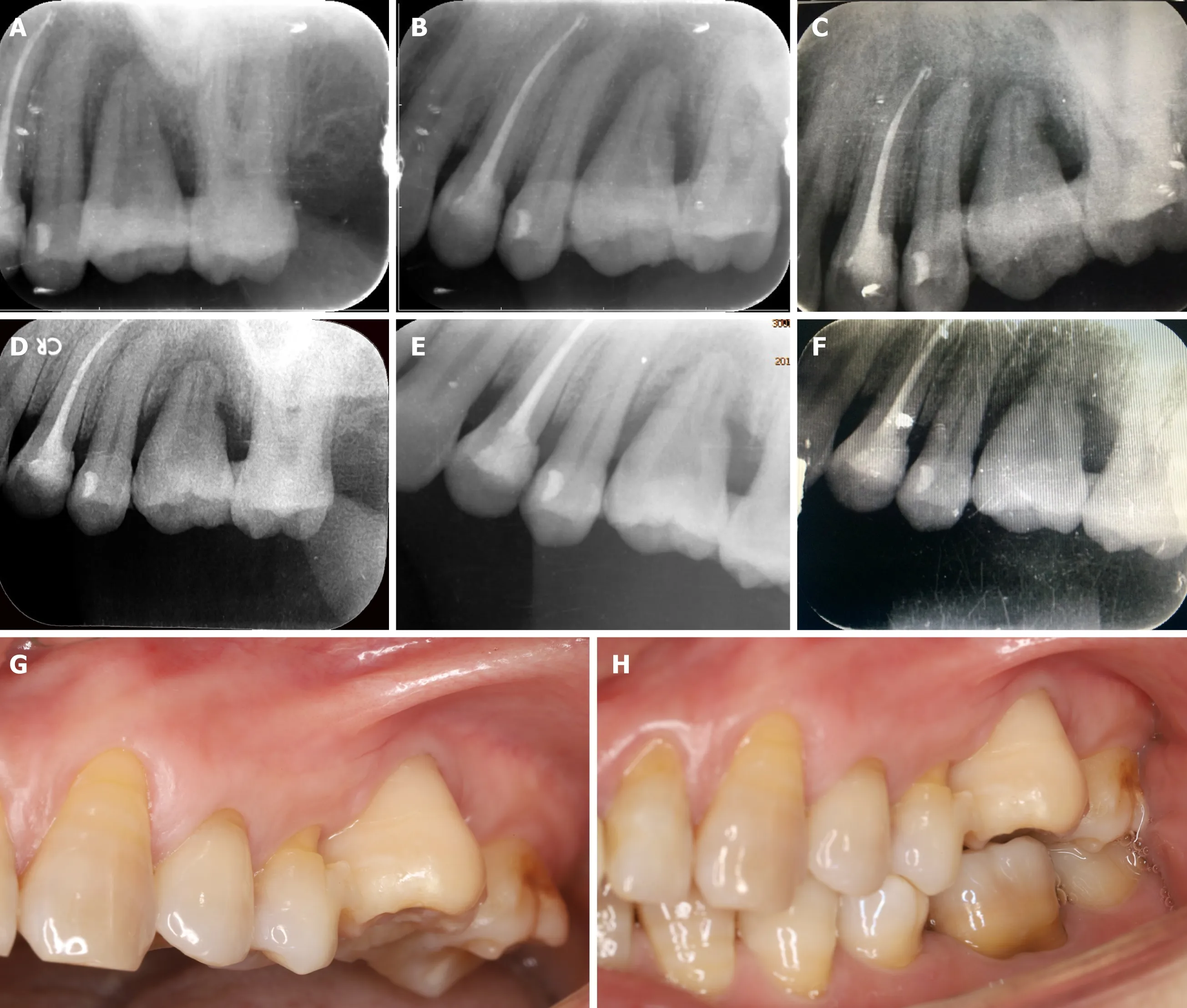
Figure4 Periapical radiographs of the transplanted tooth after surgery.
Figure4 hyaluronidase turned the tooth grafts non-immunogenic[35].These reports are quite old,yet no recent attempts have been made to further investigate if tooth transplantation can become a routine clinical procedure.
The manufacturing process of the individual drill:Direct metal laser sintering
3D printing and bioprinting are modalities of additive manufacturing.Compared to other techniques used in tissue engineering,3D printing has the advantages of accurate precision,resolution,efficiency,and accuracy[16,23].Four main 3D printing techniques exist including inkjet,laser-assisted,extrusion,and stereolithography printing[36-39].Although autogenic and allogenic tooth transplantation has a long history of use,several limitations still exist.In previous cases,since differences in root shape and length exist,surgeons have had to reposition the donor tooth back to its original socket and remodel the recipient site with a round implant bur[10].The remodeling of the recipient site adds additional time to the surgical procedure with the possibility of surgically removing more bone than needed.In the present case report,the specific design of our 3D printed bur allowed for the recipient site to better match the donor tooth.With the ability to 3D print in layers,even the protuberance of the root can be matched and 3D printed,thereby minimizing unnecessary bone loss.There are many factors that may affect the implanted tooth’s primary stability including bone quality and quantity,surgical technique utilized,and the tooth geometry[40].Since the shape of the donor’s and recipient’s roots are generally mismatching,implant stability is hard to predict and therefore the advantages of 3D printing may provide a better solution to the current standards.The advantages of 3D printing include accurate control of material distribution and sizing,fast production,scalability,and cost-effectiveness,which have made this technology successful in many areas of medicine with positive outcomes[19,20].It is therefore conceivable that since various allogenic transplantations are utilized in many areas of medicine including heart,lungs,kidneys,and other complex organs,the ability for dental clinicians to utilize this technique in the coming years should not be deemed unrealistic.With the advancements made in modern medicine and tissue engineering,future research endeavors should be geared towards utilizing this low-cost modality where 3D printing may help improve the predictability of such cases.In the present case,we report the first published attempt at utilizing 3D printing during a tooth transplantation procedure.Future research is necessary to further improve this technology,but this article offers a pioneering first attempt at such a therapy.
CONCLUSION
Our study presents a pioneering case combining 3D printing with allogenic tooth transplantation.A 3D printing system was introduced to print an individualized reamer drill for preparing the implant placement bed and the donor’s tooth as a template for the drill.With the utilization of 3D printing,the surgical trauma was minimized and the tooth implant stability was more suitable.A detailed progress and prognosis of this cases were recorded,which makes the case very useful for reference purposes since it is the first study of its kind.Other tooth treating methods such as sintering will be studied and carried out in the future.This article hopes to enhance our understanding of allogenic tooth transplantation and 3D printing,and may potentially lead to tooth transplantation being utilized more frequently - especially since transplantations are so commonly utilized in many other fields of medicine with high success rates.
杂志排行

World Journal of Clinical Cases的其它文章
- Multifocal G1-G2 gastric neuroendocrine tumors:Differentiating between Type I,II and III,a clinicopathologic review
- Attention deficit hyperactivity disorder and comorbidity:A review of literature
- Dietary manipulation and testosterone replacement therapy may explain changes in body composition after spinal cord injury:A retrospective case report
- Risk factors,clinical features,and short-term prognosis of spontaneous fungal peritonitis in cirrhosis:A matched case-control study
- Incidence of portal vein thrombosis after splenectomy and its influence on transjugular intrahepatic portosystemic shunt stent patency
- Multiplex gene expression profile in inflamed mucosa of patients with Crohn’s disease ileal localization:A pilot study