CT三维骨重建在腰椎骨折手术中的临床应用价值
2018-12-21陈志涌王文珍
陈志涌 王文珍
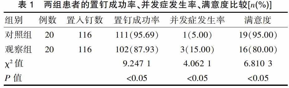
[摘要] 目的 CT三維骨重建技术在腰椎骨折手术中的临床应用价值。 方法 方便抽选2013年8月—2017年8月在该院治疗的40例腰椎骨折患者作为研究对象,随机分为两组,对照组20例,采用传统置钉术进行治疗,观察组20例,采用多排螺旋CT三维骨重建术、椎弓根螺钉固定术进行治疗,比较两组的治疗效果。 结果 观察组置钉成功率为95.69%,对照组置钉成功率为87.93%,差异有统计学意义(χ2=9.247 1,P<0.05)。观察组并发症发生率为5.00%,对照组为15.00%,差异有统计学意义(χ2=4.062 1,P<0.05)。观察组的患者满意度为95.00%,对照组仅为80.00%,差异有统计学意义(χ2=6.810 3,P<0.05)。结论CT三维重建应用于腰椎骨折手术中,清晰直观的显示骨折的位置和具体情况,为临床诊断和治疗提供科学有效的参考依据,有效提高治疗效果,减少并发症发生率,具有重要的临床研究价值,值得推广应用。
[关键词] CT三维骨重建技术;腰椎骨折;临床价值
[中图分类号] R687.3 [文献标识码] A [文章编号] 1674-0742(2018)08(c)-0188-03
The Value of CT Three-dimensional Bone Reconstruction in the Operation of Lumbar Fractures
CHEN Zhi-yong1, WANG Wen-zhen2
1.Department of Radiology, Changtai County Hospital of Fujian Province, Zhangzhou, Fujian Province, 363900 China; 2.Department of Outpatient, Health and Epidemic Prevention Station of Changtai County, Zhangzhou, Fujian Province, 363900 China
[Abstract] Objective The clinical value of CT 3D bone reconstruction in lumbar fracture surgery was analyzed. Methods 40 patients with lumbar fractures who were treated in the hospital from August 2013 to August 2017 were convenient selected and divided into two groups. The control group consisted of 20 patients. The patients were treated with traditional nail placement and 20 patients in the observation group with multi-slice spiral CT three-dimensional bone reconstruction and pedicle screw fixation to compare the therapeutic effects of the two groups. Results The success rate of nail placement in the observation group was 95.69%, The success rate of screw placement in the control group was 87.93%, and the difference was statistically significant(χ2=9.247 1, P<0.05). The incidence of complications was 5.00% in the observation group and 15.00% in the control group. The difference was statistically significant(χ2=4.062 1, P<0.05). The patient satisfaction in the observation group was 95.00%, and only 80.00% in the control group. The difference was statistically significant(χ2=6.810 3, P<0.05). Conclusion CT three-dimensional reconstruction is used in the operation of lumbar fractures. It can clearly and intuitively show the location and specific conditions of fractures. It provides a scientific and effective reference for clinical diagnosis and treatment, effectively improves the therapeutic effect, reduces the incidence of complications, and has important clinical research value, which is worth promoting.
[Key words] CT 3D bone reconstruction; Lumbar fracture; Clinical value
随着超声、MRI、CT等影像学技术的发展,对临床疾病的诊断有着十分重要的影响。X线由于能够直观完整的表达图像,加上价格比较低,成为近几年诊断骨折的主要方法,并取得良好的诊断效果。但是对于比较复杂的腰椎骨折,必须采用多排螺旋CT三维骨重建技术进行诊断,主要是由于X线难以准确诊断出特殊骨折和特殊部位[1]。该文就2013年8月—2017年8月在该院治疗的40例腰椎骨折手术患者进行CT三维骨重建的临床应用价值进行分析探讨,现报道如下。
1 资料与方法
1.1 一般资料
方便抽选在该院治疗的40例腰椎骨折患者作为研究对象,随机分为两组,对照组20例,采用传统置钉术进行治疗,其中男性患者12例,女性患者8例,年龄为20~61岁,平均年龄为(35.3±4.59)岁。交通事故致伤13例,高处坠落致伤4例,重物砸伤3例。爆裂骨折11例,屈曲压缩骨折6例,骨折脱位型3例。L1骨折15例,L2骨折5例。开始受伤到手术的时间为2~7 d,平均时间为(3.6±1.42)d。观察组20例,采用多排螺旋CT三维骨重建术、椎弓根螺钉固定术进行治疗,其中男性患者13例,女性患者7例,年龄为21~63岁,平均年龄为(35.5±4.17)岁。……
