磁共振弥散加权成像对宫颈癌盆腔淋巴结转移诊断的应用价值
2017-10-20董乐丹肖琴琴叶琼许化致杨运俊
董乐丹+肖琴琴+叶琼+许化致+杨运俊
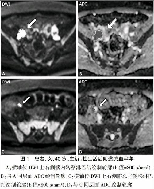
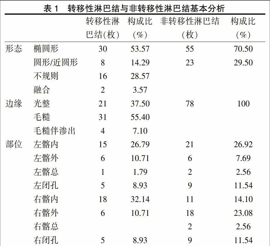
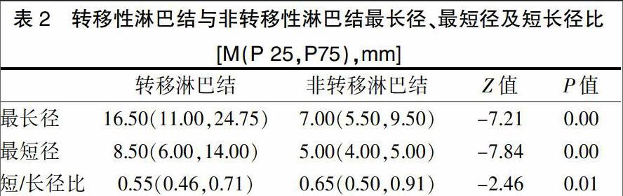
[摘要] 目的 探讨磁共振弥散加权成像对宫颈癌盆腔淋巴结转移诊断的临床应用价值。 方法 收集2015年8月~2016年10月我院收治的26例行盆腔淋巴结清扫术的宫颈癌患者的术前MRI检查资料,术后均经病理检查证实。比较分析宫颈癌原发灶、转移淋巴结及非转移淋巴结的ADC值差异;比较转移淋巴结与非转移淋巴结最长径、最短径及短/长径之比。 结果 ①26例宫颈癌患者中转移性淋巴结56枚,非转移性淋巴结78枚;②转移性淋巴结ADC均值为(874.19±158.72)×10-6 mm2/s),较非转移性淋巴结ADC均值[(921.45±232.98)×10-6 mm2/s]略降低,但两者间差异无统计学意义(P>0.05);宫颈癌原发灶ADC均值为(918.78±146.06)×10-6 mm2/s),与转移淋巴结、非转移淋巴结ADC均值两两比较,差异均无统计学意义(P>0.05);③转移性淋巴结与非转移性淋巴结最长径中位数分别为16.50 mm、7.00 mm,最短径中位数分别为8.50 mm、5.00 mm,短长径比的中位数为0.55、0.65,两两比较差异有统计学意义(P<0.05)。 结论 DWI有利于盆腔淋巴结的检出,但是ADC值测量对转移性淋巴结的临床诊断价值并不大。
[關键词] 宫颈癌;磁共振弥散加权成像;转移性淋巴结;ADC值
[中图分类号] R737.33 [文献标识码] B [文章编号] 1673-9701(2017)25-0101-04
The application value of magnetic resonance diffusion weighted imaging in the diagnosis of pelvic lymph nodes metastasis of cervical cancer
DONG Ledan XIAO Qinqin YE Qiong XU Huazhi YANG Yunjun
Department of Radiology, the First Affiliated Hospital of Wenzhou Medical University, Wenzhou 325000, China
[Abstract] Objective To explore the clinical application value of MR diffusion weighted imaging in the diagnosis of pelvic lymph node metastasis of cervical cancer. Methods The preoperative MRI examination data of 26 patients with cervical cancer who underwent pelvic lymphadenectomy in our hospital from August 2015 to October 2016 were enrolled in this study. The diagnosis of all cases was confirmed by postoperative MRI findings. The differences of ADC values among primary cervical cancer, metastatic lymph nodes and non-metastatic lymph nodes were analyzed. The longest diameter, the shortest diameter and the ratio of the short/long diameter were compared. Results ①There were 56 metastatic lymph nodes and 78 non-metastatic lymph nodes in 26 cases of cervical cancer. ②The mean value of metastatic lymph node ADC[(874.19±158.72)×10-6 mm2/s] was slightly lower than that of non-metastatic lymph node [(921.45±232.98)×10-6 mm2/s], but there was no significant difference between the two groups(P>0.05). There was no significant difference in the ADC value among the primary tumor[(918.78±146.06)×10-6 mm2/s], metastatic lymph node and non-metastatic lymph node of cervical cancer(P>0.05). ③The median of the longest diameter of the metastatic lymph nodes and the non-metastatic lymph nodes was 16.50 mm and 7.00 mm, respectively. The median of the shortest diameter was 8.50 mm and 5.00 mm, respectively. The median of short diameter/long diameter was 0.55, 0.65 respectively. There was statistically significant difference among the groups(P<0.05). Conclusion DWI is helpful for the detection of pelvic lymph nodes, but the clinical diagnostic value of ADC measurement is not significant.endprint
[Key words] Cervical cancer; Magnetic resonance diffusion weighted imaging; Metastatic lymph node; ADC value
宫颈癌是我国女性生殖系统最常见的恶性肿瘤[1]。按照国际FIGO系统,宫颈癌的临床分期与淋巴结是否转移并无直接关联[2],但是具体治疗方案、疗效及预后情况与淋巴结是否转移密切相关[3]。国内外大量文献已经证实淋巴结转移是宫颈癌患者重要的独立危险因素之一[4]。因此,盆腔淋巴结转移是判断预后及指导治疗方案的重要依据[5,6],目前临床工作中MRI诊断主要依据淋巴结形态学改变来判断是否存在转移。国内外许多的研究认为……
