鞍区软骨黏液样纤维瘤1例
2011-03-12齐悦彤
齐悦彤,宋 茜,李 莹
天津医科大学总医院放射科,天津 300052
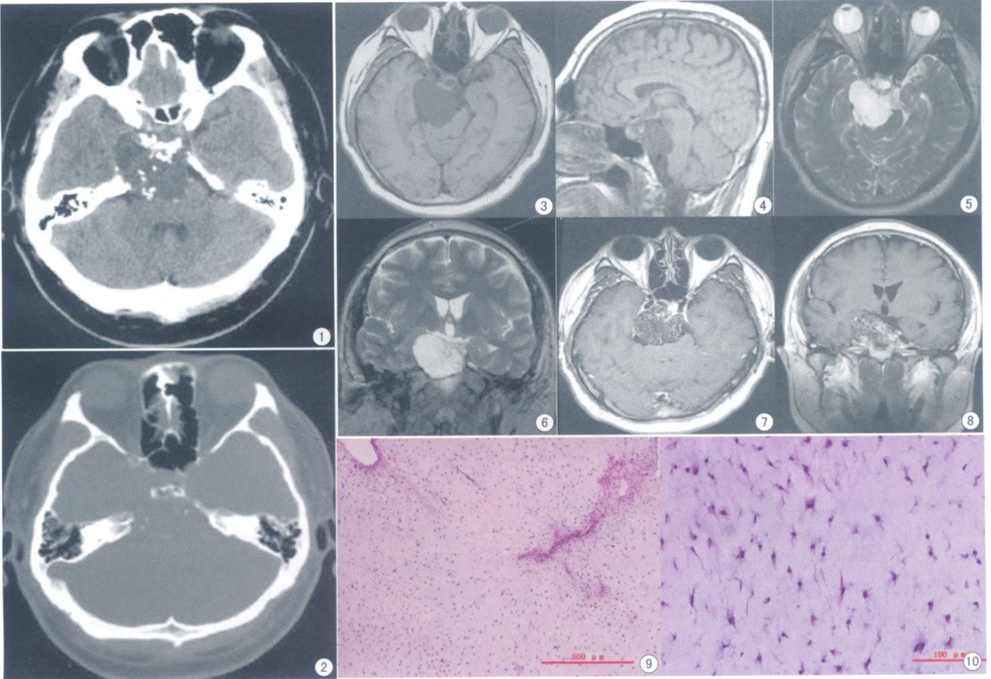
图1、2 CT示:右侧鞍区团块状混杂低密度影,其内及周边可见多发点片状钙化,边界较清晰,并可见鞍背、右侧岩骨尖溶骨性骨质破坏;图3~6 MRI示:病变呈稍长T1、稍长T2信号,其内有点状长T1、短T2信号,边界清晰。累及鞍内、鞍上、右侧鞍旁及桥前池;图7、8 T1WI增强扫描示:病灶网格样轻度强化;图9、10 瘤细胞呈梭形,并可见纤维分割,核呈梭形、卵圆形,深染、呈固缩状,细胞间有丰富的黏液样基质Fig 1,2.CT imagings:A hypodensity mass in the right side of the parasellar,with peripheral and central areas of calcifi cation,a clear boundary,the presence of a destructive lesion involving the saddle back,the right side of petrosum.Fig 3-6.MRI imagings:A longish T1 and longish T2 singal intensity mass,with long T1 and short T2 singal intensity in the cental area,a clear boundary,the mass extension to the intrasellar,the suprasellar,the right side of the parasellar and the pontine cistern.Fig 7,8.Enhanced MRI scan imagings:Mild heterogeneous latticed enhancement.Fig 9,10.Tumor cells were spindle-shaped with fiber division,the nucleus were spindle and oval shape,deep-dyed,condensation-like,with intercellular abundant myxoid matrix.
软骨黏液样纤维瘤是一种起源于成软骨结缔组织的少见良性肿瘤,其发生率不足骨肿瘤的1%。好发于长骨的干骺端或骨端,极少发生在颅骨。我们报道一例发生在鞍区的软骨黏液样纤维瘤。
患者,男,31岁,因“间断性全身乏力2个月,头痛2周”入院。查体:右瞳3.5 mm,左瞳2.5 mm,右眼裂变窄,角膜反射减弱,咽反射减弱。Babinski征(±)。未见其他阳性体征。
CT显示鞍区团块状、混杂密度影,病变以低密度为主掺杂多发点、片状钙化。病灶位置偏右侧,跨越中后颅窝,边界较清晰。MRI示病灶呈稍长T1、稍长T2信号,其内有点状长T1、短T2信号。增强后呈网格样轻度强化。本院拟诊:跨中后颅窝的颅底占位,考虑脊索瘤。
术中见右侧颅中窝近岩骨尖处,可见一个大小约3.8 cm×2.0 cm×2.0 cm的硬膜外肿物,灰白色半透明状,边缘清晰,触之质硬有弹性。术后病理诊断:鞍区软骨黏液样纤维瘤。镜下所见:瘤细胞呈梭形,其间可见纤维分隔,核呈梭形、卵圆形,深染、呈固缩状,酷似软骨细胞,细胞间有丰富的黏液样基质。免疫组化染色结果:Vimentin(+)、S-100(-)、Ki-67LI:0.03%。……