Human umbilical cord derived mesenchymal stem cells in peripheral nerve regeneration
2020-05-07ChristineBojanicKendrickToBridgetZhangChristopherMakWasimKhan
Christine Bojanic, Kendrick To, Bridget Zhang, Christopher Mak, Wasim S Khan
Christine Bojanic, Department of Plastic and Reconstructive Surgery, Cambridge University Hospitals NHS Trust, Cambridge CB2 0QQ, United Kingdom
Kendrick To, Wasim S Khan, Division of Trauma and Orthopaedic Surgery, Addenbrooke's Hospital, University of Cambridge, Cambridge CB2 0QQ, United Kingdom
Bridget Zhang, Christopher Mak, School of Clinical Medicine, University of Cambridge,Cambridge CB2 0QQ, United Kingdom
Abstract
Key words: Umbilical cord; Mesenchymal stem cells; Transplantation; Peripheral nerve regeneration
INTRODUCTION
Peripheral nerve injuries can occur as a result of trauma or disease and can lead to significant morbidity including sensory loss, motor loss and chronic pain[1]. These injuries cause life-long disability in up to 2.8% of all trauma patients[2]. Damage to peripheral nerves most commonly occurs as a result of laceration, compression,ischaemia or traction[1]. As classified by Seddon in 1943, nerve injury can range from focal demyelination termed neurapraxia, to total nerve transection termed neurotmesis[3,4]. The mechanism of recovery post-injury occurs by either branching of collateral axons or by regeneration of the damaged neuron[4,5]. In order for full neuronal recovery to occur, Wallerian degeneration, axonal regeneration and endorgan reinnervation must take place. This is driven by an array of neurotropic factors[4]. However, recovery in function following peripheral nerve injury is hindered by complex pathological mechanisms such as poor nerve regeneration,neuromuscular atrophy, and end-plate degeneration which can lead to suboptimal neuron function[6-9].
Traditionally, peripheral nerve injury can be managed conservatively or surgically with neurolysis, neural suturing, end-to-side neurorrhaphy and nerve autograft[10-12].Even with optimum surgical repair, most methods will attain partial but not full return of nerve function[10]. Certain peripheral nerve injuries, such as severe brachial plexus or long traction injuries remain inoperable[10]. Autografts have several disadvantages, including donor site morbidity, mismatch in nerve and graft size resulting in poor engraftment, and the potential for development of painful neuromas[11,13,14]. Alternative methods of treating peripheral nerve injuries may be through cell-based regenerative therapies[15].
Transplantation of mesenchymal stem cells (MSCs), given their regenerative properties and highly proliferative capacity, has been proposed as a promising therapeutic option for peripheral nerve regeneration[16,17]. MSCs are plastic-adherent,undifferentiated, multipotent cells that can be harvested from numerous sites of the body including bone marrow, adipose tissue, dental pulp, amniotic fluid and umbilical cord[17-19]. MSCs from different tissue origins can have distinct cytokine expression profiles, and thus may enable different MSCs to be particularly suited to certain clinical applications[20,21]. Owing to low immunogenicity, MSCs may be transplanted allogenically with minimal consequence[22]. The particular mechanisms through which MSCs aid nerve repair have not yet been fully characterised. MSCs from various sources such as adipose tissue and bone marrow are able to differentiate into Schwann cells[23,24]. While somein vitroexperiments suggest that transplanted MSCs may be stimulated by peripheral nerves to differentiate into Schwann cells[25],alternative findings have instead shown that transplanted MSCs encourage endogenous cells to express regenerative phenotypes[26]. Increasingly, MCSs are believed to mediate their regenerative properties predominantly through paracrine effects[27,28]. Aside from acting through soluble factors[29], MSCs have also demonstrated the ability to secrete extracellular vesicles that contain bioactive components such as miRNA and cytokines[30]. Indeed, native Schwann cells have been shown to facilitate axonal regeneration following injury through secretion of exosomes that decrease GTPase RhoA activity[31]. Similarly, human MSCs may act to achieve the same result through exosomes by upregulation of the PI3 kinase and Akt signalling cascades[32].
MSCs from umbilical cord are convenient to harvest from post-natal tissue in a non-invasive manner and possess a high capacity to expandex vivo[33]. They express low levels of HLA-DR compared to MSCs from other cell sources and therefore pose low risk of immunogenic complications following allogenic transplantation[34].Through sequential treatment with β-mercaptoethanol and various cytokines,umbilical cord derived MSCs (UCMSCs) can adopt a Schwann-like phenotype[35]. In addition, UCMSCs have been shown to possess greater paracrine effects than those of bone marrow-derived MSCs (BMMSC) and adipose-derived MSCs[17,29], and are able to potentiate axonal regeneration and peripheral nerve functional regeneration through these effects[11,17,29,36]. UCMSCs have been proposed to exert neuroprotective effects through secretion of Brain Derived Neurotrophic Factor (BDNF)[37], angiopoietin-2 and CXCL-16[38,39]. Other studies have suggested that they indirectly promote neurogenesis[40,41]. UCMSCs are also able to indirectly enhance expression of neurotransmitters such as BDNF and neurotrophin-3 (NTF3) which are postulated to aid neuro-regeneration[42,43].
To date, there have been over 400 clinical trials that explore the use of MSCs in transplantation; UCMSCs follow BMMSCs as the second most commonly used cell source[44]. In this PRISMA systematic review, we analyse the evidence for the use of human UCMSCs in peripheral nerve regeneration by examiningin vivostudies.
MATERIALS AND METHODS
A literature search was performed from conception to September 2019 using PubMed,EMBASE and Web of Science. The following search terms were used:((((((((Mesenchymal stem cells) OR mesenchymal stem cell) OR MSC) OR MSCs) OR Mesenchymal stromal cell) OR Mesenchymal cell)) AND (((((Nerve) OR Peripheral nerve) OR Peripheral nerve injury) OR damaged nerve) OR nerve injury)) AND((((((repair) OR regeneration) OR regrowth) OR regenerate) OR renew) OR restore).We adhered to the recommendations as stipulated by the Preferred Reporting Items for Systematic Reviews and Meta-Analyses (PRISMA) guidelines[45].
We included case series, case control, cohort studies and randomised controlled trials. We enrolled studies that examined peripheral nerve lesions treated with human UCMSCs inin vivohuman and animal subjects. Studies that only conductedin vitroexperiments were excluded. Studies that investigated central nervous system regeneration using UCMSCs were excluded. All included studies were published in the English language. We excluded all unpublished and retracted literature.
CB and KT carried out the search independently. RoB2 tool was used by CM and BZ to assess the risk of bias in the studies, all discrepancy in results were resolved by discussion.
RESULTS
A total of 210 studies were screened for title, abstract and the inclusion/exclusion criteria were applied (Figure 1). One retracted study was excluded. Fourteen studies were reviewed in full text. The overall bias of studies is shown in Figure 2. The summary of results is shown in Figure 3. All 14 studies were of a case control design(Table 1). Four studies obtained UCMSCs from a third-party source and the remainder were harvested directly from human subjects. Out of the 14 studies, ten involved xenogenic transplantation into sciatic nerve injury specimens that were either crushed or transected. The studies were grouped according to Seddon’s seminal nerve injury classification system, which includes axonotmesis (injury to nerve sheath alone) and neurotmesis (injury to the entire nerve)[3]. A total of 279 subjects were treated with UCMSCs. All studies reported significant improvement in UCMSC treated groups compared with the various different controls and untreated groups.
The studies did not report any significant complications.
UCMSCs in peripheral nerve axonotmesis
Four studies that included a total of 90 treated subjects assessed the use of UCMSCs in peripheral nerve axonotmesis models of sciatic nerve crush injury (Table 1). All four studies harvested UCMSCs from human subjects and transplanted the UCMSCs into murine subjects. The methods of UCMSC delivery to the crush injury varied among studies.
Studies, by Sunget al[46](2012) and Heiet al[47](2016) examined the effect of direct intralesional UCMSC injections on murine subjects with sciatic nerve crush injuries.Both studies monitored subjects up to 4 wk post-intervention. Sunget al[46](2012)found that expression of brain-derived neurotrophic factor (BDNF) and tyrosine kinase receptor B mRNA increased at 4 wk following UCMSC injection. Functional recovery was measured in terms of the sciatic function index (SFI), which showed a dramatic improvement at 4 wk in UCMSC treated groups compared to untreated groups. Retrograde axonal transport was estimated through fluoro Gold-labelled neuron counts and the UCMSC group was found to have a significantly higher neuron count. It was found that axon density was significantly greater in the UCMSC group. Heiet al[47](2016) transfected UCMSCs with a BDNF-adenovirus vector. The authors found that both UCMSC and BDNF-UCMSC groups had significant improvements in SFI, axon count and axon density at 4 wk after treatment. The BDNF-UCMSC group displayed increased peripheral nerve regeneration compared with UCMSC alone.
Gartneret al[48,49]conducted two studies published in the same year. In one study,Chitosan type III membrane was used to aid UCMSC infiltration in murine sciatic crush models[48]. The authors evaluated motor and sensory functional recovery up to 12 wk following transplantation with and without Chitosan type III wrapping. Both treatment groups showed improvement in SFI, extensor postural thrust (EPT) and withdrawal reflex latency (WRL). The control group with Chitosan type III membrane alone showed a significant improvement in post-traumatic axonal regrowth compared to the untreated control group. In a separate study, the same group examined the effect of using poly (DL-lactide-e-caprolactone) (PLC) membranes to deliver UCMSCs into sciatic nerve crush injuries[49]. Peripheral nerve regeneration was assessed in terms of SFI, EPT, and WRL at 12 wk. Undifferentiated and differentiated UCMSCs were used in different groups. Both groups showed an increase in myelin sheath thickness compared to control groups. The SFI was severely affected at week-2 postcrush injury in all experimental groups and improved gradually up to week 12 when values were indistinguishable from controls.
Studies of UCMSCs in peripheral nerve neurotmesis
Nine of the fourteen studies assessed the use of UCMSCs in peripheral nerve neurotmesis models (Table 2). All nine were case control studies. Five studies had murine subjects, two had rabbit subjects, one had canine subjects, and one had human subjects. Six studies transplanted UCMSCs into a sciatic nerve gap model. Two studies transplanted UCMSCs into tibial nerve and recurrent laryngeal nerve crush models. One study conducted allogenic transplantation in humans. A total of 151 subjects were treated. Methods of MSC delivery and transplantation varied among studies.
Several groups sought to improve nerve regeneration with UCMSCs combined with longitudinal scaffolds. Zarbakhshet al[11](2015) loaded UCMSCs on a silicone tube and interposed it into a murine sciatic nerve gap model. The authors attempted to compare the histological outcomes of human UCMSCs and rat BMMSCs in regenerating sciatic nerve gap in rats. While the author showed favourable results in nerve regeneration for both UCMSCs and BMMSC, the latter was found to produce superior results at the end point of 12 wk. The BMMSC group showed greater axon number and thicker myelin sheath diameter than the UCMSC group.
Maet al[17](2019) injected UCMSC-derived extracellular vesicles (EVs) into the tail veins of rats and sutured a silicone rubber tube into the sciatic nerve gaps of 24 rats.The authors found that UCMSC-EVs promoted motor function recovery and regeneration of axons and attenuated muscle atrophy. SFI analysis was used to assess the functional improvements. At 8 wk, UCMSC-EV group had similar SFI values to normal rats.
总之,几种教学方法,只有根据小学语文阅读课的教材实际,根据所带学生的认知特点,进行有主有次,科学合理的组织,综合的运用,才能收到卓越的功效。
Matsuseet al[35](2010) combined UCMSCs and Matrigel into transpermeable tubes and transplanted it into transected murine sciatic nerve tissue specimens. The authors induced UCMSCs into cells with Schwann cell properties by using βmercaptoethanol, all-trans-retinoic acid and various cytokines. Subsequently, Matsuseet al[35]examined the effect of these induced UCMSCs and used two control groups; a positive control of human Schwann cells and a negative control of Matrigel alone. Thegroup assessed SFI values and compared immunoelectron micrographs. They concluded that the treatment group with Schwann Cell-UCMSCs group was equivalent to treatment with Schwann cells based on histological criteria and functional recovery.
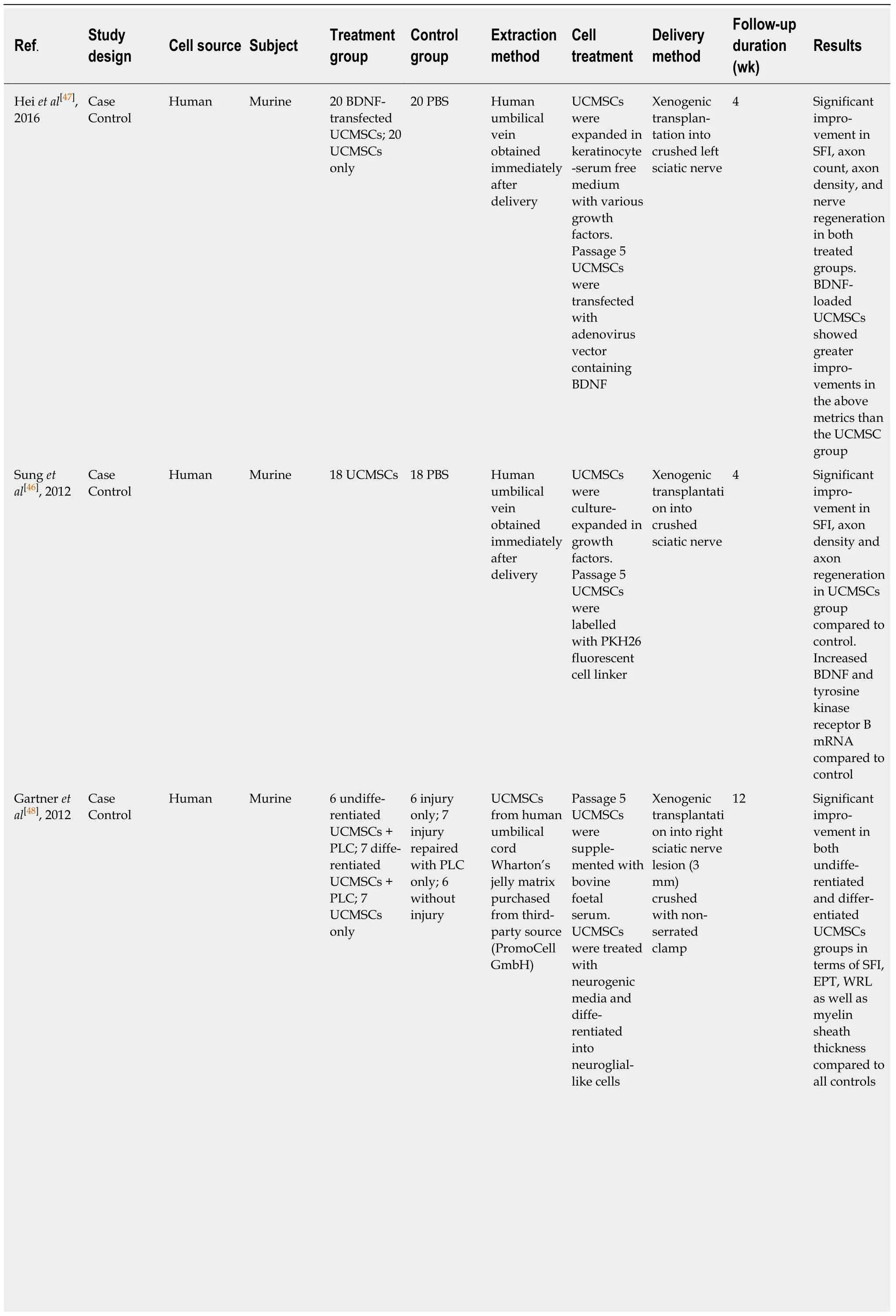
Table 1 Studies of umbilical cord derived mesenchymal stem cells in peripheral nerve axonotmesis and diabetic neuropathy in vivo

UCMSCs: Umbilical cord derived mesenchymal stem cells; BDNF: Brain-derived neurotrophic factor; SFI: Sciatic function index; PBS: Phosphate buffered saline; PLC: Poly (DL-lactide-ε-caprolactone); WRL: Withdrawal reflex latency; EPT: Extensor postural thrust.
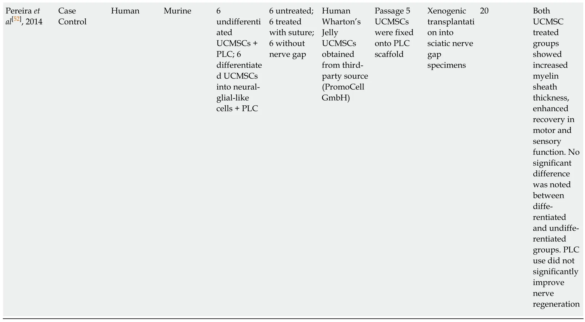
Cuiet al[14](2018) and Panet al[50](2017) delivered UCMSCs using a collagen conduit. Cuiet al[14](2018) transplanted human UCMSCs into canine sciatic nerve gap models via a longitudinally orientated collagen conduit embedded with UCMSCs.Compound muscle action potential (CMAP) was found to be statistically greater in the UCMSC treated group compared with the collagen conduit only group. Panet al[50](2017) appraised the use of UCMSCs with a heparinised collagen conduits in transected rabbit recurrent laryngeal nerves. The authors assessed the effectiveness of passage-4 UCMSCs loaded on heparinised scaffold that released Nerve growth factor(NGF). Electromyograms at 8 wk revealed that treated lesions recovered normal nerve function. Biological markers of neurogenesis, including calcium-binding protein S100,neurofilament and AChE, were expressed at a greater level following treatment. Xiaoet al[51](2015) undertook a study exploring the effect of UCMSCs in a chitosan conduit interposed into the tibial nerve of a rabbit model. Xiaoet al[51]found that nerve conduction velocity was significantly higher in the treatment group. The myelin sheath thickness and the growth of axis bud were both increased in the UCMSC group. Pereiraet al[52](2014) used PLC as a conduit for UCMSCs in murine sciatic nerve crush models. The group compared differentiated and undifferentiated UCMSCs. They established no difference in the degree of nerve regeneration between UCMSC that were differentiated into neural-glial-like cells and undifferentiated UCMSC groups. Both UCMSC groups showed increased myelin sheath thickness and enhanced recovery in motor and sensory function.
Two groups sought to investigate the use of UCMSCs embedded on a human amniotic membrane scaffold[5,53]. Liet al[53](2012) found significant improvements in SFI, CMAP and gastrocnemius muscle diameter in UCMSC-loaded scaffolds group compared to cell-free scaffolds. Liet al[6](2013) analysed how UCMSCs loaded on a human amniotic membrane scaffold affected the repair of a transected radial nerve in human subjects. Thirty-two patients with radial nerve injuries from radial shaft fractures were included in the study; twelve patients received neurolysis to remove neural scar tissue, and transplantation of UCMSCs on an amniotic membrane. The remainder 18 patients received neurolysis only. At 12 wk, the electrophysiological function of the UCMSC-treated group had improved electromyography readings. The muscular power, touch sensation and pain sensation were also significantly improved as compared to the neurolysis group.
Studies of UCMSCs in diabetic neuropathy
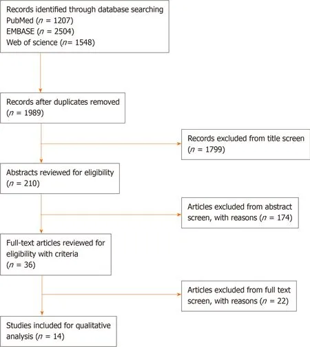
Figure 1 PRlSMA Flow diagram.
One study explored the role of UCMSCs in femoral nerve neuropathy[54](Table 1). The authors modelled diabetic neuropathy in murine subjects by inducing diabetes with streptozotocin and created a dorsal hind foot ulcer through empyrosis. UCMSCs were delivered intravascularly through the femoral artery in the treatment group. Saline injections were used in the control group. Serum NGF and neurofilament 200 (NF-200) were measured by Enzyme Linked Immunosorbent Assay (ELISA). The results demonstrated that serum NGF and NF-200 increased in the UCMSC treated rats.Additionally, functional studies using electroneurogram showed that femoral nerve conduction was improved in the UCMSC subjects.
DISCUSSION
The studies in this review reported compelling positive outcomes for the use of human UCMSC to repair peripheral nerve lesions. None of the studies reported immunogenic nor significant complications. While the source cell utilised was consistent among the studies, there were significant variability in cell treatment and methods of transplantation with variable effectiveness as determined by several different outcome measures. There was also moderate heterogeneity in thein vivomodels used. It is therefore difficult to draw conclusions on the optimal method of cell delivery to nerve lesions. Nevertheless, it does imply that UCMSCs are a useful cell source.
The process of cell harvest did not vary greatly between the studies. Human umbilical cord and umbilical cord blood are generally considered medical waste and so there are minimal ethical barriers to tissue sampling[55]. This provides a practical advantage for the use of UCMSCs and may explain why it is commonly used in tissue engineering experiments. The biochemical properties of UCMSCs may also be of advantage as studies comparing different source cells for MSCs have found UCMSCs to possess a greater ability to proliferateex vivoand express a higher level of Vascular Endothelial Growth Factor (VEG-F) and Human Growth Factor (HGF) at late passages[56,57]. One study in this review compared UCMSCs to BMMSCs in sciatic nerve regeneration and found BMMSCs to produce superior results. The authors however, evaluated cell architecture on microscopy but did not carry out nerve conduction studies or functional analysis which may better inform clinical relevance[11]. UCMSCs also appear to have a different multilineage differentiation profiles to other MSC, and is able to be induced into neuron-like cellsin vitro, which may favour applications in nerve regeneration[58]. There is some evidence to suggest that characteristics of the donor affect the ability of UCMSCs to differentiate. For example, undifferentiated UCMSCs obtained from patients with pre-eclampsia may produce greater levels of neuronal markers[59]. Therefore, exploration of different patient and gestational characteristics, such as age could help determine optimal source conditions.
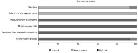
Figure 2 Summary of overall bias.
There is no set protocol for theex vivoexpansion of UCMSCs in the literature. In our article, UCMSCs were generally transplanted between the third and fifth passage.Indeed, the gene expression profile of UCMSC is known to vary according to the number of passages, with some studies showing that UCMSCs do not express CD105,a defining marker for MSCs, until passage-5[60]. In view of this, it would be meaningful to identify and investigate the expression of important neurogenic markers as a function of stages of passage in future experiments. Some of the studies in this review pre-treated UCMSCs in order to induce them into particular cellular phenotypes prior to transplantation. Pereiraet al[52]and Gartneret al[49]utilised a similar culture protocol and pre-treated UCMSCs with neurogenic media. They observed a neuroglial-like morphology on microscopy and a transcriptomic profile showing upregulation of neuroglial genes including Glial Fibrillary Acidic Protein (GFAP), Growth Associated Protein 43 (GAP43) and Neuronal Specific Nuclear Protein (NeuN). Both studies found differentiated MSCs to be more effective than undifferentiated UCMSCs. Aside from gene expression, it would be important to clarify the exact mechanism through which these differentiated cells act to promote nerve regeneration. The optimal protocol for differentiation is also ill-defined as Matsuseet al[35]used a different set of culture condition to induce UCMSCs into Schwann-like cells and found the latter to be more effective. Furthermore, there is a lack of agreement on the primary mode of action of MSCs in promoting nerve regeneration. It is unclear whether transplanted cells directly replicate and replace cells in the lesion. Some experiments of optic nerve lesions suggest that transplanted MSCs remain local and replicate[61]. Emergingin vitroevidence points towards paracrine effects as the predominant mechanism of action. It appears that pre-treatment of Schwann cells with UCMSC conditioned media increases BDNF and NGF expression which are surrogate measures of neurogenic potential[29]. In our review, Maket al[17]examined the effectiveness of UCMSC-derived exosomes in nerve repair. Through peripheral intravenous injection of UCMSC EVs,they demonstrated that it could act systemically to encourage nerve regeneration at a nerve gap without off-site complications. As the use of EVs in this endeavour gains attention, further studies would be required to establish a dose-response relationship and the best method for delivering EVs to lesions.
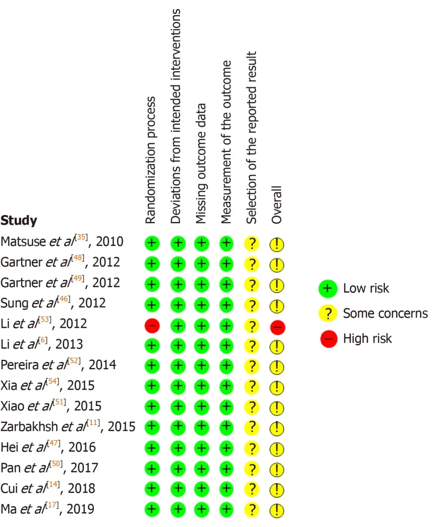
Figure 3 Risk of bias in individual studies.
It is difficult to determine the best UCMSC implantation method. Our review has captured studies that directly implanted UCMSCs and reported good outcomes.Studies investigating the use of conduits to guide nerve regeneration suggest that this is superior to direct implantation of MSCs alone[14,50]. The use of conduit that elude growth factors such as NGF along with UCMSC implantation appear to confer additional benefit[50]. Additionally, intravenous injection of UCMSC-EVs at a peripheral site also produce positive outcomes[17]. Interestingly, a comparison of local and intravenous BMMSC administration in sciatic nerve injury models suggest that systemic treatment provides a more significant improvement in nerve conduction,whereas local treatment improved neuronal fibre counts[62]. Other experiments have shown that peripherally injected MSCs localise to nerve lesions in murine models of sciatic nerve injury[63]. It could be inferred from these findings that there are differing mechanisms and sites of action for the two methods of implantation, suggesting that a treatment regime including both delivery methods concomitantly may produce the best outcome.
There are several issues pertaining to translating the findings derived fromin vivoanimal models for therapeutic application in humans. The majority of studies in our review employed a murine surgical sciatic nerve defect model to assess nerve regeneration. The critical nerve gap length, defined as a gap across which regeneration would not occur without nerve grafting or bridging is considered to be greater in humans than murine subjects[64,65]. Therefore, studies assessing murine nerve gaps may overestimate the therapeutic potential of treatments. Furthermore, it may be difficult to scale-up effective concentrations of transplanted UCMSCs to humans. In a study of rat nerve defects treated with tacrolimus, functional recovery tapers off at 9 wk following treatment and becomes indistinguishable from untreated rats at 10 wk[66]. Therefore, at later time points, which are more relevant to clinical presentations of nerve injury, the regenerative biology of murine nerve appears to differ from that of humans. Our interpretation from thein vivoanimal studies in this review is complicated by the use of the sciatic nerve, which possesses a sensory and motor component, and thus renders functional analysis difficult. It is conceivable for sensory loss to mask a post-surgical motor defect on gait analysis, similarly, it may be possible for loss in motor function to cause underestimation of sensory recovery.Owing to the heterogeneity in starting points for different functional measures, a pooled analysis of quantitative outcomes could not be performed in this review.Therefore, clinically relevant and robust quantifiable outcome measures remain a significant barrier to the reliability of animal studies. One study in this review assessed UCMSC transplantation in human radial nerve defects and reported improved motor and sensory function and electrophysiological measures[53]. The group however, delivered the MSCs through a scaffold, and did not compare outcomes with a control group of the scaffold alone.
According to the results of our risk of bias analysis, 13 of 14 studies had a moderate risk of bias, and one study had a high risk of bias (Figure 1). The reporting of outcome measures contributed to an increased risk of bias in all studies, as most of the studies reported improvement in some but not all outcomes yet concluded that UCMSCs were effective overall. This could be owing to the significant heterogeneity in cell treatment and delivery methods which as the literature suggests, could contribute to different aspects of nerve regeneration.

Table 2 Studies of umbilical cord derived mesenchymal stem cells in Peripheral Nerve Neurotmesis in vivo

Li et al[53],2012 Case Control Human Murine 40 amnion tube with UCMSCs 40 amnion tube with saline implant Human umbilical cords obtained from fullterm deliveries Passage 3-4 UCMSCs were cultured and loaded on an amniotic scaffold Xenogenic transplantati on into transected sciatic nerve tissue specimens 20 Significant improvement in SFI and CMAP in UCMSC group compared to control.Gradual improvement in threshold stimulus and maximum stimulus intensity in UCMSC group compared to control Li et al[6],2013 Case Control Human Human 12 neurolysis followed by 10 mL UCMSCs injection of 1.75 × 10⁷ cells 20 neurolysis only Human umbilical cords obtained from fullterm deliveries Passage 2 UCMSCs were loaded on an amniotic membrane scaffold.Both groups received 3 days of oral cephalosporin Allogenic transplantati on into radial nerve injury following radial shaft fracture 12 Significant improvement in muscular strength,touch and pain sensations in UCMSC group compared to control.Improved electrophysiologica l function in UCMSC group as compared to control Matsuse et al[35], 2010 Case Control Human Murine 6 UCMSCs;10 Induced UCMSC 6 negative control; 5 induced UCMSC Wharton’s Jelly extracted from umbilical cords of fullterm caesarean deliveries Passage 3 UCMSCs were induced into Schwannlike cells Xenogenic transplantation into transected sciatic nerve tissue specimens 3 Significant improvement in SFI in all treated as compared to control with the greatest improvement in UCMSC group Xiao et al[51],2015 Case Control Human Rabbit 10 chitosan conduit anastomosis bridge filled with UCMSCs 10 chitosan conduit anastomosis only; 10 untreated Not specified UCMSCs were loaded into a chitosan conduit Xenogenic transplantati on into tibialcommon peroneal nerve endto-side anastomosis 12 Significant improvement in myelin sheath thickness,Schwann cell growth,growth of axis bud and growth velocity of regenerated fibre in UCMSC group compared to controls. No significant difference observed between either control groups
UCMSCs: Umbilical cord derived mesenchymal stem cells; LOCC: Longitudinally orientated collagen conduit; SFI: Sciatic function index; NGF: Nerve growth factor; PBS: Phosphate buffered saline; HC: Heparinized collagen; PLC: Poly (DL-lactide-ε-caprolactone); EV: Extracellular vesicle.
In conclusion, while there are homeostatic responses that promote nerve regeneration following injury, the body’s natural capacity is inadequate for the recovery of satisfactory nerve function. The evidence summarised in this systematic review supports the notion that UCMSC transplantation is an effective treatment option for nerve injury. Several barriers must be overcome before these findings can be translated into the clinical setting. Importantly, development of a reliablein vivoanimal model, and a standardised method of assessing nerve regeneration would allow the optimal method of cell transplantation to be determined.
ARTICLE HIGHLIGHTS
Research background
Peripheral nerve injury can be a debilitating condition. Traditional treatment options are often ineffective. There is an urgent need for new treatment modalities. Mesenchymal stem cell (MSC)transplantation holds promise as a cell-based regenerative approach in treating nerve lesions.MSCs can be sourced from various tissues, and this may affect their regenerative capacity. Here,we appraise thein vivoevidence for the use of human umbilical cord-derived MSCs (UCMSCs)in peripheral nerve regeneration.
Research motivation
There is contention regarding the optimal cell-source for the harvest of MSCs. Some evidence suggests that MSCs from certain tissue types have superior neurogenic capacity. It is critical that we determine the best cell-source for nerve repair, in order to facilitate an efficient production protocol and maximise clinical benefit.
Research objectives
To investigate whether UCMSCs are effective in nerve regeneration inin vivomodels of nerve injury.
Research methods
We performed a systematic literature review according to the PRISMA statement. A search was conducted on three databases (PubMed, EMBASE and Web of Science) by two independent investigators from inception to September 2019 for studies examining the use of UCMSCs inin vivomodels of nerve injury. The evidence was appraised using Cochrane’s RoB 2.0 Tool.
Research results
A total of 14 studies were included in the review, with a total of 279 subjects. The studies reported that transplantation of human umbilical cord MSCs were effective in regenerating nerve lesions. There were general improvements in histological and functional outcomes. The studies did not report significant complications.
Research conclusions
Human umbilical cord-derived MSCs were effective in repairing nerve lesions in both animal and human models of nerve injury. Additional studies are required to correlate histological outcomes with functional improvements, as not all studies assessed both. More human studies are necessary to inform the efficacy in humans. High quality randomized controlled trials would be instructive in this case. Long-term follow up in these types of study will help inform the safety of MSC transplantation.
Research perspectives
There is limited evidence examining the use of MSCs derived from other tissues in their capacity to regenerate nerve lesions. Further studies comparing different tissue cell-source directly would be highly informative.In vitrostudies of MSC-biomaterial scaffolds may aid the development of more efficient MSC delivery methods. As the nature of nerve injury can vary significantly, the approach to transplantation, such as dose delivery may need to be catered to the individual lesion. Studies comparing the effect of MSCs on differentin vivomodels could help delineate this.
猜你喜欢
杂志排行

World Journal of Stem Cells的其它文章
- Bone marrow-derived products: A classification proposal - bone marrow aspirate, bone marrow aspirate concentrate or hybrid?
- Overview of noncoding RNAs involved in the osteogenic differentiation of periodontal ligament stem cells
- lnsights of stem cell-based endogenous repair of intervertebral disc degeneration
- Ability of human umbilical cord mesenchymal stem cells to repair chemotherapy-induced premature ovarian failure