Clinicohistopathological implications of phosphoserine 9 glycogen synthase kinase-3β/ β-catenin in urinary bladder cancer patients
2019-08-26NiharikaMauryaRinniSinghApulGoelAtinSinghaiUdayPratapSinghVinitaAgrawalMinalGarg
Niharika Maurya, Rinni Singh, Apul Goel, Atin Singhai, Uday Pratap Singh, Vinita Agrawal, Minal Garg
Niharika Maurya, Rinni Singh, Minal Garg, Department of Biochemistry, Lucknow University,Lucknow 226007, India
Apul Goel, Department of Urology, King George Medical University, Lucknow 226003, India
Atin Singhai, Department of Pathology, King George Medical University, Lucknow 226003,India
Uday Pratap Singh, Department of Urology and Renal Transplantation, Sanjay Gandhi Post Graduate Institute of Medical Sciences, Lucknow 226014, India
Vinita Agrawal, Department of Pathology, Sanjay Gandhi Post Graduate Institute of Medical Sciences, Lucknow 226014, India
Abstract
Key words: Pathobiology; Target genes; Tumor stage and grade; Wnt/β-catenin signal cascade; Urothelial carcinoma of bladder
INTRODUCTION
Urothelial carcinoma of bladder (UCB) is the fourth most common cancer in men and it ranks ninth most frequent cancer in women worldwide[1]. Intravesical immuno or chemotherapy and surgical resections are commonly employed modalities to treat non-muscle invasive cancer (NMIBC) and locally advanced cancer. Nevertheless,these tumors are prone for relapse and disease progresses with 50% to 90% rate of recurrence. Twenty five percent of patients, who are initially presented as muscle invasive bladder cancer (MIBC), undergo either radical cystectomy or radiochemotherapy but fail to achieve complete therapeutic response[2]. Owing to heterogeneous clinical outcome of two distinct types of diseases (NMIBC and MIBC);limitations of standard cytopathology for low grade urothelial malignancies; and lack of mechanism for detecting early recurrence in high risk NMIBC and progression in MIBC, intense investigation of tumors using molecular diagnostics is a favorable model for urologic oncologists.
碾压混凝土浇筑过程中的温度控制同样是防比温度裂缝的关键。温度控制措施有控制温升、采用低热水泥、增加活性掺合料用量、采取保温措施、在适宜的气候下浇筑、埋设冷却水管以及预冷却等方法。因碾压混凝土浇筑仓面大,受周围气候的影响大,预冷却法的效果不明显,所以预冷却法已经基本不用。一般来说,碾压混凝土施工气温在3~25℃较为合适。
Wnt/β-catenin signaling plays pivotal role in the development of urogenital systemand hence, its abnormal activation plays significant role in bladder cancer pathogenesis[3]. β-catenin is a multifunctional protein, exists at the cell membrane in a complex with E-cadherin and α-catenin and is involved in cell-cell adhesion;maintenance of epithelial cell integrity; normal cell growth/differentiation, embryonic development, wound healing and apoptosis[4]. In normal cells, cytoplasmic β-catenin is usually maintained at low level; constitutively captured in a destruction complex[composed of adenomatous polyposis coli (APC), glycogen synthase kinase-3β (GSK-3β) and Axin]; phosphorylated by GSK-3β and casein kinases 1α (CK1α); and degraded via the ubiquitin proteasome pathway[5].
GSK-3 is a multifunctional serine/threonine kinase and is one of the essential components of Wnt/β-catenin signal cascade[6]. It is involved in multicellular processes, including proliferation, differentiation, cell cycle regulation, glycogen metabolism, cell mobility and survival. Its two isoforms: (1) GSK-3α and (2) GSK-3β,exhibit 98% identity within their kinase domain, nevertheless, their functions are different[7]. Site-specific phosphorylations at tyrosine-216 (active form of GSK-3β) and at serine-9 (inactive form of GSK-3β) are responsible for enhanced and reduced kinase activity of GSK-3β respectively. This makes GSK-3β, an important molecule and allows it to function as either tumor promoter or tumor suppressor[8].
Phosphorylation of GSK-3β at serine 9 (pS9GSK-3β) attenuates its enzyme activity and its increased cytosolic levels prevent it from phosphorylating its downstream targets including β-catenin. This leads to stabilization and cytosolic accumulation of β-catenin[9]. Subsequently, it is shuttled to the nucleus, forms complexes with members of the T-cell factor (TCF)/lymphoid enhancer factor (LEF) family of transcription factors, and thereby activates target genes including Cyclin D1, Snail and Slug. These genes are regulators of cell proliferation and are involved in tumorigenesis[10]. Cyclin D1 is an important regulator of cell cycle progression and contributes to the neoplastic transformation of cells[11]. Snail and Slug belong to Snail family of zinc-finger transcription factors, share highly conserved zinc finger domains and regulate the transcription and translation of molecules that orchestrate the process of epithelial-to-mesenchymal transition (EMT)[12,13]. EMT renders tumor cells to transiently adapt altered morphological traits with reduced requirements of cellcell contact, immune evasion and allows tumor cells to undergo series of events characterized by metastatic cascade[14].
An aberrant activation of pS9GSK-3β has been reported to trigger on Wnt/βcatenin signal cascade and has been associated with cell proliferation, differentiation and increased aggressiveness in several major human cancers including pancreatic,colorectal, ovarian, prostate, cervical, breast and bladder cancer[15-18]. Nevertheless,functional implications of pS9GSK-3β in the pathogenesis/prognosis of bladder cancer remain unclear. The present study has been taken up to examine the quantitative expression and localization of pS9GSK-3β protein in urinary bladder tumors. Aberrant accumulation of stabilized β-catenin and transcriptional induction of target genes (Cyclin D1, Snail and Slug) via activated β-catenin signaling pathway were evaluated for their clinical significance in well-defined cases of bladder cancer.
Findings of the present study suggest that pS9GSK-3β may be involved in urothelial tumorigenesis by stabilizing/regulating β-catenin via Wnt/β-catenin signal cascade. Association of aberrantly co-expressed pS9GSK-3β and β-catenin proteins,with tumor stage, tumor type, smoking/tobacco chewing status and shorter overall survival probabilities of patients validates them as potential markers of clinical relevance in the pathobiology of UCB. Histopathological association validates Cyclin D1 as a marker of clinical significance, nevertheless, these results rule out the possible post translational activation of target genes by pS9GSK-3β/β-catenin /TCF/LEF transcriptional complex in urothelial tumorigenesis.
MATERIALS AND METHODS
Study population
The study population consisted of ninety North Indian patients diagnosed with UCB were enrolled in the Department of Urology, King George Medical University(KGMU), Lucknow, India and Department of Urology, Sanjay Gandhi Post Graduate Institute of Medical Sciences (SGPGIMS), Lucknow, India during 2014-2017 (Figure 1).All the patients examined with symptoms of hematuria followed by urinary frequency/irritative symptoms were assessed for primary tumor, which included bimanual examination under anesthesia before and after transurethral resection.Frozen tissue specimens were obtained from the patients at the time of transurethral resection/radical cystectomy. Matched formalin fixed and paraffin embedded tissue sections were archived from the Department of Pathology, KGMU and SGPGIMS,Lucknow, India. Studies were approved by the appropriate institutional research ethics committee and were performed in accordance with the ethical standards as laid down in the 1964 Declaration of Helsinki and its later amendments or comparable ethical standards. Ethical clearance was obtained from Bioethics Cell, Institutional Ethics Committee (IEC), KGMU (Reference No. 77thECM II A/P14) and SGPGIMS(Reference no. 2014-166-CP-80), Lucknow, India.
Out of 90 patients, 48 were diagnosed with non-muscle invasive (stage pTa-pT1)and 42 had muscle invasive (stage pT2-pT4) tumors. Eighty three tumor tissue specimens were obtained from the patients who underwent transurethral resection of the bladder (TURB) and seven tumor tissue samples from the patients who underwent radical cystectomy. Ten tissue specimens of normal bladder mucosa were collected from patients with benign prostate hyperplasia after taking their signed and informed consent. These patients showed no obvious malignancy and had cold cup bladder biopsy. A small fraction of the excised bladder tumor or normal tissue was immersed in RNA later solution (Ambion; Thermo Fisher Scientific Life Sciences,Waltham, MA), and stored at -800C until further use.
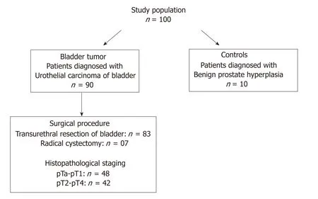
Figure 1 Flow chart of the number and selection of patients/controls in the study population.
The diagnoses and histopathological classification of the bladder tumor tissues were performed independently by pathologists according to WHO-ISUP, 2004 classification system[19]. Pathological stage was examined on the basis of degree of invasion of tumorous mass in lamina propria while, histological grade was determined by cell architecture, polarity, thickness and cohesiveness.
Study design
Quantitative expression and cellular localization analysis at protein level:Immunohistochemistry staining: Immunohistochemical (IHC) staining for putative biomarkers involved in GSK-3β and β-catenin signaling was performed using Ultra Vision Quanto detection system, horseradish peroxidase-diaminobenzidine (Thermo Fisher Scientific Life Sciences), according to the manufacturer's instructions. Primary antibodies were used to stain biomarker proteins and these include antibodies to: (1)β-catenin (14; 224M-15; Cell Marque, Rocklin, CA) (1:40 dilution); (2) pS9GSK-3β(phospho S9; EPR2286Y; ab75814; Abcam, Cambridge, CA) (1:100 dilution); (3) Cyclin D1 (SP4; 241R-15; Cell Marque) (1:100 dilution); (4) Snail (Ab180714; Abcam,Cambridge, CA) (1:100 dilution); and (5) Slug (H-140; sc-15391; Santa Cruz Biotechnology, Santa Cruz, CA) (1:50 dilution). Three micron thick formalin fixed and paraffin embedded tumor and normal tissue sections from each sample were fixed on poly-L-lysine coated glass slides by heating at 700C for 3 h. Tissue sections were dewaxed using xylene and then subsequently washed with 100%, 70%, 50%, and 20%alcohol for 5 min each. After a brief wash, slides were then subjected to antigen retrieval with citrate buffer [low pH (citrate) buffer or high pH (Tris ethylene diamine tetra acetic acid) buffer] for 4 cycles of 5 min at full efficiency microwave system.Choice of buffer depends on the type of primary antibody. Sections were washed after blocking the activity of endogenous peroxidase by peroxide block (provided with the kit) for 10 min. Following this treatment, sections were incubated with the primary antibodies overnight at 40C followed by their incubation with primary antibody amplifier for 10 min at room temperature. Signals were detected by adding horseradish peroxidase polymer and diaminobenzidine, followed by counterstaining with Mayers' hematoxylin for 1 min. Slides were dehydrated and mounted in dibutyl phthalate xylene medium, and visualized for the brown reaction product. Evaluation of IHC putative biomarkers for their cellular localization and expression at protein level was done using a Nikon Eclipse 50i light microscope (Nikon, Tokyo, Japan).
IHC score
A total of 1000 cells were counted in consecutive fields at a magnification of 400× bytwo independent observers. Immunoreactivity of the tumor/normal/control tissue was analyzed as strong, weak/reduced, or no expression. Marker proteins were examined for their membranous, nuclear, and or cytoplasmic localization. Staining intensity was assessed on the scale of 1, 2 and 3 where 1 was assigned as reduced expression, 2 as moderate expression and 3 as strong expression. Overall IHC score was calculated by multiplying the percent positive cells stained and its staining intensity. Based on percent positive cells and their staining intensity, IHC score was assigned as low as 0 to as high as 300.
Nuclear immunostaining was examined for Cyclin D1, Snail and Slug.PhosphoS9GSK-3β and β-catenin were evaluated for no expression/low membranous expression; nuclear expression; cytoplasmic and; high membranous expression.
Quantitative expression analysis at transcriptome level: RNA extraction and real time-quantitative polymerase chain reaction:Total cellular RNA extraction was done from frozen tumor and control tissue specimens using 1 mL/mg TRIzol reagent(Ambion; Thermo Fisher Scientific Life Sciences). Extracted RNA was quantified by measuring its absorbance at 260 nm and purity was checked by taking its absorbance ratio at 260 nm and 280 nm. One microgram of RNA from each tissue/sample was reverse transcribed to cDNA using verso cDNA synthesis kit (Thermo Fisher Scientific Life Sciences) according to the manufacturer's instructions. Subsequently, 10 ng of cDNA in 5 µL reaction mix was subjected to real-time quantitative polymerase chain reaction (RT-qPCR) for each tumor and normal tissue sample. RT-qPCR was performed in a Quant Studio 7 Flex Real-Time PCR platform (Applied Biosystems;Thermo Fisher Scientific Life Sciences) using Light Cycler 480 Syber green I master mix (TaKaRa, Clontech). Following primers were used to amplify the specific genes/putative biomarkers under study: (1) β-catenin, Forward:5 ' G C T T G G A A T G A G A C T G C T G A 3 ' a n d R e v e r s e :5'C T G G C C A T A T C C A C C A G A G T 3'; (2) C y c l i n D 1, F o r w a r d:5 ' T G C C A C A G A T G T G A A G T T C A T T 3 ' a n d R e v e r s e :5'C A G T C C G G G T C A C A C T T G A T 3'; (3) S l u g, F o r w a r d:5'TCGGACCCACACATTACC3' and Reverse: 5'CCGAGATGGGGTTGATAATG3';(4) Snail, Forward: 5'TCGTCCTTCTCCTCTACTTC3' and Reverse:5'TTCCTTGTTGCAGTATTTGC3'; and (5) β-actin as a housekeeping gene, Forward:5 ' G G A C T T C G A G C A A G A G A T G G 3 ' a n d R e v e r s e :5'AGCACTGTGTTGGCGTACAG3'.
RT-qPCR cycling conditions include, initial denaturation at 950C for 5 min;followed by 35 cycles of denaturation at 940C for 30 s, annealing at 560C for 30 s,extension at 720C for 1 min; and final extension at 720C for 10 min. Relative levels of mRNA expression were quantified as ΔCt values by comparing it with the mean Ct values of β-actin. β-actin was taken as reference/endogenous control gene (ΔCt = Cttarget- Ctreference) to normalize any possible difference in the amount of total RNA.Furthermore, ΔΔCt for each target gene in tumor sample was calculated using ΔCt of normal tissue against respective gene (ΔΔCt = ΔCttumor- ΔCtnormal). Average fold change expression was then calculated by applying 2-ΔΔCt. Ct values for all the genes in each tissue sample were assayed and analyzed in triplicates.
Statistical analysis
Quantitative expression of putative biomarkers involved in β-catenin signaling at transcriptome and protein level was statistically correlated with clinical features including smoking/tobacco chewing status of patients and presence or absence of hematuria; and histopathological variables including tumor stage, tumor grade,tumor type (primary/recurrence), presence or absence of metastasis and lymph node involvement using SPSS, version 20.0 software; Microsoft, Redmond, WA. Kaplan-Meier along with log-rank test was employed to evaluate probable relation between biomarker expression and overall survival (OS)/progression free survival (PFS) of patients. Period of assessment for OS of patients includes the time period between the date of surgery/cystoscopy as an entry date and the date of last follow up/date of patients' demise due to cancer. Time period between the date of surgery/cystoscopy as an entry date and date of tumor progression on last follow up/date of patients'demise due to tumor progression was used for PFS studies. Quantitative data are presented as the mean ± SE. The P values less than 0.05 were considered as statistically significant.
RESULTS
Clinical and histopathological summary of patients
Present study was performed on a cohort of 90 patients with UCB and 10 normaltissues. Median age of these patients was 60 years, ranging from minimum of 38 years to maximum of 82 years. Eleven percent (10/90) patients were female. Of total, 80%patients (72/90) were examined with gross painless hematuria as the primary clinical symptom. Smoking/tobacco chewing history of 76 patients was available; out of which 64.5% were regular smokers or tobacco chewers and 35.5% were non-smokers.Fifty three percent (48/90; stage pTa-pT1) patients were presented with non-muscle invasive disease, while 46.7% patients (42/90; stage pT2-pT4) were diagnosed with muscle invasive disease. Histologically, 34.4% tumors were classified as low grade carcinomas and 65.6% patients were presented with high grade carcinomas. Among the total 90 patients, 43.3% (39/90) patients were diagnosed with the tumor of recurrent type. As per clinicohistopathological records, only 6.7% patients were examined with lymph node metastasis that developed over a mean period of 42 mo since initial diagnosis. Follow up data was available in 71 patients. Of 71 patients, 69%patients survived and 28.2% patients showed progression over an average time line of 36 mo. Clinicohistopathological profiles of the patients included in the study are summarized in Table 1.
pS9GSK-3β and its diagnostic/ prognostic relevance in human bladder tumors
IHC staining was done to detect the expression of pS9GSK-3β in a cohort of 90 human bladder tumor sections and 10 normal bladder tissue sections. High membranous expression of pS9GSK-3β was observed in all the sections obtained from control bladder tissues. However, 51.1% (46/90) tumors exhibited its aberrant expression which includes low membranous (20% tumors with IHC score ≤ 200),nuclear/cytoplasmic (6.7% tumors with IHC score ≥ 1) or no expression (24.4%tumors IHC score = 0) (Figure 2A and Figure 3). Aberrant expression of pS9GSK-3β was observed in 52% (25/48) non-muscle invasive (low membranous: 20.8%,cytoplasmic/nuclear: 12.5% and no expression: 18.8%) and in 50% (21/42) muscle invasive tumors (low membranous: 19%, cytoplasmic/nuclear: 0% and no expression:31%) (P = 0.01; Mann Whitney U test). Immunostaining of pS9GSK-3β was observed in 45.2% (14/31) low grade (low membranous: 25.8%, cytoplasmic/nuclear: 6.4% and no expression: 12.9%) and 54.2% (32/59) high grade tumors (low membranous: 16.9%,cytoplasmic/nuclear: 6.8% and no expression: 30.5%) (P = 0.04; Mann Whitney U test).Aberrant expression of pS9GSK-3β was noted in 59% (23/39) recurrent tumors (low membranous: 20.5%, cytoplasmic/nuclear: 12.8% and no expression: 25.6%); 49%(24/49) patients with smoking/ tobacco chewing habit (low membranous: 20.4%,cytoplasmic/nuclear: 8.2% and no expression: 20.4%); and 54.2% (39/72) patients with hematuria (low membranous: 22.2%, cytoplasmic/nuclear: 8.3% and no expression:23.6%) (Table 2). Forty four tumors out of 90 cases exhibited strong membranous expression of pS9GSK-3β but did not show any statistical relevance with clinicohistopathological variables (data not shown).
β-catenin and pS9GSK-3β proteins: Diagnostic/ prognostic relevance in human bladder tumors
Immunostaining was done to check β-catenin expression at protein level and its subcellular localization in 90 bladder tumors (Figure 2B and Figure 3). Its strong membranous expression was seen in control/normal urothelium while out of 90 tumors, 44 showed aberrant expression of β-catenin. Out of these 44 tumors, 23 showed low membranous (25.6% with IHC score ≤ 200); 12 exhibited nuclear/cytoplasmic (13.3% with IHC score > 0); and 9 did not show any expression(10% with IHC score = 0). Forty four percent (21/48) non-muscle invasive (low membranous: 20.8%; cytoplasmic/nuclear: 16.7%; no expression: 6.2%); 54.8% (23/42)muscle invasive tumors (low membranous: 30.9%; cytoplasmic/nuclear: 9.5%; no expression: 14.3%); 67.7% (21/31) low grade (low membranous: 32.3%;cytoplasmic/nuclear: 25.8%; no expression: 9.7%); and 39% (23/59) high grade tumors(low membranous: 22%; cytoplasmic/nuclear: 6.8%; no expression: 10.3%) were examined to aberrantly express β-catenin (Figure 2B). Its altered expression was determined in 53.8% (21/39) recurrent tumors (low membranous: 38.5%;cytoplasmic/nuclear: 10.3%; no expression: 5.1%); 53.1% (26/49) patients with smoking/tobacco chewing history (low membranous: 30.6%; cytoplasmic/nuclear:12.2%; no expression: 10.2%); and 45.8% (33/72) patients with hematuria (low membranous: 25%; cytoplasmic/nuclear: 12.5%; no expression: 8.3%).
Out of the total 90 tumors examined, 28 (31.1%) tumors co-expressed altered protein levels of pS9GSK-3β and β-Catenin proteins (Figure 2C). These proteins were observed to be aberrantly co-expressed in 67.9% (19/28) muscle invasive tumors (P =0.01; Mann Whitney U test); 78.6% (22/28) high grade tumors; 60.7% (17/28) recurrent tumors (P = 0.02; Mann Whitney test); 60.7% (17/28) frequent smokers/tobacco chewers and 82.1% (23/28) tumors from hematuric patients (P = 0.04; Mann Whitney U test) (Table 3).
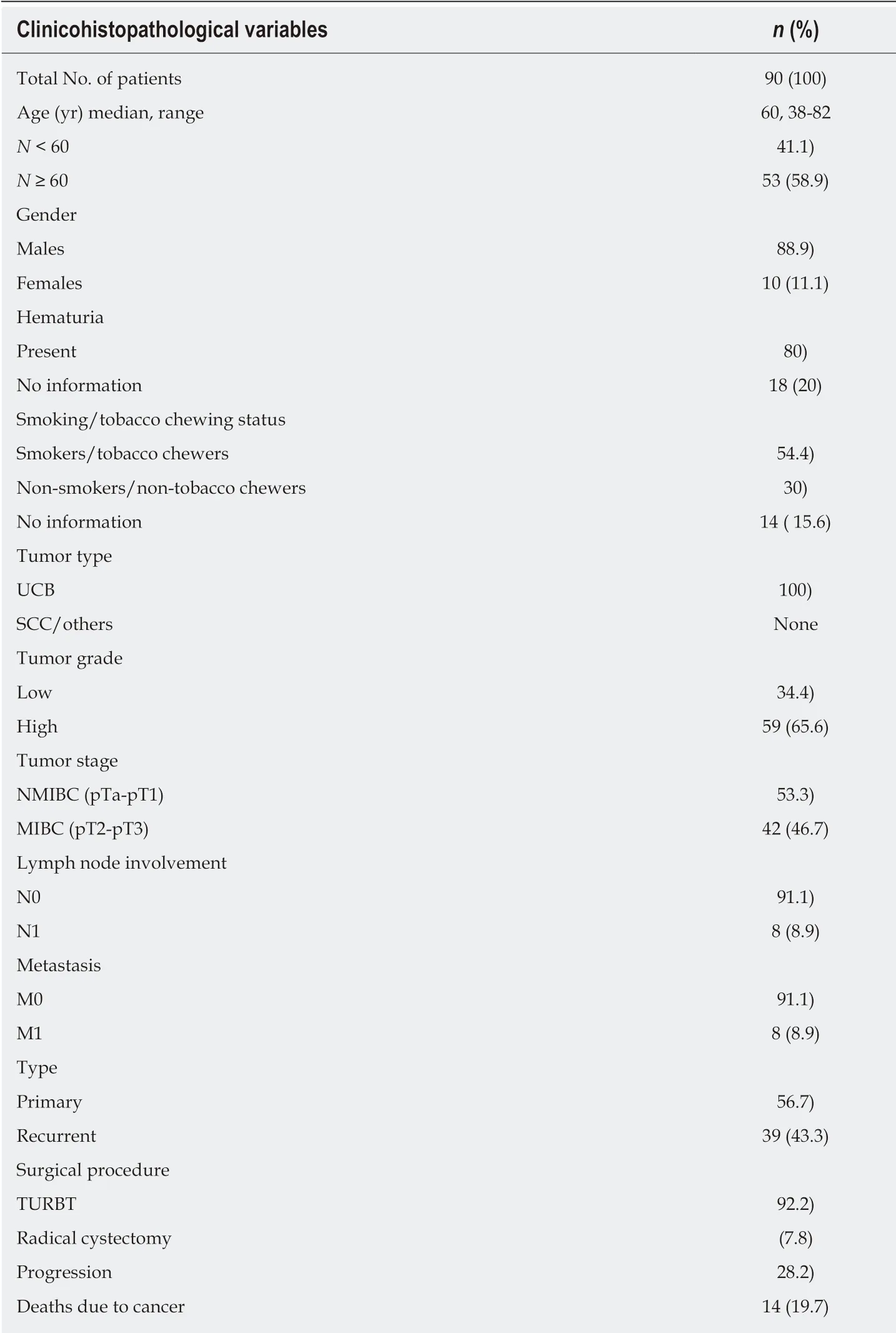
Table 1 Clinicohistopathological summary of patients with urothelial carcinoma of bladder
Effect of altered levels of proteins in combination was investigated on patients'survival. Follow up data of 71 patients was available over an average time period of 36 months. Progression and mortality due to cancer was observed in 28.2% (20/71)and 19.7% (14/71) patients, respectively.
Kaplan Meier analysis along with log rank test identified the aberrantly coexpressed pS9GSK-3β and β-catenin, as significant prognostic factor for a short OS (P= 0.02) (Figure 4). However, PFS analysis failed to deduce any significant correlation in a given cohort.
Target genes’ expression and pS9GSK-3β/β-catenin levels: Their clinical implications
RT-qPCR study was done to evaluate total β-catenin expression at transcriptome level in 90 bladder tumors and its expression as fold change was calculated by comparing its levels in control bladder tissues (Figure 5A and B). Of 90 bladder tumors, 63.3% (57/90) tumors were analyzed to exhibit strong expression of totalβ-Catenin. These tumors were examined for quantitative expression of target genes including Cyclin D1, Slug and Snail at transcriptome level (Figure 5A, B and Table 4).Out of 57 tumors, 39 tumors showed strong expression of Cyclin D1 and found to be significantly correlated with increased total β-catenin (P = 0.017; Kendall's tau-b correlation test). Excluded group revealed no significance. RT-qPCR results demonstrated stronger expression of Snail in 70.2% (40/57) tumors compared to normal urothelial tissue. Seventy four percent (42/57) tumors were found to express higher levels of Slug. Statistical studies showed strong correlation between higher levels of Slug and total β-catenin (P = 0.001; Pearson correlation test).
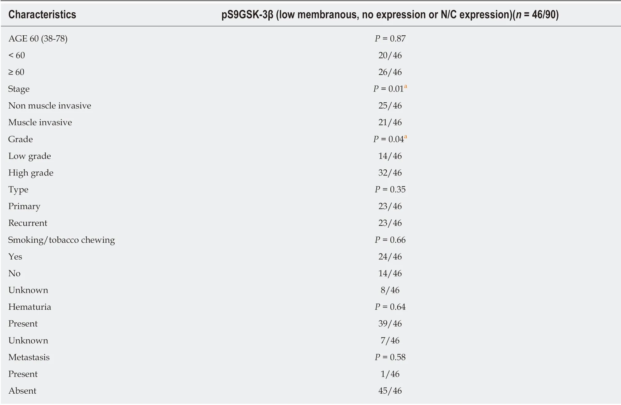
Table 2 Association of aberrant expression of pS9GSK-3β protein with clinicohistopathological factors
At protein level, immunostaining analysis identified nuclear staining of Cyclin D1(IHC score > 0) in 32% (29/90) tumors and its association with aberrant levels of βcatenin with respect to tumor recurrence (P = 0.017; Kendall's tau-b correlation test).Strong nuclear expression of Cyclin D1 was examined to be statistically associated with the aberrant expressions of either pS9GSK-3β or β-catenin with respect to tumor stage (P = 0.05; Mann Whitney U test) and tumor grade (P = 0.01; Mann Whitney U test) (Figure 2D, E, Figure 3 and Table 5).
Elevated levels of nuclear Snail (IHC score > 0) were observed in 27 out of 90 tumors (Figure 2F and Figure 3). Out of 61 tumors with aberrant expressions of either pS9GSK-3β or β-catenin, 29.5% tumors showed increased nuclear expression of Snail(Figure 2G). None of the clinicohistopathological variables showed significance with the expression levels of these proteins.
Ninety tumors were examined for nuclear immunostaining of Slug (IHC score > 0).Out of 90, 20 tumors were observed with strong expression (Figure 2H and Figure 3).Out of 61 tumors with aberrant immunoexpressions of either pS9GSK-3β or β-catenin,only 19.7% (12/61) tumors were found to exhibit nuclear expression of Slug in urothelium but no statistical association was observed (Figure 2I and Table 5).
DISCUSSION
Diverse cellular processes including proliferation, differentiation, glycogen metabolism, motility, cell survival are regulated by GSK-3β. Many important cellular
functions of GSK-3β are known to be mediated by its influence on Wnt/β-catenin signal cascade. Being the essential component of the protein complex, GSK-3β phosphorylates and targets β-catenin for subsequent ubiquitin-proteasomal degradation, thereby maintains the normal cell physiology[20].
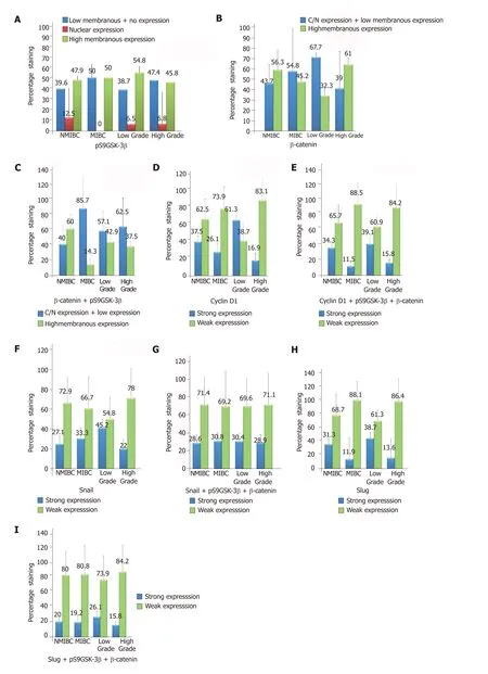
Figure 2 Graphical representation of percent immunohistochemical expression of marker proteins in 90 tissues of low stage (non-muscle invasive bladder cancer)/high stage (muscle invasive bladder cancer) and low grade/high grade tumors. A: pS9GSK-3β [Normal expression: strong membranous expression in urothelium; Aberrant expression: low membranous, strong nuclear and cytoplasmic, no expression in urothelium]; B: β-Catenin [Normal expression: strong membranous expression in urothelium; Aberrant expression: low membra nous, strong nuclear and cytoplasmic, no expression in urothelium]; C: β-Catenin + pS9GSK-3β; D: Cyclin D1 [Normal expression: strong nuclear expression in mesenchymal area; Aberrant expression: strong nuclear expression in mesenchymal area]; E:Cyclin D1 + pS9GSK-3β + β-Catenin; F: Snail [Normal expression: strong nuclear expression in mesenchymal area; Aberrant expression: strong nuclear expression in mesenchymal area]; G: Snail + pS9GSK-3β + β-Catenin; H: Slug [Normal expression: strong nuclear expression in mesenchymal area; Aberrant expression: strong nuclear expression in mesenchymal area]; I: Slug + pS9GSK-3β + β-Catenin. Numbers mentioned at the top of bars represent percentage staining of target proteins.IHC expression: Immunohistochemical expression; MIBC: Muscle invasive bladder cancer; NMIBC: Non-muscle invasive bladder cancer.
Serine 9 phosphorylation of GSK-3β leads to dephosphorylation of glycogen synthase; increases glycogen synthesis; and promotes protein and lipid biosynthesis.Its expression is restricted to umbrella cells of urinary tract, epithelial cells of breast and pancreaticobiliary duct, fallopian tube, epididymis, secretory cells of prostatic gland, distal nephron of kidney, gastrointestinal tract[20]. Expression of GSK-3β is known to be associated with differentiation of ectodermal derived tissues. Expression of phosphorylated form of GSK-3β (pS9GSK-3β) in epithelial cells of endodermal derived tissues in humans may validate its function to serve as an IHC marker for epithelial cells[20].
Strong membranous staining of pS9GSK-3β suggests functional and physiologic requirement of its expression in normal bladder urothelium/umbrella cells[20]. Normal urothelial cells were examined for strong membranous expression of pS9GSK-3β.Strong intensity of membranous pS9GSK-3β may indicate its association with the barrier function (barrier to urine, toxin and pathogen) of umbrella cells[21].
Phosphorylation of GSK-3β at Serine 9 inhibits its activity, causes dysregulation of the Wnt/β-catenin pathway; prevents β-catenin from degradation, allows its stabilization and accumulation in nucleus and cytosol, thus positively regulates cell proliferation and survival[22]. Aberrant expression of pS9GSK-3β examined by loss/no membranous expression or its nuclear/cytosolic accumulation was observed in 51.1%urothelial tumors and these tumors showed significant association with tumor grade and stage.
Activation of Wnt/β-catenin signaling pathway is known to be associated with altered pS9GSK-3β and cytosolic/nuclear accumulation of stabilized β-catenin in several human cancers[9]. Total β-catenin expression was noted in 63.3% (57/90)tumors at transcriptome level whereas 49% (44/90) tumors showed either its loss of membranous immunostaining or appearance of nuclear/cytosolic β-catenin protein.In accordance with earlier reports, strong membranous staining was confined to nonmalignant group/normal urothelial cells[4,10]. Present study examines the comparative aberrant expression of β-catenin in non-muscle invasive (43.7%) and muscle invasive(53.8%) tumors, however, 67.7% low grade tumors and 39% high grade tumors showed its altered levels. Study by Elserafy et al[4]reports statistical association between nucleocytoplasmic expression of β-catenin with poor prognostic parameters(tumor grade, stage, lymph node metastasis, vascular and perineural invasion,mitosis, and microvessel density). Another study by Li et al[23], investigates the expression of either cytoplasmic or nuclear β-catenin as an independent prognostic factor for overall survival in non-small-cell lung cancer. Nevertheless, in concordance with the results of the work by Bilim et al[24], 2000, current study failed to derive any independent prognostic relevance of β-catenin in bladder tumorigenesis.
Tumors co-expressing aberrant levels of pS9GSK-3β and β-catenin proteins were examined and identified to exhibit statistical association with tumor stage; tumor type; smoking/tobacco chewing status of patients and poor overall survival probabilities. These results indicate that low membranous pS9GSK-3β and cytosolic/nuclear pS9GSK-3β participate in neoplastic transformation by stabilizing βcatenin during urothelial tumorigenesis and could be considered as potential prognostic marker.
Tumor promoting effects of nuclear pS9GSK-3β have been reported to be mediated by activating cell cycle and survival via inducing the expression of critical cell cycle regulators, such as Cyclin D1[25,26]. Increased cytosolic amount of β-catenin facilitates its translocation to nucleus where it binds to T-cell-specific transcription factors, TCF and LEF and stimulates the transcription of Cyclin D1[27]. Research studies reveal the clinical functions of overexpressed Cyclin D1 via activation of pS9GSK-3β/β-catenin signal cascade in tumor development in major human cancers[10,26,28-30]. Increased Cyclin D1 levels in bladder tumor tissues showed significant association with total βcatenin in recurrent tumors at transcriptome level. Immunostaining of Cyclin D1 in tumors with altered levels of either pS9GSK-3β or β-catenin proteins and its association with tumor stage and grade may validate it as a marker of prognosticsignificance but does not explain its possible post translational activation by pS9GSK-3β/β-catenin/TCF/LEF transcriptional complex in bladder cancer progression.

Figure 3 Representative images of immunohistochemical staining of marker protein in urinary bladder tumors at the magnification of 400×. A: Controls: a:Positive control for pS9GSK-3β showing strong membranous expression in normal urothelium; b: Positive control for β-catenin showing high membranous expression in normal urothelium; c: Negative control for Cyclin D1; d: Negative control for Snail; e: Negative control for Slug; B: Immunohistochemical (IHC) staining in nonmuscle invasive bladder cancer cases; f: Nuclear/cytoplasmic expression of pS9GSK-3β; g: Nuclear expression of β-catenin (few tumor cells are circled); h: Cyclin D1 nuclear immunoexpression in 60%-70%; i: Snail nuclear immunopositivity in 80-90% cells; j: Slug nuclear expression in 60%-70% cells; C: IHC staining in muscle invasive bladder cancer cases; k: Loss of membranous pS9GSK-3β expression; l: Loss of β-catenin membranous expression; m: 100% nuclear staining for Cyclin D1 in urothelium; n: 100% nuclear immunoexpression of Snail in urothelium; o: 100% nuclear immunopositivity of Slug (Snail and Slug showed both nuclear and nonspecific cytoplasmic staining, however, only nuclear expression was taken into account during analysis).
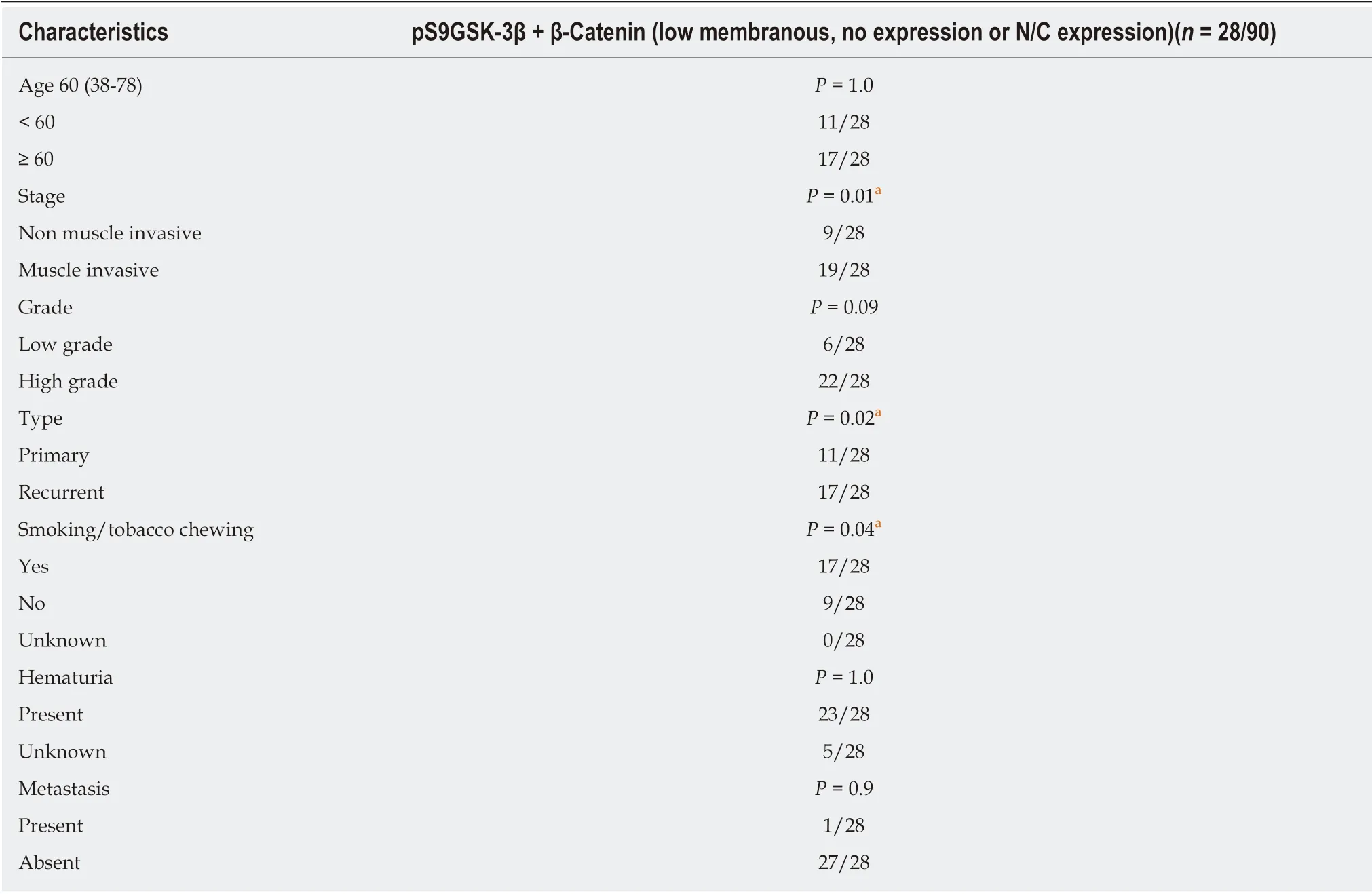
Table 3 Association of aberrantly co-expressed pS9GSK-3β and β-Catenin proteins with clinicohistopathological factors in patients with urothelial carcinoma of bladder
Membranous β-catenin complexes with E-Cadherin and α-catenin and maintains cell-cell adhesion and cell polarization[31]. Loss of membranous β-catenin results in dissociation of cells and promotes invasion and metastasis[32]. pS9GSK-3β mediated βcatenin stabilization followed by activation of β-catenin/TCF/LEF transcriptional complex is known to induce EMT and enhance cell invasion/migration by upregulating the expression of Snail and Slug transcription factors[31,33]. Experimental studies demonstrate the effect of β-catenin, Snail and Slug on EMT promoted cell invasion during bladder tumorigenesis and correlate them with poor prognosis[13,31,34,35]. Induced nuclear expressions of Snail and β-catenin by inactive form of GSK-3β and their functional interaction have been shown to regulate fibroblasts migration, EMT activation in non-tumorigenic breast epithelial cells and lung epithelial cells and correlate with poor prognosis in patients with gastric cancer and colorectal cancer[36-38]. Present study reports an inter-correlation between total β-catenin levels and increased Slug expression with tumor stage at transcriptome level. In disagreement with other studies, immunostaining results fail to exhibit any possible correlation between increased nuclear Snail/Slug and aberrant levels of pS9GSK-3β/β-catenin and their prognostic significance in a given cohort of bladder tumors. Conflicts in the results could be due to different sample size and their source and therefore, multicenter clinical studies are required for reliable conclusion.
Findings of the present study strongly support the involvement of pS9GSK-3β in urothelial tumorigenesis by stabilizing β-catenin via β-catenin signal cascade.Statistical association of aberrantly co-expressed pS9GSK-3β and β-catenin proteins with tumor stage, type, smoking/tobacco chewing status and shorter overall survival probabilities of patients validates them as potential markers of clinical relevance in the pathobiology of bladder cancer.
Phosphorylated GSK-3β mediated stabilization and activation of β-catenin signal cascade and its cross talk with other key pathways have been reported to be associated with cell proliferation, invasion, angiogenesis, and increased aggressiveness in several major human cancers. To validate the clinical functions ofpS9GSK-3β and its downstream effects through β-catenin signaling in the pathobiology of bladder cancer, expression levels of pS9GSK-3β, stabilized/activated β-catenin and its target genes, Cyclin D1, Snail and Slug were examined in welldefined cases of bladder cancer. Results based on the association/correlation studies between the expression levels of marker proteins and clinicohistopathological variables indicate that aberrantly expressed pS9GSK-3β participate in neoplastic transformation by stabilizing β-catenin during urothelial tumorigenesis and could be considered as potential prognostic marker. Histopathological association of nuclear Cyclin D1 in tumors expressing either pS9GSK-3β or β-catenin validates it as a prognostic marker in urothelial tumorigenesis. Nevertheless, absence of any significance rules out the possible post translational activation of Cyclin D1, Snail or Slug by pS9GSK-3β/β-catenin/TCF/LEF transcriptional complex in bladder cancer progression.
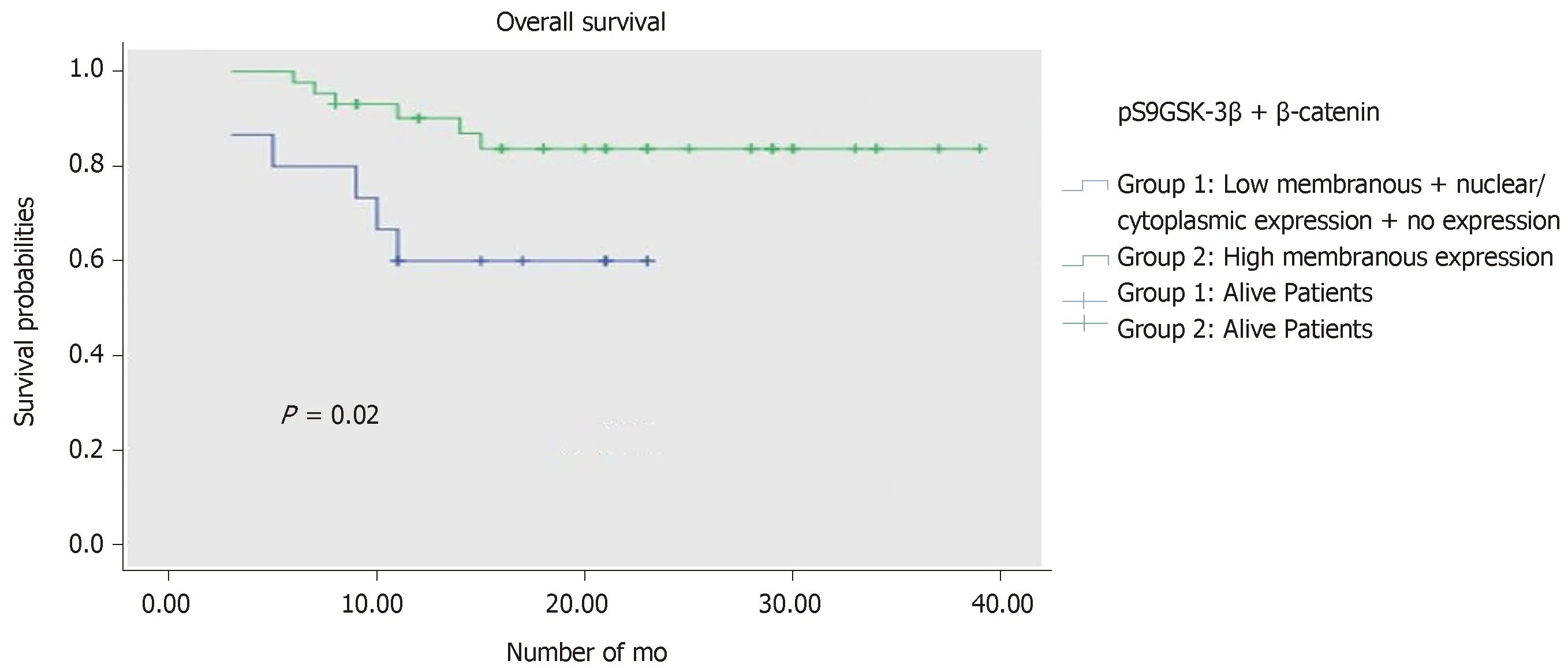
Figure 4 Kaplan-Meier overall survival curve analysis in patients with urothelial carcinoma of bladder.
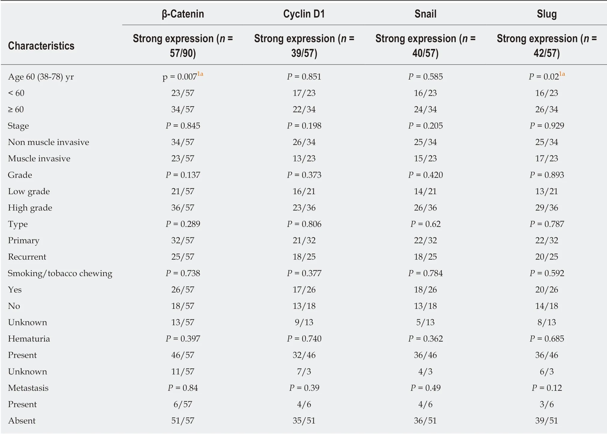
Table 4 Statistical analysis for correlation between expression of target genes at transcriptome level in tumor tissues and clinicohistopathological parameters
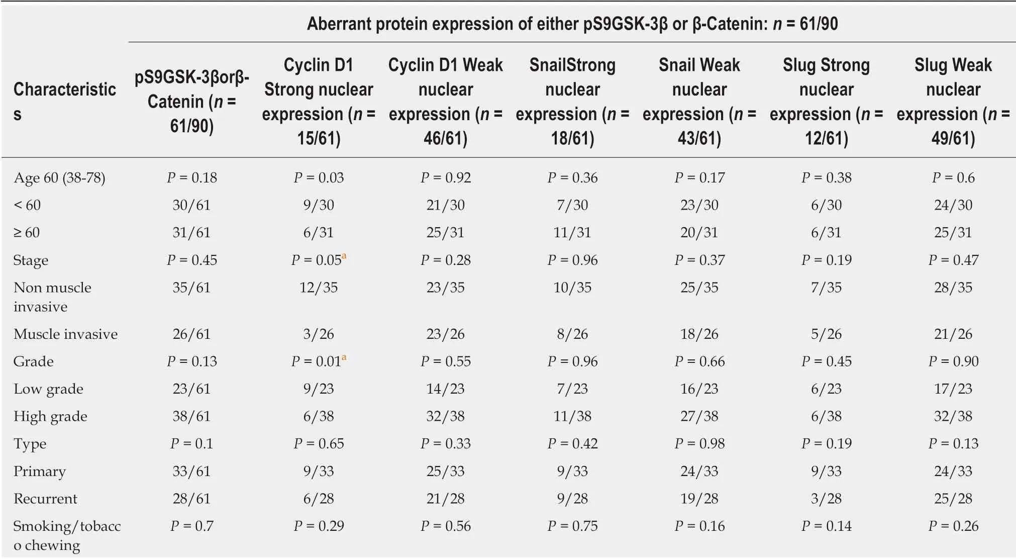
Table 5 Clinicohistopathological association of nuclear immunoexpressions of Cyclin D1, Slug and Snail proteins in tumors expressing aberrant levels of either pS9GSK-3β or β-catenin proteins
Non parametric Mann Whitney U test was used to determine the P-values;aP values ≤ 0.05 are considered as significant.
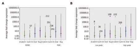
Figure 5 Box plot representation of quantitative expression profile of β-catenin and its target genes (Cyclin D1, Snail and Slug) in a cohort of 90 urinary bladder tumors at transcriptome level in non-muscle invasive bladder cancer and muscle invasive bladder cancer, and low grade and high grade tumors.Boxes represent the 25th to 75th percentile, horizontal lines indicate median values, and stars represent the maximum value. A: Non-muscle invasive bladder cancer and muscle invasive bladder cancer; B: Low grade and high grade tumors. The Y axis was logarithmically scaled in all cases. MIBC: Muscle invasive bladder cancer;NMIBC: Non-muscle invasive bladder cancer.
ARTICLE HIGHLIGHTS
Research background
Aberrant activation of phosphorylated form of glycogen synthase kinase-3β [pS9GSK-3β (Serine 9 phosphorylation)] is known to trigger Wnt/β-catenin signal cascade.
Research motivation
The clinicohistopathological implications of glycogen synthase kinase-3β [pS9GSK-3β (Serine 9 phosphorylation)]/β-catenin in bladder carcinogenesis remain unknown.
Research objectives
The aim of the present study is to investigate the diagnostic and prognostic relevance of expressions of pS9GSK-3β, β-catenin and its target genes in the pathobiology of bladder cancer.
Research methods
Cellular localization and the expression of pS9GSK-3β proteins were examined by immunohistochemical (IHC) staining in ninety tumor tissues resected from patients diagnosed with bladder cancer. Real time-quantitative polymerase chain reaction and IHC were done to check the expression of β-catenin, Cyclin D1, Snail and Slug at transcriptome and protein level respectively. Expressions of the markers were statistically correlated with these variables to determine their significance in clinical setting.
Research results
Aberrant (low or no membranous/high nuclear/high cytoplasmic) expression of pS9GSK-3β was noted in 51% patients and found to be significantly associated with tumor stage and tumor grade. Thirty one percent tumors exhibited aberrant co-expression of pS9GSK-3β and β-catenin proteins and showed strong statistical association with tumor stage (P = 0.01), tumor type (P =0.02), smoking/tobacco chewing status (0.04) and shorter overall survival probabilities of patients (P = 0.02).
Research conclusions
Findings of the present study strongly support the involvement of pS9GSK-3β in urothelial tumorigenesis by stabilizing β-catenin. Statistical association of aberrantly co-expressed pS9GSK-3β and β-catenin proteins with patients' variables validates them as potential markers of clinical relevance in the pathobiology of bladder cancer.
Research perspectives
Results based on the association/correlation studies between the expression levels of marker proteins and clinicohistopathological variables indicate that aberrantly expressed pS9GSK-3β participates in neoplastic transformation by stabilizing β-catenin during urothelial tumorigenesis and could be considered as potential prognostic marker.
ACKNOWLEDGEMENTS
Authors, Niharika Maurya and Rinni Singh are thankful to University grant commission (UGC), Govt. of India for providing research fellowship.
