Evaluation of right ventricular volume and systolic function in normal fetuses using intelligent spatiotemporal image correlation
2019-08-14JiaXingSunAiLuCaiLiMeiXie
Jia-Xing Sun,Ai-Lu Cai,Li-Mei Xie
Abstract
Key words: Ultrasonography; Fetus; Intelligent spatiotemporal image correlation; Right ventricular volume; Cardiac function
INTRODUCTION
Heart defects are the most common congenital malformations in fetuses with an incidence six times greater than chromosome abnormalities and four times greater than neural tube defects[1,2].The diagnosis of congenital heart disease is a challenge using prenatal ultrasound and is the focus of perinatal medical research.Congenital heart disease can be divided into structural and functional abnormalities.Indeed,fetal cardiac structure and function abnormalities often lead to changes in ventricular volume.As ventricular volume is an important index for evaluating fetal cardiovascular development,an effective and reliable method for measuring fetal ventricular volume and cardiac function is necessary for accurate ultrasonic diagnosis and effective clinical treatment of congenital heart disease.
Numerous studies have focused on fetal heart function in an effort to evaluate cardiac function in the healthy fetus and in those in states of cardiac decompensation[3].The accuracy of traditional two-dimensional (2D) ultrasound in the quantitative evaluation of fetal heart function has not been widely accepted[4-15].Spatiotemporal image correlation (STIC) technology overcomes many of the shortcomings of conventional 2D ultrasound in the measurement of fetal ventricular volume and evaluation of fetal cardiac function.Recently,a number of studies have used STIC technology in combination with organ computer-aided analysis software to measure fetal ventricular volume and evaluate heart function with proven accuracy and feasibility[4-7,16-27].However,STIC still has some limitations and the imaging principle determines that STIC is not a real-time three-dimensional (3D) imaging technology.One-way scanning using the sensors during the scanning process execution is slow,which leads to relatively long image acquisition time.Therefore,STIC is vulnerable to the effects of fetal and maternal respiration,resulting in degradation of image quality[21,23].
The new intelligent STIC (iSTIC) technology adopts an electronic matrix type of probe,which is composed of thousands of vibrating bits,thus generating real-time 3D images with acquired data.The new iSTIC technology acquires high-resolution volumetric images of one cardiac cycle in only 2 s,thus reducing the effects of fetal movement on the image.iSTIC realizes real-time 3D visualization of the fetal heart,which has been shown to have many advantages[24,25,27-29].In the present study,the iSTIC technique was used to measure right ventricular volume in normal fetuses and to evaluate right ventricular systolic function to provide a new method for more accurate and convenient evaluation of fetal heart function.
相对于例(24),例(23)较为明显地表现出了广告宣传的意味,而例(24)更容易引起读者兴趣。广告似乎是大家最不愿意理会的帖子,但是通过“确认过眼神X”构式表达出来,却增添了一丝风趣。如例(24)借助“确认过眼神X”构式,勾起了原本不愿理会的读者的好奇心,反而想“一睹芳容”。
MATERIALS AND METHODS
General information
One hundred twenty-three healthy gravidas with singleton gestations who visited our hospital for prenatal ultrasonography between October 2014 and September 2015 were included in the study.The maternal age range was 22-36 years (average,26.60 ±3.54 years).The gestational age range was 22-35+6wk (mean,28 ± 3.79 wk),including 20 cases at 22-23+6wk,24 cases at 24-25+6wk,20 cases at 26-27+6wk,16 cases at 28-29+6wk,18 cases at 30-31+6wk,15 cases at 32-33+6wk,and 10 cases at 34-35+6wk.The inclusion criteria were as follows:(1) singleton pregnancy; (2) fetal biological measurement indicators consistent with the corresponding gestational age; (3) no obvious fetal anatomic abnormalities shown on 2D ultrasound examination; (4)pregnant women were informed of the purpose of the evaluation,and informed consent was obtained before image acquisition; and (5) fetal heart image acquired using real-time 3D iSTIC technology was free of artifacts.
Instruments
An IU22 3D color Doppler ultrasound diagnostic instrument from Philips Company(Bothell,WA,USA) was used with a 2-4 MHz electronic matrix probe.QLAB software was used for image analysis and post-processing.
Data acquisition
The information of the last menstrual period and the gestational age in the pregnant women was obtained and recorded.The women were asked to lie flat,and a fetal echo program was begun with transabdominal scanning using an X6-1 probe.They were advised to hold their breath during the acquisition process to avoid interference of motion artifacts.Using a 3D/4D iSTIC technology model for a 4-chamber view of the fetal heart,the 3D/4D sampling frame location was adjusted and the sampling range assured 3D/4D volume data acquisition.The sampling frame included the entire fetal heart.The fetal heart rate was measured and recorded after completion of data acquisition.All images were stored in a built-in hard disk in the machine for later offline analysis.
Data analysis
(1) Post-processing of 3D image.The image stored in the built-in hard disk in the machine was extracted for analysis using QLAB software.The atrioventricular valve opening and closing framing at end systole and diastole in the entire cardiac cycle were observed to measure the fetal right ventricular volume.End systole was defined as the moment before atrioventricular valve opening,and end diastole was defined as the moment immediately after the atrioventricular valve closed.The center point of the central position of the right ventricle was adjusted using stacked contours to draw a line along the endocardium of the right ventricular apex to the edge of the tricuspid valve,the software was then automatically stratified followed by manual outlining of the contour of each layer of the lining of the heart at the short axis view of the heart including the trabecular muscle and regulating bundle.Computer software automatically calculated the volume of the right ventricle (Figure 1).The right ventricular end-diastolic volume (REDV) and the right ventricular end-systolic volume (RESV) were measured by the same observer three times to obtain the average value.
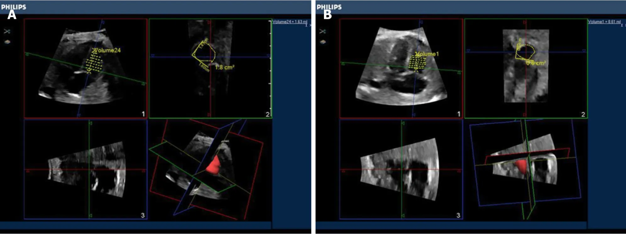
Figure 1 Fetal right ventricular end diastolic volume obtained by intelligent spatiotemporal image correlation technique.
(2) The heart function index was calculated as follows:The right stroke volume(RSV) = REDV- RESV; the right cardiac output (RCO) = RSV × fetal heart rate; the right ejection fraction = RSV/REDV.
(3) The paired Student'sttest was performed by randomly retrieved images from 30 cases with the fetal right ventricular volume independently measured by two observers (twice for each observer or twice by the same observer) to evaluate the consistency of the measurement.The average value by each observer was compared.
Statistical analysis
Data input and processing were carried out using SPSS 23.0 statistical software.The quantitative data conforming to normal distribution were presented as x ± s.Spearman rank correlation analysis was used to determine the relationship between fetal right ventricular volume and cardiac function parameter changes with gestational age.In addition,the paired Student'sttest was used to evaluate the consistency of the measurement by the same observer and between different observers.Differences withP< 0.05 were considered statistically significant.
RESULTS
Spearman rank correlation analysis
The changes in normal fetal REDV,RESV,RSV,and RCO with gestational age showed significant linear correlations,and the correlation coefficients were 0.903,0.874,0.866 and 0.865,respectively (P< 0.01,Figure 2,Table 1).Right ejection fraction did not change as gestational age increased,was relatively constant throughout the entire pregnancy,and was not significantly correlated with gestational age (P> 0.05,Table 1).Measurement of fetal right ventricular volume and cardiac function was carried out using the iSTIC technique (Table 2).
Paired Student's t test
The intraclass correlation coefficients (ICC) of REDV measured by the same observer was 0.989 with a 95% confidence interval (CI) of 0.978-0.995.The ICC of RESV was 0.978 with a 95%CI of 0.955-0.989.The ICC of REDV measured by different observers was 0.988 with a 95%CI of 0.975-0.944.The ICC of RESV was 0.988 with a 95%CI of 0.900-0.977.There was no statistical difference between the same observer and different observers when applying the iSTIC technique to measure RESV and REDV using the consistency test,and the correlation coefficients were 0.989,0.951,0.990,and 0.980,respectively.The results showed that there was particularly good consistency between the same observer and different observers (P< 0.001,Table 3).
DISCUSSION
Fetal cardiac function can be affected by a variety of conditions and prenatal interventions.Accurate assessment of fetal cardiac function could help to understand the process of fetal heart disease,improve the accuracy of diagnosis,and determine prenatal interventions[2,5,6,16,19,20,22,23,26,30-39].Two-dimensional ultrasound is still thetraditional gold standard for diagnosis of fetal congenital heart diseases[17,20,21]but has a number of limitations in the quantitative evaluation of fetal ventricular volume and cardiac function.When abnormal fetal heart structure causes significant cardiac geometric morphology changes,accurate quantitative assessment of fetal ventricular volume and cardiac function by 2D ultrasound is difficult[3,34].
Table 1 The relationship between normal fetal right ventricular end-diastolic volume,right ventricular end-systolic volume,right stroke volume,right cardiac output,and right ejection fraction with gestational ages from 22 to 35+6 wk
Application of the STIC imaging technique in fetal heart disorders has been studied extensively,which adds the time factor into the process of 3D data acquisition with continuous unidirectional scanning of the target area by the sensor to obtain the 3D volume data composed of each 2D slice.After completion of volume data acquisition,the software can automatically group 2D sections at the same time to reorganize the image of the entire fetal cardiac cycle,with end-systolic and end-diastolic phases of the cardiac cycle image defined according to opening and closing of the atrioventricular valve in post-processing[3,16,17,20-23,29,36,38-42].Such technology overcomes the imaginary heart geometry by 2D ultrasound along with a relatively simple image acquisition process and fewer requirements for operator experience.The imaging principle determines that STIC is not a real-time imaging technology but a delayed signal.Therefore,when an abnormality of the fetal heart leads to ventricular desynchrony,the maximum and minimum volume of the fetal ventricle cannot be accurately estimated using this method.
In contrast,iSTIC can generate real-time 3D images with an electronic matrix probe used vertically,and a horizontally-arranged 2D probe to conduct matrix volume imaging.The acoustic beam can automatically rotate for large-range scanning,which can significantly reduce the acquisition time of 3D image data and improve work efficiency.The acquisition time of one image by STIC is usually 8-12 s,but iSTIC only requires 2 s.With the imaging time significantly shortened,iSTIC has solved the contradiction between slow STIC imaging and a rapid fetal heart rate and reduced the effects of fetal and maternal respiration and other factors on the quality of the image.The image resolution is thus increased[29,43,44].In theory,accuracy of the iSTIC technique in the measurement of fetal ventricular volume is higher than STIC,which also reduces the exposure time of the fetus to ultrasound.
In this study,iSTIC technology was used to evaluate the heart function of 123 normal fetuses.The results showed that the fetal right ventricular volume,right ventricular stroke output,and RCO increased as gestational age increased,thus showing a good correlation with gestational age.Right ejection fraction did not change as the gestational age increased,which was relatively constant throughout the pregnancy and consistent with a previous report[23].During the entire process of fetal growth and development,the circulatory system plays a critical role.Unlike adults,the fetal right ventricle has the main circulating function.As the right ventricular volume continues to increase,the increased RCO with gestational age can meet the oxygen and nutrient demands for fetal development.In contrast,due to the anatomic and structural characteristics of the right ventricle,which has an irregular shape with the regulation bundle at the apex as well as a rougher endocardial surface compared with the left ventricle,it is particularly important to find an accurate and reliable method to measure fetal right ventricular volume and cardiac function indices.
试验地于上一年秋收后旋耕,当年春季南北起垄,垄宽0.60米。田间管理按照农民的习惯,苗前用旱田除草剂进行封闭,生育中期不进行铲趟作业。在6月中旬施放赤眼蜂防治玉米螟。
This study had some shortcomings,such as small sample size and the manual delineation of the lining of the heart in post-processing,which requires clear endocardial imaging and multiple measurements to obtain an average value and reduce measurement errors as far as possible.Due to the rough endocardium surface of the right ventricle,measurement error is inevitable.In addition,there is currently no gold standard for the assessment of fetal heart function to compare the data obtained by iSTIC technology.
In summary,iSTIC technology can be used for the quantitative measurement of fetal right ventricular volume and the evaluation of right ventricular systolic function,which has shown a number of advantages,and the application of iSTIC technology inprenatal diagnosis is worthy of further study and discussion.
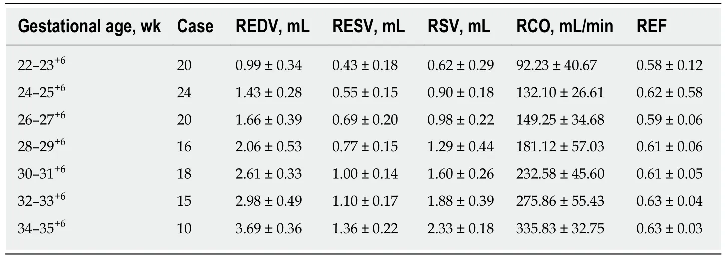
Table 2 Measured normal fetal right ventricular end-diastolic volume,right ventricular endsystolic volume,right stroke volume,right cardiac output,and right ejection fraction values with gestational ages between 22 and 35+6 wk
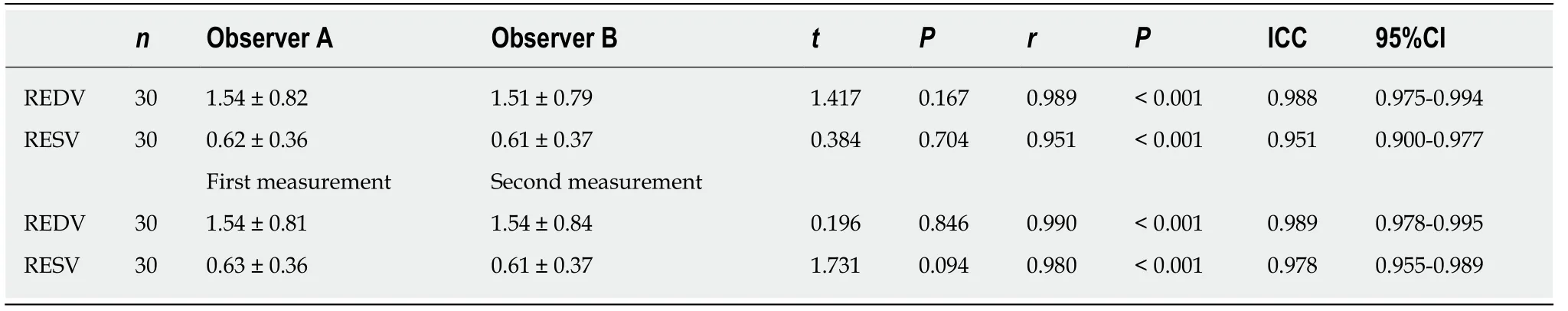
Table 3 Consistency between the same observer and different observers
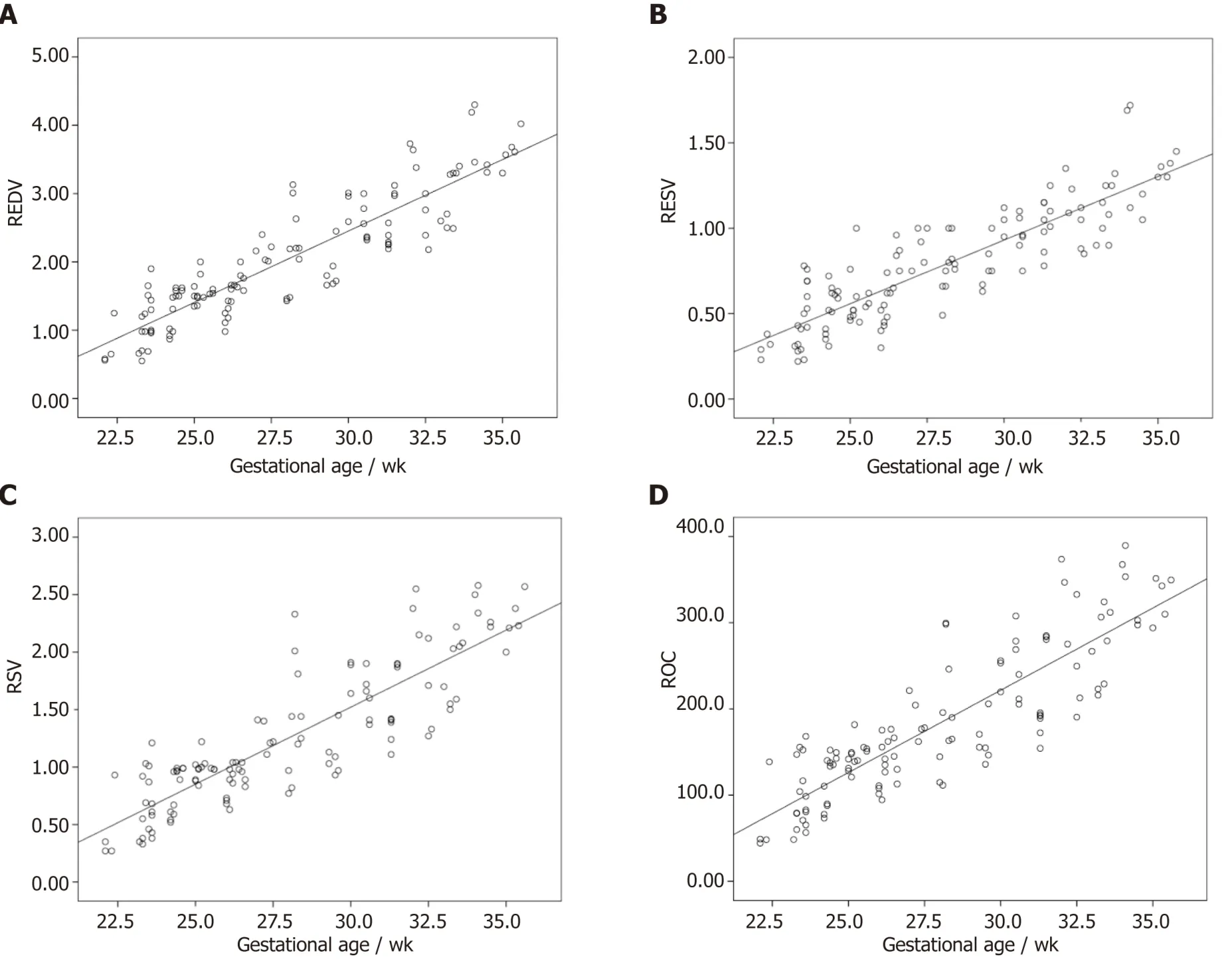
Figure 2 Correlations.
ARTICLE HIGHLIGHTS
Research background
Heart defects are the most common congenital malformations in fetuses.Fetal cardiac structure and function abnormalities lead to changes in ventricular volume.As ventricular volume is an important index for evaluating fetal cardiovascular development,an effective and reliable method for measuring fetal ventricular volume and cardiac function is necessary for accurate ultrasonic diagnosis and effective clinical treatment.The new intelligent spatiotemporal image correlation (iSTIC) technology acquires high-resolution volumetric images.Numerous studies have focused on fetal heart function which have not been widely accepted.The iSTIC technique was used to measure right ventricular volume in 123 normal fetuses,and to evaluate right ventricular systolic function to provide a new method for more accurate and convenient evaluation of fetal heart function.
Research motivation
The iSTIC technique was used to provide a new method for more accurate and convenient evaluation of fetal heart function.
Research objectives
One hundred twenty-three healthy gravidas with singleton gestations who visited our hospital for prenatal ultrasonography between October 2014 and September 2015 were included in the study.
Research methods
The women were asked to lie flat,and a fetal echo program was begun with transabdominal scanning using an X6-1 probe.Using a 3D/4D iSTIC technology model for a 4-chamber view of the fetal heart,the 3D/4D sampling frame location was adjusted and the sampling range assured 3D/4D volume data acquisition and the sampling frame included the entire fetal heart.The fetal heart rate was measured and recorded after completion of data acquisition.
Research results
The changes in normal fetal right ventricular end-diastolic volume,right ventricular end-systolic volume,right stroke volume,and right cardiac output with gestational age showed significant linear correlations.Right ejection fraction did not change as gestational age increased,was relatively constant throughout the entire pregnancy,and was not significantly correlated with gestational age.
Research conclusions
iSTIC technology can be used for the quantitative measurement of fetal right ventricular volume and the evaluation of right ventricular systolic function.
Research perspectives
iSTIC can generate real-time 3D images,only requires 2 s,and is the best method now.The direction of the future research is to improve the scanning time.
猜你喜欢
杂志排行

World Journal of Clinical Cases的其它文章
- Bone alterations in inflammatory bowel diseases
- Extrahepatic hepcidin production: The intriguing outcomes of recent years
- Neoadjuvant endocrine therapy: A potential strategy for ER-positive breast cancer
- Vestigial like family member 3 is a novel prognostic biomarker for gastric cancer
- HER2 heterogeneity is a poor prognosticator for HER2-positive gastric cancer
- Changes in corneal endothelial cell density in patients with primary open-angle glaucoma