立体心电图空间QRS-T夹角对冠心病患者心功能不全的评估价值
2019-01-17张松文汪岚
张松文 汪岚
【摘要】目的 研究立体心电图空间QRS-T夹角对冠心病患者心功能不全的评估价值。方法 选择我院2017年4月~2019年2月在心内科住院并做过做立体心电图的冠心病患者60例,其中心功能正常者45例,心功能不全者15例,比较两组不同心功能患者间的一般临床资料、不同空间QRS-T 夹角,并分析左室射血分数(LVEF)和QRS-T夹角相关性。结果 空间QRS-T夹角<50°、50°≤QRS-T 夹角<100°及QRS-T夹角≥100°在心功能正常和心功能不全组间比较,差异有统计学意义(P<0.05),表明随着LVEF值的降低,QRS-T夹角越大,空间QRS-T 夹角和左室射血分数呈负相关关系。当左室射血分数≥50%时,相关系数r=-0.663,P<0.001,当左室射血分数<50%时,相关系数r=-0.538,P<0.05。结论 冠心病患者的立体心电图空间QRS-T夹角和LVEF值呈负相关关系,LVEF值越小,空间QRS-T 夹角越大。
【关键词】冠心病;立体心电图;空间QRS-T夹角;心功能不全
【中图分类号】R541.4 【文献标识码】A 【文章编号】ISSN.2095.6681.2019.25..02
【Abstract】Objective To study the value of the three-dimensional electrocardiogram(3-D ECG) spatial QRS-T angle in the evaluation of cardiac insufficiency in patients with coronary heart disease(CHD).Methods A retrospective study was done to compare the general clinical data and proportion of different spatial QRS-T angle in 60 cases with coronary heart disease,who visited 3-D ECG in our hospital during April 2017 to February 2019,including 40 patients with normal cardiac function,and 15 patients with cardiac insufficiency,and the correlation between LVEF and spatial QRS-T angle was analyzed.Results There was difference in proportion of spatial QRS-T angle<50°,50°≤QRS-T angle<100°and QRS-T angle≥100°between cardiac insufficiency group and normal cardiac function group,the difference being statistically significant(P<0.05),indicating that with the decrease of LVEF,the spatial QRS-T angle increased and the spatial QRS-T angle was negatively correlated to LVEF value.When LVEF≥50%,the correlation coefficient r =-0.663,P<0.001,and when LVEF<50%,the correlation coefficient r =-0.538,P<0.05.Conclusion There was a negative correlation between the spatial QRS-T angle and LVEF value in patients with CHD,which is defined as the lower LVEF value,the bigger spatial QRS-T angle.
【Key words】Coronary heart disease;Three-dimensional electrocardiogram;Spatial QRS-T Angle;Cardiac insufficiency
冠心病是嚴重危害人类健康的最常见的疾病之一,在我国的发病率呈递增趋势。而冠心病最终都会导致心功能不全的发生,影响患者的生活质量及预后。本研究主要探讨立体心电图空间QRS-T夹角与冠心病患者心功能不全的相关性分析。
1 资料与方法
1.1 一般资料
选择2017年4月~2019年2月期间在我院心内科住院并经冠状动脉造影明确诊断为冠心病患者60例(排除既往有心脏外科手术史、恶性肿瘤、心脏瓣膜病、心肌病等其他心脏疾病),男性31例,女性29例,年龄(61~88)岁。并根据超声心动图中LVEF值,将LVEF值≥50%的患者分为心功能正常组45例,平均(70.75±6.33)岁,男性占48.89%,LVEF<50%的患者分为心功能不全组15例,平均(73.47±5.66)岁,男性占60%。
1.2 研究方法
记录所有入选患者的性别、年龄、合并症(高血压病、糖尿病、血脂异常),并记录超声心动图测定的LVEF及立体心电图相关数据。立体心电图均由北京卡迪斯医疗科技有限公司生产的3D-立体心电图仪采集。心电向量图泪点(时标)间距为2.5 ms。对比分析各组间LVEF值与空间QRS-T夹角间的相关性。
1.3 统计学方法
所有统计均采用SPSS13.0软件进行分析,计量数据均服从正态分布,用均数±标准差表示,两组间采用独立样本t检验。计数数据采用x2检验,不同心功能患者间LEVF值与空间QRS-T夹角的相关性比较采取偏相关分析。以P<0.05为差异有统计学意义。
2 结 果
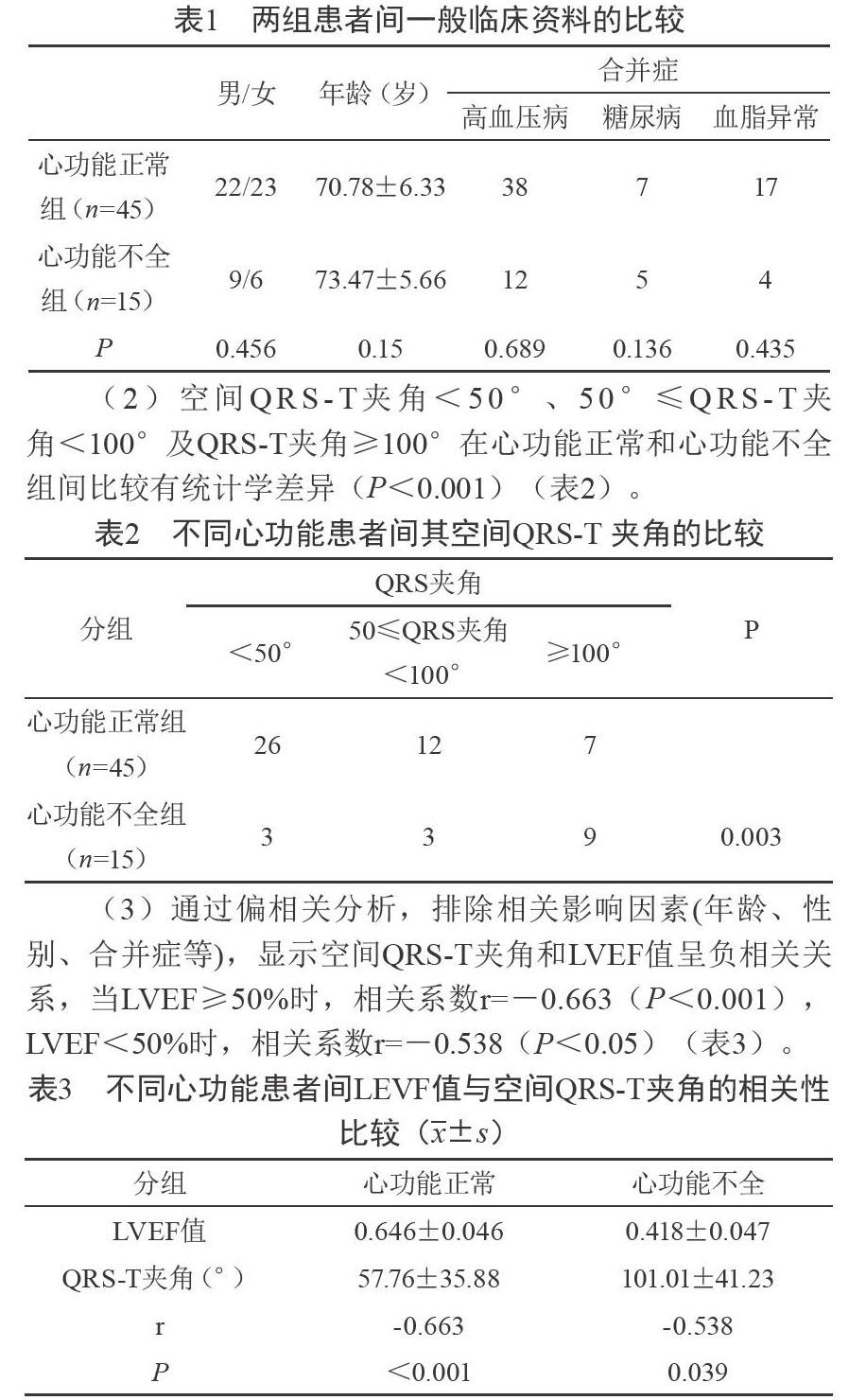
(1)两组患者的年龄、性别及合并症等均未发现统计学差异(P>0.05)(表1)。
3 讨 论
心功能不全是因为各种因素降低了心肌收缩力及心前向性排血,导致体循环或肺循环中的血液淤滞所引起的症状。其并不是单纯的血流动力学障碍,而是有多种神经体液因子参与的持续发展的综合临床症状。QRS-T夹角分为平面QRS-T夹角及空间QRS-T夹角,代表心脏部分电活动,是心室除极向量与复极向量间的夹角,受心脏功能和结构改变的影响。空间QRS-T夹角是指空间QRS波向量与空间T波向量之间的夹角。研究显示空间QRS-T夹角可作为除了急性冠脉综合征以外的很多疾病死亡率的预测因子[1-2]。空间QRS-T夹角已作为心源性死亡、新出现心力衰竭、冠状动脉心脏病事件和总死亡率的预测因子[3-7]。
有学者研究发现在预测冠心病事件和评估全因死亡率的价值方面平面QRS-T夹角与空间QRS-T夹角之间并无差别,表示平面QRS-T夹角可以替代空间QRS-T夹角[8]。但大多学者认为,在预测疾病的发生率及死亡率方面,空间QRS-T夹角比平面QRS-T夹角更准确,平面QRS-T夹角不可以替代空间QRS-T夹角[9]。这可能是由于平面QRS-T夹角是通过体表心电图计算所得,而心电图在二次投影过程中常常会有信息丢失,所以空间QRS-T夹角所反映的心脏电活动比平面QRS-T夹角更真实、准确[10]。
目前关于空间QRS-T夹角正常范围的多项研究结果均不一致,可能与计算方法和研究对象的不同有关。有研究发现男性和女性的空间QRS-T夹角的正常范圍分别为(80±24)°和(66±23)°[11]。也有研究发现空间QRS-T夹角<105°为正常,105°~135°为临界值,>135°为异常[12]。QRS-T夹角受种族差异影响,而国外相关文献中多以白人或黑人作为研究对象,因此所得的空间QRS-T夹角的正常范围不适用于中国人群。
有学者研究发现:在455例非缺血性心肌病患者中心功能越差,平面QRS-T夹角越大,随着心功能的改善,平面QRS-T夹角逐渐变小,心功能从NYHAⅢ/Ⅳ级恢复到Ⅰ级,平面QRS-T夹角减小17.5°±8.7°[13]。进而也可认为QRS-T夹角与LVEF值呈负相关。而本研究显示:空间QRS-T夹角<50°、50°≤QRS-T夹角<100°及QRS-T夹角≥100°在心功能正常和心功能不全组间比较差异有统计学意义(P<0.05),表明随着LVEF值的降低,QRS-T夹角越大,空间QRS-T夹角和LVEF值呈负相关关系,当LVEF≥50%时,相关系数r=-0.663,P<0.001,当LVEF<50%时,相关系数r=-0.538,P<0.05。与文献报道基本相同。
空间QRS-T夹角反映心肌细胞变化情况,影响其增大的因素很多,但是在临床诊治冠心病过程中,如果发现立体心电图空间QRS-T夹角有增大趋势,应及时关注该患者的心脏功能及冠状动脉供血情况,应用合理的治疗方案,改善预后。因此,空间QRS-T夹角对冠心病患者的诊治和预后具有一定价值。
参考文献
[1] Yamazaki T,Froelicher VF,Mugers J,et al.Spatial QRS-T angle predicts cardiac death in a clinical population[J].Heart Rhythm,2005,2(1):73-78.
[2] Rautaharju PM,Kooperberg C,Larson JC,et al.Electrocardiographic predictors of incident congestive heart failure and all-cause mortality in postmenopausal women:the womens health initiative[J].Circulation,2006,113(4):481-489.
[3] Morrow DA,Antman EM,Giugliano RP,et al.A simple risk index for rapid initial triage of patients with ST-elevation myocardial infarction:an InTIME II substudy[J].Lancet,2001,358(9293):1571–1575.
[4] Dorsch MF,Lawrance RA,Sapsford RJ,et al.A simple benchmark for evaluating quality of care of patients following acute myocardial infarction[J].Heart,2001,86(2):150–154.
[5] Granger CB,Goldberg RJ,Dabbous O,et al.Predictors of hospital mortality in the global registry of acute coronary events[J].Arch Intern Med,2003,163(19):2345–2353.
[6] Eagle KA,Lim MJ,Dabbous OH,et al.A validated prediction model for all forms of acute coronary syndrome:estimating the risk of 6-month postdischarge death in an international registry[J].JAMA,2004,291(22):2727–2733.
[7] Westerhout CM,Fu Y,Lauer MS,et al.Short-and long-term risk stratification in acute coronary syndromes:the added value of quantitative ST-segment depression and multiple biomarkers[J].J Am Coll Cardiol,2006,48(5):939–947.
[8] Zhang ZM,Prineas RJ,Case D,et al.Comparison of the prognostic significance of the electrocardiographic QRS/T angles in predicting incident coronary heart disease and total mortality(from the atherosclerosis risk in communities study)[J].Am J Cardiol,2007,100(5):844-849.
[9] Brown RA,Schlegel TT.Diagnostic utility of the spatial versus individual planar QRS-T angles in cardiac disease detection[J].J Electrocardiol,2011,44(4):404-409.
[10] 李俊伟,王建理,王红宇.立体心电向量图心室复极参数的分析[J].中国医药指南,2012,10(17):1-2.
[11] Scherptong RWC,Henkens IR,Man SC,et al.Normal limits of the spatial QRS-T angle and ventricular gradient in 12-lead electrocardiograms of young adults:dependence on sex and heart rate[J].J Electrocardiol,2008,41(6):648-655.
[12] Kors JA,Kardys I,Van IM,et al.Spatial QRS-T angle as a risk indicator of cardiac death in an elderly population[J].J Electrocardiol,2003,36(1):113-114.
[13] Behzad B,Matthew B,Haris S,et al.Prognostic value and temporal behavior of the planar QRS-T angle in patients with nonischemic cardiomyopathy[J].Circulation,2008,117(25):3181-3186.
本文編辑:吴 卫
