Guiding the migration of grafted cells to promote axon regeneration
2016-12-01Xiao-bingYuan,ChristopherHaas,ItzhakFischer
PERSPECTIVE
Guiding the migration of grafted cells to promote axon regeneration
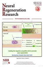
A promising therapeutic strategy to promote the regeneration of injured axons in the adult central nervous system (CNS) is the transplantation of cells or tissues that can modify the local host environment and support the growth of regenerating axons. Growth-supportive cells that have been successfully used in experimental transplantation therapy of spinal cord injury (SCI) include Schwann cells, mesenchymal stromal cells, olfactory ensheathing cells, genetically modified fibroblasts, and neural stem/progenitor cells (Huang et al., 2010). Cells derived from the embryonic spinal cord and peripheral nerve grafts have been shown to promote the regeneration of injured axons, due largely to the presence of growth-supportive cells such as glial progenitors and Schwann cells, respectively (Cote et al., 2011; Haas and Fischer, 2013). These transplants generate a permissive environment for axon growth by secreting growth factors and forming an adhesive extracellular matrix to overcome the inhibitory environment of the injured tissue. However, the value of these transplants to promote axon regeneration is limited by the fact that most regenerating axons are trapped inside the permissive environment generated by the transplants, failing to grow out of the graft (Figure 1A, B) (Haas and Fischer, 2013). While this strategy can be effective for building functional relays via graft-derived neurons (Haas and Fischer, 2014), this approach can not be generalized to other cell types. Therefore, a remaining challenge for therapeutic cell transplantation in CNS injury, in the context of long distance regeneration and connectivity, is to develop strategies to promote axonal growth beyond the graft into putative target areas to form functional synaptic connections. Currently the nature of the “graft trap” of regenerating axons is not fully understood. One possibility is that the regenerating axons stay inside the graft, which expresses much higher levels of attractive guidance factors, i.e., neurotrophic factors, and much lower levels of inhibitory/repulsive factors, i.e., chondroitin sulfate proteoglycan (CSPG), compared to the adjacent host tissue. Another possibility is that although adult CNS axons maintain their growth potential and can regenerate in an optimized environment, their intrinsic growth capability is much lower than axons of embryonic neurons, and thus not suitable for long-distance regeneration. Targeting these mechanisms, several strategies have recently been applied to overcome the “graft trap” in transplantation-based therapy of SCI. One strategy is to further modify host spinal tissue, making the host tissue less inhibitory and thus allowing some of the regenerating axons inside the graft to exit into the host tissue. As an example, Tom et al. (2009) showed that in an experimental model of grafting a peripheral nerve bridge at the site of the injured spinal cord, application of chondroitinase (Chase) at the distal graft/host interface to reduce CSPG-mediated inhibition promoted modest improvement in host-entry of regenerating axons, which would otherwise stop at the distal graft/host junction. Another strategy to promote axonal growth beyond the graft focuses on genetic modification of injured neurons to enhance their intrinsic growth potential using regeneration associated genes (Ma and Willis, 2015). For example, overexpressing the constitutively active form of the Rheb GTPase (downstream of the mTOR pathway) has been shown to enhance the intrinsic growth potential of adult neurons (Wu et al., 2015).
Recently, we sought to explore an alternative strategy for promoting axon regeneration by inducing the directional migration of grafted cells (Yuan et al., 2016). We hypothesized that controlled migration of grafted cells could be beneficial to axon regeneration and functional recovery by expanding the permissive environment and directing axon growth. However, following transplantation into the injured spinal cord, most grafted cells remain at the injury site, with few grafted cells showing long-distance migration without rostral or caudal directional selectivity (Lankford et al., 2008; Ekberg et al., 2012; Yuan et al., 2016). An intriguing but yet untested question is whether we can promote axon regeneration beyond the injury/graft site by guiding the migration of grafted cells toward the putative target region of regenerating axons. Theoretically this is feasible, because if a large cohort of grafted cells can be guided to migrate out of the injury/graft site toward the original target area of injured axons, the migratory stream of these growth-supportive cells is very likely to form a corridor for the advance of regenerating axons beyond the injury/graft site toward the target area. Moreover, migration of grafted cells may even enhance axon growth by towing of growth cones, like the towing of embryonic sensory axons by migrating target cells during embryonic development (Gilmour et al., 2004).
To begin testing whether this novel strategy is feasible, we needed to establish a reliable method to induce the directional migration of grafted cells in the adult spinal cord, as highlighted by one of our recent research projects (Yuan et al., 2016). We first used a variety of cell culture-based assays to screen for factors that may be attractive or repulsive to the migration of glial-restricted progenitors (GRPs) derived from embryonic spinal cord, a promising cell type to support axon regeneration in transplantation-based therapy of SCI (Haas et al., 2012; Haas and Fischer, 2013; Hayakawa, 2016). Next, we used a cervical dorsal column lesion model of SCI in adult rats, a well-characterized in vivo nerve injury model, for transplantation of GRPs and application of lentivirus coding for candidate guidance factors rostral to the injury/graft site to test the guidance of GRP migration by candidate factors in vivo. Although GRPs for transplantation exhibit active migration in vitro, we observed limited migration of grafted GRPs in adult spinal cord, with or without injury. This limited migration of grafted GRPs may indicate the presence of endogenous factors that restrict/inhibit the migration of grafted GRPs in the adult spinal cord, and that effective guidance of GRP migration may depend on the removal of this restrictive/ inhibitory signal. CSPG is a well-characterized axon growth inhibitor in the adult CNS that is present in the gliotic scar following CNS injury. As GRPs express receptor tyrosine phosphatase sigma (PTPRS), one of the major receptors of CSPG, it is likely that these cells can also respond to this inhibitory signal. Indeed, when coated on culture substrate, CSPG strongly inhibits the adhesion and migration of cultured GRPs. Injection of lentivirus vectors encoding Chase rostral to the injury/graft area induced the preferential migration of grafted GRPs toward the injection site. These in vitro and in vivo findings support the notion that CSPG is a major endogenous factor that restricts the migration of grafted GRPs in the adult CNS. We also observed that basic fibroblast growth factor (bFGF) is an attractive migration factor for GRPs, as lenti-bFGF injection also induced directional migration of a fraction of grafted GRPs toward the injection site in vivo, similar to the effect of lenti-Chase. These findings suggest that an effective way of guiding the directional migration of grafted cells is the lentivirus-mediated delivery of factors that can either remove the restrictive/inhibitory effect of the host tissue or actively promote cell migration. An interesting future question is whether simultaneous application of these two types of factors - one relieving the inhibition and the other directly attracting - results in synergistic activity and stimulates the migration of greater numbers of grafted cells toward the putative target. The combination of the in vitro screening system together with the in vivo injury model that disrupts sensory axons described in our study (Yuan et al., 2016) can be used to test the effects of additional molecules on the migratory properties of other cells. It is also important to further explore whether directional migration of a large cohort of grafted cells can support axon regeneration beyond the injury/graft site. Moreover, guided migration of grafted cells can be further combined with other therapeutic interventions to improve axon regeneration and ultimately recovery of function. In this context, the additional advantage of using lenti-Chase to guide the migration of grafted GRPs is that this treatment also benefits the growth of regenerating axons. Thus, a therapeutic strategy that focuses on the application of a guidance factor that can promote both the extension of regenerating axons and the migrationof grafted cells may be the best option for a combined effect. For the chemotropic factor, it is unclear whether bFGF, which we found to be attractive to GRPs, is also directly attractive to regenerating axons. If a common attractant for both regenerating axons and grafted cells is not available, one potential option is to transplant cells genetically engineered to express the specific receptor for the attractant that can effectively guide the extension of regenerating axons, so that grafted cells gain sensitivity to the same attractant.

Figure 1 Guiding the migration of grafted cells to promote axon regeneration.
It is generally accepted that the glycosaminoglycan chains in CSPG mediate the inhibitory effect of CSPG on axon growth, and that Chase treatment is a widely used method in experimental therapy of SCI to alleviate CSPG-mediated inhibition by digestion of the glycosaminoglycan chains (Bradbury et al., 2002). Consistent with Chase-mediated CSPG digestion, we observed that Chase treatment completely blocked the inhibitory effect of CSPG on the attachment of GRPs to cell culture substrate. However, in a “stripe assay” designed to evaluate the guidance effect of substrate-bound CSPG on GRP migration, we noticed that Chase-treatment mildly mitigated, but did not completely block, the repulsive action of CSPG stripes on GRPs (Yuan et al., 2016). This observation indicates the existence of CSPG inhibition that is independent of glycosaminoglycan chains, and underscores the importance of developing novel ways that can effectively mitigate this Chase-insensitive inhibitory action of CSPG in the scenario of long distance regeneration of injured axons. Basic research to clarify the structural basis of this Chase-independent inhibitory action of CSPG will be the key for this solution in the near future.
In summary, we have established a framework of inducing the directional migration of grafted GRPs in a SCI model using lentivirus-mediated expression of two types of guidance factors (Figure 1C). A similar strategy can be applied when other cell types are used in transplantation-based therapy of SCI, and can be applied in combination with other therapeutic interventions to improve axon regeneration.
This work was supported by NIH NS055976 and Craig H. Neilsen Foundation 280850.
Xiao-bing Yuan*, Christopher Haas, Itzhak Fischer
Hussman Institute for Autism, Baltimore, MD, USA; Department of
Anatomy and Neurobiology, University of Maryland School of
Medicine, Baltimore, MD, USA (Yuan XB)
Spinal Cord Research Center, Department of Neurobiology and
Anatomy, Drexel University College of Medicine, Philadelphia, PA, USA (Haas C, Fischer I)
*Correspondence to: Xiao-bing Yuan, Ph.D., xyuan@hussmanautism.org.
Accepted: 2016-07-14
orcid: 0000-0002-1632-8460 (Xiao-bing Yuan)
0000-0003-3187-8740 (Itzhak Fischer)
How to cite this article: Yuan XB, Haas C, Fischer I (2016) Guiding the migration of grafted cells to promote axon regeneration. Neural Regen Res 11(8):1224-1225.
References
Bradbury EJ, Moon LD, Popat RJ, King VR, Bennett GS, Patel PN, Fawcett JW, McMahon SB (2002) Chondroitinase ABC promotes functional recovery after spinal cord injury. Nature 416:636-640.
Cote MP, Amin AA, Tom VJ, Houle JD (2011) Peripheral nerve grafts support regeneration after spinal cord injury. Neurotherapeutics 8:294-303.
Ekberg JA, Amaya D, Mackay-Sim A, St John JA (2012) The migration of olfactory ensheathing cells during development and regeneration. Neurosignals 20:147-158.
Gilmour D, Knaut H, Maischein HM, Nusslein-Volhard C (2004) Towing of sensory axons by their migrating target cells in vivo. Nat Neurosci 7:491-492.
Haas C, Fischer I (2013) Human astrocytes derived from glial restricted progenitors support regeneration of the injured spinal cord. J Neurotrauma 30:1035-1052.
Haas C, Fischer I (2014) Transplanting neural progenitors to build a neuronal relay across the injured spinal cord. Neural Regen Res 9:1173-1176.
Haas C, Neuhuber B, Yamagami T, Rao M, Fischer I (2012) Phenotypic analysis of astrocytes derived from glial restricted precursors and their impact on axon regeneration. Exp Neurol 233:717-732.
Hayakawa K, Haas, C. and Fischer, I. (2016) Examining the properties and therapeutic potential of glial restricted precursors in spinal cord injury. Neural Regen Res 11:529-533.
Huang H, Chen L, Sanberg P (2010) Cell therapy from bench to bedside translation in CNS neurorestoratology Era. Cell Med 1:15-46.
Lankford KL, Sasaki M, Radtke C, Kocsis JD (2008) Olfactory ensheathing cells exhibit unique migratory, phagocytic, and myelinating properties in the X-irradiated spinal cord not shared by Schwann cells. Glia 56:1664-1678.
Ma TC, Willis DE (2015) What makes a RAG regeneration associated? Front Mol Neurosci 8:43.
Tom VJ, Sandrow-Feinberg HR, Miller K, Santi L, Connors T, Lemay MA, Houle JD (2009) Combining peripheral nerve grafts and chondroitinase promotes functional axonal regeneration in the chronically injured spinal cord. J Neurosci 29:14881-14890.
Wu D, Klaw MC, Connors T, Kholodilov N, Burke RE, Tom VJ (2015) Expressing constitutively active rheb in adult neurons after a complete spinal cord injury enhances axonal regeneration beyond a chondroitinase-treated glial scar. J Neurosci 35:11068-11080.
Yuan XB, Jin Y, Haas C, Yao L, Hayakawa K, Wang Y, Wang C, Fischer I (2016) Guiding migration of transplanted glial progenitor cells in the injured spinal cord. Sci Rep 6:22576.
10.4103/1673-5374.189169
杂志排行
中国神经再生研究(英文版)的其它文章
- Secondary parkinsonism induced by hydrocephalus after subarachnoid and intraventricular hemorrhage
- Prospects for bone marrow cell therapy in amyotrophic lateral sclerosis: how far are we from a clinical treatment?
- Uncoupling protein 2 in the glial response to stress: implications for neuroprotection
- Selective neuronal PTEN deletion: can we take the brakes off of growth without losing control?
- TRPV1 may increase the effectiveness of estrogen therapy on neuroprotection and neuroregeneration
- Tamoxifen: an FDA approved drug with neuroprotective effects for spinal cord injury recovery