正常妊娠中晚期胎儿脐静脉及静脉导管血流灌注量的检测及分析
2015-08-29项宇识
项宇识
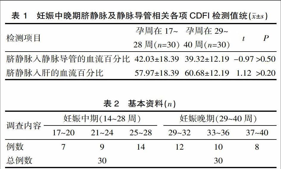
[摘要] 目的 该研究检测孕中晚期胎儿脐静脉及静脉导管的血流速度和血流量,以彩色多普勒超声辅助判定胎儿的发育生长的水平。 方法 对2013年10月—2014年10月产前检查的正常妊娠17~40周的健康孕妇60例,应用彩色多普勒超声检探测胎儿一般发育状况的同时检测胎儿脐静脉及其末支静脉导管的管腔内径、血流速度及血流量。 结果 妊娠中期与妊娠晚期胎儿的脐静脉和静脉导管的各项血流参数的均值进行t检验,差异有统计学意义(P<0.001)。妊娠中、晚期胎儿静脉导管血流量与脐静脉血流量比及脐静脉肝内灌注量与脐静脉血流量比均值进行t检验,差异无统计学意义(P>0.05)。结论 妊娠中晚期胎儿脐静脉及静脉导管血流速度、灌注量随着孕龄的增大而增加。对应脐静脉肝内灌注血流量也随孕龄增大而增加,妊娠中晚期分别为57.97%及60.68%,对维持胎儿的发育生长起优先作用。
[关键词] 静脉导管;脐静脉;彩色多普勒;血流灌注
[中图分类号] R596.11 [文献标识码] A [文章编号] 1674-0742(2015)01(c)-0007-03
[Abstract] Objective To detect the blood flow velocity and blood flow of fetal umbilical vein and venous catheter in the second and third trimester of pregnancy so as to determine the level of growth and development of the fetus supplemented by color Doppler ultrasound. Methods Color Doppler ultrasound was used to detect the general fetal development and the lumen diameter, blood flow velocity and blood flow of fetal umbilical vein and venous catheters of its terminal branches in the 60 normal healthy pregnant women with the gestational age of 17 to 40 weeks undergoing the prenatal examination from October 2013 to October 2014. Results There were significant differences in the mean values of blood flow parameters of fetal umbilical vein and the venous catheter between the second and the third trimester of pregnancy, by t test, P<0.001. The difference in the mean ratio of fetal venous catheter blood flow to fetal umbilical vein blood flow, and that in the mean ratio of intrahepatic umbilical vein perfusion to fetal umbilical vein blood flow between the second and the third trimester of pregnancy were not significant, by t test, P>0.05. Conclusion The blood flow velocity and perfusion of fetal umbilical vein and venous catheters in the second and third trimester of pregnancy increase with the increase of gestational age. The intrahepatic umbilical vein perfusion also rises with the increase of gestational age, which increases by 57.97%, 60.68%, in the second trimester and third trimester of pregnancy, respectively, playing preferential interaction in maintaining the development and growth of the fetus.
[Key words] Venous catheter; Umbilical vein; Color Doppler; Perfusion
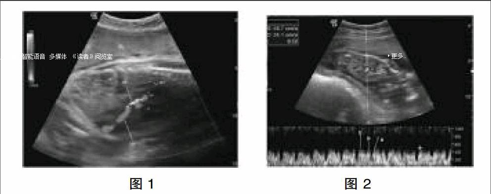
脐静脉携带来自胎盘含有80%氧气和营养物质较丰富的血液,经胎儿腹前壁进入肝脏[1]。脐静脉是把富含氧和营养物质的血从胎盘运送到胎儿体内的唯一通道。来自胎盘的血液进入胎儿体内分为3支:1支直接入肝,1支与门静脉汇合入肝,此2支血液经肝静脉入下腔静脉,另1经静脉导管直接入下腔静脉[2]。通过多普勒超声技术测量2013年10月—2014年10月在该院就诊的孕妇,孕20 ~40周胎儿大约20%~30%脐静脉血流入静脉导管[3,4]。脐静脉70%~80%的血流量灌注到肝脏,为灌注的第一个器官[1],这表明了胎儿在宫内生长发育肝脏的重要性,现报道如下。
1 资料与方法
