Expression oflamino acid transport system 1 and analysis of iodine-123-methyltyrosine tumor uptake in a pancreatic xenotransplantation model using fused high-resolution-micro-SPECT-MRI
2011-07-07CorinnavonForstnerMaazZuhayraOleAmmerpohlYiZhaoSanjayTiwariOlavJansenHolgerKalthoffEberhardHenzeandJanHendrikEgberts
Corinna von Forstner, Maaz Zuhayra, Ole Ammerpohl, Yi Zhao, Sanjay Tiwari, Olav Jansen, Holger Kalthoff, Eberhard Henze and Jan-Hendrik Egberts
Kiel, Germany
Expression oflamino acid transport system 1 and analysis of iodine-123-methyltyrosine tumor uptake in a pancreatic xenotransplantation model using fused high-resolution-micro-SPECT-MRI
Corinna von Forstner, Maaz Zuhayra, Ole Ammerpohl, Yi Zhao, Sanjay Tiwari, Olav Jansen, Holger Kalthoff, Eberhard Henze and Jan-Hendrik Egberts
Kiel, Germany
BACKGROUND:The specificity in discriminating pancreatitis is limited in the positron emission tomography (PET) using Fluorine-18-fluorodeoxyglucose. Furthermore, PET is not widely available compared to the single photon emission computed tomography (SPECT). Since amino acids play a minor role in metabolism of inflammatory cells, the potential of the SPECT tracer, 3-[123I]iodo-L-α-methyltyrosine (123I-IMT), for detecting pancreatic cancer was examined in xenotransplantation models of human pancreatic carcinoma in mice.
METHODS:123I-IMT was injected to eight mice inoculated with subcutaneous or orthotopic pancreatic tumors. Fused high-resolution-micro-SPECT (Hi-SPECT) and magnetic resonance imaging were performed. The gene expression level oflamino acid transport-system 1 (LAT1) was analyzed and correlated with tumor uptake of123I-IMT.
RESULTS:A high uptake of123I-IMT was detected in all tumor-bearing mice. The median tumor-to-background ratio (T/B) was 12.1 (2.0-13.2) for orthotopic and 8.4 (1.8-11.1) for subcutaneous xenotransplantation, respectively. Accordingly, the LAT1 expression in transplanted Colo357 cells was increased compared to non-malignant controls.
CONCLUSIONS:Our mouse model could show a high123I-IMT uptake in pancreatic cancer. Fused MRI scans facilitate precise evaluation of uptake in the specific regions of interest. Further studies are required to confirm these findings in tumors derived from other human pancreatic cancer cells. Since amino acids play a minor role in the metabolism of inflammatory cells, the potential for application of123I-IMT to distinguish pancreatic tumor from inflammatory pancreatitis warrants further investigation.
(Hepatobiliary Pancreat Dis Int 2011; 10: 30-37)
pancreatic ductal adenocarcinoma; iodine-123-methyltyrosine; high-resolution-micro-SPECT;lamino acid transport-system 1; xenotransplantation model
Introduction
The radiolabeled amino acid 3-[123I]iodo-L-αmethyltyrosine (123I-IMT) which derives from the natural acid amino tyrosine was introduced by Biersack et al in 1989.[1]Since then the tracer has become a successful radiotracer in the examination of brain tumors using single photon emission computed tomography (SPECT).[1]Furthermore, several studies have suggested that imaging with these radio-labeled amino acids visualizes protein synthesis and amino acid transport phenomena.[2-4]These processes are generally accelerated in dividing cells and cancer cells with high metabolism.[5]
To date the most frequently used tracer for tumor imaging is Fluorine-18-fluorodeoxyglucose (18F-FDG). This tracer is avidly taken up by almost all kinds of tumor cells, representing increased glucose metabolism, but it also accumulates in inflammatory tissues.[6,7]For pancreatic cancer, positron emission tomography (PET) with18F-FDG has been demonstrated to be useful in the evaluation of indeterminate pancreatic masses, staging of pancreatic cancer, detection of metastatic disease, and differentiation of viable tumors from posttherapeutic processes like necrosis or scar tissue.[8,9]However, the differentiation of pancreatic malignancy from focal pancreatitis involving the pancreatic head is still a challenge by means of18F-FDG PET.[10]Since amino acids play a minor role in the metabolism of inflammatory cells, the application of123I-IMT might be more tumor specific than18F-FDG.[11,12]
In a recent study with a murine orthotopic xenotransplantation model for pancreatic cancer, we compared different PET tracers (18F-FDG, 3-deoxy-3-18F-fluorothymidine (18F-FLT)) and demonstrated18F-FLT to be the PET tracer with the highest and most consistent tumor uptake in various human pancreatic tumors.[13]Consistent with these findings the imaging results could be explained by gene expression patterns of membrane transporters and enzymes for tracer uptake and retention as measured by gene array analysis and quantitative polymerase chain reaction in the respective cell lines.
As PET is expensive and not available in every nuclear medicine department, the question arises whether another tracer for conventional and more widely available SPECT can be used alternatively for imaging of pancreatic cancer. SPECT using18F-FDG or18F-FLT also is an option, but aside from the limited specificity, clinical oncologic application is hampered by detection difficulties such as a limited resolution, a low sensitivity for small lesions and a septal penetration in ultra-high-energy collimators.[14]
Jager et al[15]demonstrated the feasibility of tumor imaging using whole-body123I-IMT scintigraphy in a heterogeneous group of extracranial tumor entities in 1998. Referring to this study we applied123I-IMT in a mouse xenotransplantation model of pancreatic carcinoma. To date, this special tracer has not been investigated in a tumor model comparing subcutaneous with orthotopic tumor-localization.
It is well known that uptake of123I-IMT is mediated by thelamino acid transport system 1 (LAT1), which has increased expression in tumor cells.[16]LAT1 works bidirectionally with 1∶1 stoichiometry, exchanging an intra- for an extra-cellular amino acid. Recent in vitro studies showed a relative fast transport rate via LAT1 for123I-IMT.[17]Amino acid tracers are currently being investigated predominantly for the diagnosis and followup of brain tumors such as gliomas.[18,19]
In this study we investigated the uptake of123I-IMT in a fused high-resolution-micro-SPECT and magnetic resonance imaging (Hi-SPECT-MRI) in orthotopically and subcutaneously inoculated xenotransplantation models of pancreatic carcinoma in mice. The examination of tumors at different anatomical sites will further determine the utility of this tracer for early tumor detection in the clinical setting. To correlate tracer uptake with transporter expression, gene expression of LAT1 was also examined using gene array analyses of pancreatic ductal adenocarcinoma (PDAC) tumor samples.
Methods
Chemicals
Na123I was supplied by GE Health care, and L-αmethyltyrosine was from Merck Chemical Co. All other chemicals including iodogen (1, 3, 4, 6-tetrachloro-3a, 6a-diphenylglycoluril) were supplied from Merck Chemical Co. and Sigma Aldrich.
Chromatography
The quality control and labelling efficiency of123I-IMT was performed with high-performance liquid chromatography (HPLC). A low-pressure gradient HPLC system (HP 1100) quaternary pump was used with a diode array UV detector and a syringe injector equipped with a 0.1 mmol loop and a nucleosil 100-5 C18-HD column (Macherey-Nagel). For preparative procedures the eluted fractions were collected manually. The flow rate was set at 1.0 mmol/min. The column was eluted with 5% ethanol in saline. UV detection was achieved at 260 nm and radioactive detection with a NaI detector (Gabi, Raytest).
Preparation of radiopharmaceuticals
123I-IMT was synthesized using iodogen (1, 3, 4, 6-tetrachloro-3a, 6a-diphenylglycoluril) as an oxidizing agent. Prior to radiolabeling, 1 mg of iodogen in 1 ml of CH2Cl2solution was allowed to evaporate forming a thin solid layer on the wall of the reaction vials. Radiolabelling was performed using these coated vials.
A total of 0.08 ml of a saturated solution of IMT in borate puffer (pH of 8) was added to the vial. Then the labelling was started by the addition of 185 MBq (5 mCi)/0.02 ml of Na123I and the mixture was gently mixed and incubated for 5 minutes at room temperature. The labelled compound was then purified on a semipreparative Nucleosil C18-HD high-performance liquid chromatography column (Macherey-Nagel). The column was eluted with a mobile phase of 5% ethanol in saline.The final product had a specific activity of 2-5 GBq/mmol, with a radiochemical purity of more than 99.9%.
Laboratory animals
Four-week-old female severe combined immunodeficient (SCID) beige mice weighing 14-19 g were obtained from Harlan-Winkelmann. The mice were allowed to become acclimatized for 10 days and were housed in a sterile environment in which bedding, food, and water had been autoclaved (Scantainer). Animal experiments and animal care were in accordance with the guidelines of institutional committees and approved by local ethics and radiation safety authorities.
Tumor cell preparation, orthotopic and subcutaneous xenotransplantation
The human ductal pancreatic adenocarcinoma cell line Colo357 was investigated. The origin of Colo357 from lymph node metastasis was investigated. The primary histological grade was G1-G2, and genetic aberration was published elsewhere.[20]The cells were routinely cultured in RPMI-1640 medium, supplemented with 10% fetal calf serum (PAN-Systems) and 2 mmol glutamine (Life Technologies). The cell line was maintained at 37 ℃in a humid atmosphere with 5% CO2. For orthotopic inoculation, the cells were trypsinized and suspended in Matrigel (BD Bioscience) at a concentration of 106 cells/ml and stored on ice. The viability of the cells was confirmed by the trypan blue dye exclusion test.
Hi-SPECT-MRI
Imaging was performed 28 days after orthotopic or subcutaneous inoculation of tumor cells. Mice were fed ad libitum, and imaged on the same day for direct comparison of tracer uptake. 15-20 MBq123I-IMT was injected via the tail vein, with the total injected volume being no more than 200 μl. In vitro uptake studies using adenocarcinoma cell line Colo357 showed a maximum uptake of123I-IMT in the cells after incubation for 90 minutes. Therefore the animals were anesthetized, 90 minutes after tracer injection, by intraperitoneal injection of a combination of fentanyl, midazolam, and medetomidine at dosages of 0.05, 5, and 0.5 mg/kg, respectively. Each mouse was separately positioned on a special mouse bed composed of polyvinyl chloride and suitable for consecutive MRI and Hi-SPECT without repositioning of the animal.
The animals were examined with a MRI scanner (Achieva, 3 T, Philips, The Netherlands). For signal exploitation, the anesthetized animals were placed in a supine position, placing the animal's body into the bore of a small loop RF-coil with an inner diameter of 4.7 cm (Microscopy coil 47, Philips, Best, The Netherlands).
After initial localization of the mice tumor (orthotopically and subcutaneously), coronal T2-weighted imaging [TR/TE=3171/120 msec, matrix 128×128, field of view (FOV) 64 mm ] was performed. The total scanning time was 8 minutes and 24 seconds.
After MRI, the mouse bed with the anesthetized animal was transferred to the Hi-SPECT unit and positioned on an acryl glass-animal-bed for Hi-SPECT.
120 minutes after injection, imaging was performed for whole-body-data with the ECAM (Siemens) equipped with the detector configuration 180° and a mouse-full body-equipment with multi-pinhole-collimators (Scivis) (full width at half maximum <1.2 mm). A total of 15 projections were acquired in a 256×256 acquisition matrix with per 90 seconds per projection. After the imaging an iterative reconstruction followed with the Osem-Algorithm (Scivis GmbH).
The MRI and SPECT scans were coregistered and fused by rigid body transformations using PMOD software (version 2.8; PMOD Technologies). The perpendicular long- and short-axis diameters of the tumors were measured on MR images, and tumor volume was calculated on the basis of an ellipsoid model by PMOD software. Only the solid parts of the tumors were used for diameter measurements; cystic portions of the tumors were excluded.
We chose the visceral region as background regionof-interest (ROI) according to the results of Tisljar et al[22]demonstrating minor visceral uptake.
For each image, tracer activity in the tumor tissue and in the soft tissue background localized in the visceral-colon region (excluding the tracer activity in the normal pancreas tissue) for both subcutaneously and orthotopically transplanted mice was measured by the ROI technique using the transversal SPECT view.[22]From these data, the geometric mean for each ROI was calculated after correction for different acquisition times and radioactive decay.
According to a simplified 2-compartment model, soft-tissue activity in the visceral region was compared with the tumor-tissue activity 120 minutes after injection.
After the ROI based picture evaluation, the tumor uptake analysis was followed by calculation of the tumor/(visceral) background quotient (T/B).
Gene array analyses
To investigate whether aberrant expression of LAT1 (SLC7A) has an impact on tracer uptake in PDAC we analyzed and compared the expression data obtained from a previously published study based on Affymetrix microarray analysis[23,24]specifying the gene expression profiles from 19 micro-dissected PDAC directly obtained from patients, 13 micro-dissected normal pancreatic ducts, and numerous pancreatic carcinoma cell lines including Colo357. Student's t test was performed for statistical evaluation of gene array analysis using GraphPad Prism 4.02 (GraphPad Software).
Results
Tumor tracer uptake in orthotopic and subcutaneous tumors
In mouse no. 1 with orthotopic tumor, a part of the tracer accidentally did not enter the tail vein due to paravascular injection. Excluding the data of this mouse the pancreatic tumor could be visualized (100%) in seven of eight mice.
In both categories of xenotransplanted pancreatic tumors, the specific uptake was high. Interestingly, the tumor-to-back ground ratio in the orthotopic tumor tissue was higher, compared to subcutaneous pancreatic tumor tissue. The later tumor model is widely used, yet, clearly the orthotopic model is preferable, since the specific micro-environment is of utmost importance, particularly in the stroma-rich PDAC.[25]The median tumor to background ratio in orthotopically transplanted mice was 12.1 (2.0-13.2;P=0.01) and in subcutaneously transplanted mice was 8.4 (1.8-11.1) (P=0.001) (Fig. 1).
Biodistribution of123I-IMT

Fig. 1. Tumor uptake of123I-IMT in orthotopic (n=3, P=0.01) and subcutaneous (n=4, P=0.001) localized pancreatic tumors (Colo357) versus normal pancreas (n=4, P=0.7) uptake.
123I-IMT revealed physiologic tracer uptake in the kidneys and urinary tract. In the brain, diffuse uptake was noted after 2 hours. No uptake was observed in the thoracic region. Moderate uptake was present in the liver without the typical pattern of hepatobiliary clearance (no gallbladder or bile duct visualization).
MRI and image fusion
MRI and image fusion were mandatory for exact tumor delineation and, thus, for Hi-SPECT image interpretation. Fused images were also helpful for orthotopic pancreas in order to distinguish it from the liver which also exhibited a moderate tracer update. For example, in it is hard to define the exact extension of tumor uptake in the Hi-SPECT images (Fig. 2). Fused images, however, allow the delineation of the tumor and its differentiation from surrounding tissue. The cystic morphology within the pancreatic tumor, both in orthotopical and in subcutaneous settings, look like white bubbles in the T2-weighted images (Figs. 2 and 3). In Fig. 3 the localization of the tumor could be easily detected in the images of Hi-SPECT and also in the MRI.
Gene array analyses
The LAT1 expression in transplanted Colo357 cells was increased compared to non-malignant controls, including immortalized human pancreatic duct cells. This was in accordance with expression profiling of LAT1 in micro-dissected human pancreatic tissues, exhibiting an increased LAT1 level in malignant duct cells compared to normal ductal cell samples.

Fig. 2. Tumor uptake of123I-IMT in mouse 3 with a Colo357 orthotopic pancreatic tumor (row 1: MRI; row 2: Hi-SPECT; row 3: Hi-SPECT-MRI). Tumor is readily visible in the abdomen on MR images (arrows). Red hot spots indicate123I-IMT activity. Orange ROI represents tumor localization, and green ROI represents visceral region. Because of slightly different positioning of the mice in SPECT and MRI the tracer uptake in the orthotopic tumor and in the bladder are not exactly congruent with the MRI images.
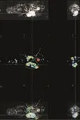
Fig. 3. Tumor uptake of123I-IMT in mouse 2 with a Colo357 subcutaneous pancreatic tumor (row 1: MRI; row 2: Hi-SPECT; row 3: Hi-SPECT-MRI). Tumor is readily visible in the right flank on MR images (arrows). Hot spot is of123I-IMT activity. Orange ROI represents tumor localization, and green ROI represents visceral region. Because of slightly different positioning of the mice in Hi-SPECT and MRI the tracer uptake in the subcutaneous tumor and in the bladder are not exactly congruent with the MRI images.

Fig. 4. Expression of LAT1 (SLC7A) in A) micro-dissected PDAC samples (n=19) compared with the expression in normal pancreatic ducts (n=13), and B) PDAC cell lines (n=10) compared with non-malignant growing cell lines (n=3) by microarray analysis. The median expression level is marked by a line. No significant difference in the gene expression of LAT1 was found (P>0.05; t test) since two exceptional high values were found in the normal ductal cell samples. The expression value corresponding to Colo357 cells is indicated by the upper of the two circles.
Comparing the expression of LAT1 in either microdissected PDAC sample tissues or PDAC cell lines with the expression in their particular corresponding nonmalignant counterparts did not reveal significant differences (P>0.05; Fig. 4). However, there was a strong trend and an additional more quantitative approach like q-RT-PCR was required to ascertain differences in expression. Expression of LAT1 in Colo357 cells used in this study was increased by nearly 4 folds compared with the median expression level in PDAC derived cell lines or non-malignant growing controls.
Discussion
In this study we utilized tumor uptake of123I-IMT in human pancreatic tumor cell line Colo357. All subcutaneous and orthotopic tumors were detected following successful injection of the radioactive tracer into SCID mice. In order to improve imaging we fused Hi-SPECT and MRI. These results fit with the findings of the gene array analyses that show a strong trend for high expression of the LAT1 transporter in this pancreatic tumor cell line.
Carnochan et al[26]demonstrated that the biokinetics of123I-IMT are not only influenced by LAT1, but also strongly depend on blood flow and on diffusion of the tracers into the tissue and that tumor growth status may not be closely associated with amino acid uptake alone. In this underlying study, there was a remarkable high uptake particularly in orthotopic pancreatic tumors. This finding may gain impact, since unpublished studies compared blood flow of subcutaneous and orthotopic tumors using ultrasound sonography (Heneweer C and Kalthoff H, personal communications). Here, a hypervascularisation of tumors in the subcutaneous site was observed, which is not to be seen in the orthotopic tumors. Yet, orthotopic tumors more closely resemble human tumors because nearby blood vessel system and other supporting tissues better mirror the tumor's microenvironment.[27]The remarkable high uptake of123I-IMT in orthotopic (hypovascularized) tumors as observed in our study clearly points to a robust tumor cell-dependent labeling. The use of123I-IMT in measuring pancreatic tumor growth therefore also has important application in establishing a ‘gold standard' for the assay of drug activity in clinically adapted, orthotopic mouse models of human pancreatic cancer.[28]
Accumulation of123I-IMT in the tumor is correlated with LAT1 expression in the PDAC cells.[23,24]A significantly higher expression of the LAT1 appears in the Colo357 cell line used in this study. Therefore the high tracer uptake is most likely caused by the increased LAT1 of the tumor cells themselves rather than by other cells of the tumor stroma, which even in SCIDmice may influence tumor growth.[29]To extend the findings on LAT1 to clinical samples, analyses of LAT1 expression was also performed on PDAC tumor samples from patient material (micro-dissected pancreatic tumor epithelium versus normal pancreatic tissue). The finding indicated an apparent although not significant trend towards higher expression of LAT1 in tumor cells. Statistical significance was hampered by 2 out of 13 non-malignant specimens, which revealed an unusual high expression level of LAT1 relative to all others. Evaluating the corresponding clinical data, we found no reasonable explanation for these two outliers. Further studies are required with a more quantitative technique such as q-PCR in order to confirm the significance of these results. Additional animal experiments including murine pancreatic carcinoma and pancreatitis models are planned to substantiate that123I-IMT specifically accumulates in tumor cells and not or less in the inflammatory microenvironment.
PET with18F-FDG has been demonstrated to be useful in evaluating indeterminate pancreatic masses, staging pancreatic cancer, detecting metastatic disease, and differentiating viable tumor from post-therapeutical changes.[30-32]However, although critical, the differentiation of pancreatic malignancy from focal pancreatitis, as well as tumor detection in cases of coexisting pancreatitis, is still a challenge by means of18F-FDG-PET.[24,33]
Metabolic imaging of tumors using PET with radiolabeld amino acids such as L-(1-[11C])tyrosine or O-(2-[18F]fluoroethyl)-L-tyrosine is possible and has demonstrated a potential clinical usage including tumor staging and evaluation of therapy.[4,34]As amino acids play a minor role in the metabolism of inflammatory cells, these tracers might be more “tumor specific”than18F-FDG and therefore may be more effective in monitoring therapy response. Due to the still limited availability and high costs of PET there is a need for similar radiopharmaceuticals that are suitable for conventional nuclear medicine.[15]
In the underlying study Hi-SPECT fused images with MRI easily allowed the delineation of the tumor and improved its differentiation from surrounding tissue.
Ruf et al[35]described the impact of18F-FDGPET/MRI image fusion on the detection of pancreatic cancer in 32 patients. He observed an improvement in the anatomical assignment and interpretation of18F-FDG foci by PET/MRI fusion compared to the side by side analysis. However, in this study the specificity for cancer detection by18F-FDG-PET was only 41% for visual and 58% for semi-quantitative analysis whereas MRI achieved 76%. The differentiation of pancreatic malignancy from focal pancreatitis involving the pancreatic head is still critical, because inflammatory tissue takes up18F-FDG avidly, and chronic pancreatitis is recognized as the most common reason for false positive18F-FDG-PET findings, indicating the need for developing more specific tracers. Referring to these results in our study we demonstrated the feasibility of an in vivo imaging model in which subcutaneously and orthotopically growing PDAC tumors in mice could be detected by123I-IMT-Hi-SPECT and consecutive MRI. This model allows a systematic uptake comparison of the various anatomical tumor localizations, which can be further allocated by performing a body transformation image fusion (Hi-SPECT/MRI fusion). This combination enables the visualization of anatomical and functional information within one set of 3D-images without applying any further radiation.
In conclusion, in our xenotransplantation SCID mouse model, a high123I-IMT uptake was seen in pancreatic cancer. The data on overall increased LAT1 expression in micro-dissected PDAC epithelia support this finding. The imaging with123I-IMT would be an additional option for detecting pancreatic cancer and potentially distinguishing it from inflammatory pancreatitis. However, further research is needed to clarify whether these results are also applicable to different kinds of human pancreatic cancer cells. In addition to molecular analysis of tumor cells and Hi-SPECT imaging, the use of combined functional Hi-SPECT and morphological high-resolution MRI helped to improve tumor imaging and image interpretation in our mouse model of pancreatic cancer.
Funding:This project was supported in part by a BMBF grant (TOMCAT) given to H.K. and by the Molecular Imaging North Competence Center (MOIN-CC).
Ethical approval:Not needed.
Contributors:FC and ZM contributed equally to this manuscript. KH and HE proposed the study. FC, ZM and ZY performed the MRI and SPECT analysis. AO did the molecular biological evaluation for LAT1. EJH performed animal experiments supported by TS. JO supervised MRI analysis. FC, ZM and EJH contributed to the design and interpretation of the study and wrote the first draft, which was edited by KH. ZM is the guarantor.
Competing interest:No benefits in any form have been received or will be received from a commercial party related directly or indirectly to the subject of this article.
1 Biersack HJ, Coenen HH, Stöcklin G, Reichmann K, Bockisch A, Oehr P, et al. Imaging of brain tumors with L-3-[123I]iodoalpha-methyl tyrosine and SPECT. J Nucl Med 1989;30:110-112.
2 Vaalburg W, Coenen HH, Crouzel C, Elsinga PH, Långström B, Lemaire C, et al. Amino acids for the measurement of protein synthesisin vivoby PET. Int J Rad Appl Instrum B 1992;19:227-237.
3 Kubota K, Ishiwata K, Kubota R, Yamada S, Takahashi J, Abe Y, et al. Feasibility of fluorine-18-fluorophenylalanine for tumor imaging compared with carbon-11-L-methionine. J Nucl Med 1996;37:320-325.
4 Willemsen AT, van Waarde A, Paans AM, Pruim J, Luurtsema G, Go KG, et al.In vivoprotein synthesis rate determination in primary or recurrent brain tumors using L-[1-11C]-tyrosine and PET. J Nucl Med 1995;36:411-419.
5 Isselbacher KJ. Sugar and amino acid transport by cells in culture--differences between normal and malignant cells. N Engl J Med 1972;286:929-933.
6 Kubota R, Yamada S, Kubota K, Ishiwata K, Tamahashi N, Ido T. Intratumoral distribution of fluorine-18-fluorodeoxyglucosein vivo: high accumulation in macrophages and granulation tissues studied by microautoradiography. J Nucl Med 1992;33: 1972-1980.
7 Strauss LG. Fluorine-18 deoxyglucose and false-positive results: a major problem in the diagnostics of oncological patients. Eur J Nucl Med 1996;23:1409-1415.
8 Arnér ES, Eriksson S. Mammalian deoxyribonucleoside kinases. Pharmacol Ther 1995;67:155-186.
9 Shields AF, Grierson JR, Dohmen BM, Machulla HJ, Stayanoff JC, Lawhorn-Crews JM, et al. Imaging proliferationin vivowith [F-18]FLT and positron emission tomography. Nat Med 1998;4:1334-1336.
10 Barthel H, Cleij MC, Collingridge DR, Hutchinson OC, Osman S, He Q, et al. 3'-deoxy-3'-[18F]fluorothymidine as a new marker for monitoring tumor response to antiproliferative therapyin vivowith positron emission tomography. Cancer Res 2003;63:3791-3798.
11 Kubota K, Matsuzawa T, Fujiwara T, Sato T, Tada M, Ido T, et al. Differential diagnosis of AH109A tumor and inflammation by radioscintigraphy with L-[methyl-11C]methionine. Jpn J Cancer Res 1989;80:778-782.
12 Kubota R, Kubota K, Yamada S, Tada M, Takahashi T, Iwata R, et al. Methionine uptake by tumor tissue: a microautoradiographic comparison with FDG. J Nucl Med 1995;36:484-492.
13 von Forstner C, Egberts JH, Ammerpohl O, Niedzielska D, Buchert R, Mikecz P, et al. Gene expression patterns and tumor uptake of18F-FDG,18F-FLT, and18F-FEC in PET/ MRI of an orthotopic mouse xenotransplantation model of pancreatic cancer. J Nucl Med 2008;49:1362-1370.
14 Martin WH, Delbeke D, Patton JA, Sandler MP. Detection of malignancies with SPECT versus PET, with 2-[fluorine-18]fluoro-2-deoxy-D-glucose. Radiology 1996;198:225-231.
15 Jager PL, Franssen EJ, Kool W, Szabó BG, Hoekstra HJ, Groen HJ, et al. Feasibility of tumor imaging using L-3-[iodine-123]-iodo-alpha-methyl-tyrosine in extracranial tumors. J Nucl Med 1998;39:1736-1743.
16 Yanagida M, Hayano T, Yamauchi Y, Shinkawa T, Natsume T, Isobe T, et al. Human fibrillarin forms a sub-complex with splicing factor 2-associated p32, protein arginine methyltransferases, and tubulins alpha 3 and beta 1 that is independent of its association with preribosomal ribonucleoprotein complexes. J Biol Chem 2004;279:1607-1614.
17 Lahoutte T, Mertens J, Caveliers V, Franken PR, Everaert H, Bossuyt A. Comparative biodistribution of iodinated amino acids in rats: selection of the optimal analog for oncologic imaging outside the brain. J Nucl Med 2003;44:1489-1494.
18 Samnick S, Bader JB, Hellwig D, Moringlane JR, Alexander C, Romeike BF, et al. Clinical value of iodine-123-alphamethyl-L-tyrosine single-photon emission tomography in the differential diagnosis of recurrent brain tumor in patients pretreated for glioma at follow-up. J Clin Oncol 2002;20:396-404.
19 Keyaerts M, Lahoutte T, Neyns B, Caveliers V, Vanhove C, Everaert H, et al. 123I-2-iodo-tyrosine, a new tumour imaging agent: human biodistribution, dosimetry and initial clinical evaluation in glioma patients. Eur J Nucl Med Mol Imaging 2007;34:994-1002.
20 Sipos B, Moser S, Kalthoff H, Torok V, Löhr M, Klöppel G. A comprehensive characterization of pancreatic ductal carcinoma cell lines: towards the establishment of anin vitroresearch platform. Virchows Arch 2003;442:444-452.
21 Egberts JH, Schniewind B, Sipos B, Hinz S, Kalthoff H, Tepel J. Superiority of extended neoadjuvant chemotherapy with gemcitabine in pancreatic cancer: a comparative analysis in a clinically adapted orthotopic xenotransplantation model in SCID beige mice. Cancer Biol Ther 2007;6:1227-1232.
22 Tisljar U, Kloster G, Ritzl F, Stocklin G. Accumulation of radioiodinated L-alpha-methyltyrosine in pancreas of mice: concise communication. J Nucl Med 1979;20:973-976.
23 Grützmann R, Pilarsky C, Ammerpohl O, Lüttges J, Böhme A, Sipos B, et al. Gene expression profiling of microdissected pancreatic ductal carcinomas using high-density DNA microarrays. Neoplasia 2004;6:611-622.
24 Pilarsky C, Ammerpohl O, Sipos B, Dahl E, Hartmann A, Wellmann A, et al. Activation of Wnt signalling in stroma from pancreatic cancer identified by gene expression profiling. J Cell Mol Med 2008;12:2823-2235.
25 Müerköster SS, Werbing V, Koch D, Sipos B, Ammerpohl O, Kalthoff H, et al. Role of myofibroblasts in innate chemoresistance of pancreatic carcinoma--epigenetic downregulation of caspases. Int J Cancer 2008;123:1751-1760.
26 Carnochan P, Deehan B, Trivedi M, Tombs A, Sandle J, Ott R. Uptake of radiolabelled tyrosine and iodo-methyl tyrosine in experimental rat tumours: influence of blood flow and tumour growth rate. J Nucl Biol Med 1994;38:92-95.
27 Olive KP, Jacobetz MA, Davidson CJ, Gopinathan A, McIntyre D, Honess D, et al. Inhibition of Hedgehog signaling enhances delivery of chemotherapy in a mouse model of pancreatic cancer. Science 2009;324:1457-1461.
28 Tepel J, Kruse ML, Kapischke M, Haye S, Sipos B, Kremer B, et al. Adjuvant treatment of pancreatic carcinoma in a clinically adapted mouse resection model. Pancreatology 2006;6:240-247.
29 Egberts JH, Cloosters V, Noack A, Schniewind B, Thon L, Klose S, et al. Anti-tumor necrosis factor therapy inhibits pancreatic tumor growth and metastasis. Cancer Res 2008; 68:1443-1450.
30 Delbeke D, Rose DM, Chapman WC, Pinson CW, Wright JK, Beauchamp RD, et al. Optimal interpretation of FDG PET in the diagnosis, staging and management of pancreatic carcinoma. J Nucl Med 1999;40:1784-1791.
31 Lemke AJ, Niehues SM, Hosten N, Amthauer H, Boehmig M, Stroszczynski C, et al. Retrospective digital image fusion of multidetector CT and18F-FDG PET: clinical value in pancreatic lesions--a prospective study with 104 patients. J Nucl Med 2004;45:1279-1286.
32 Sendler A, Avril N, Helmberger H, Stollfuss J, Weber W, BengelF, et al. Preoperative evaluation of pancreatic masses with positron emission tomography using18F-fluorodeoxyglucose: diagnostic limitations. World J Surg 2000;24:1121-1129.
33 Pakzad F, Groves AM, Ell PJ. The role of positron emission tomography in the management of pancreatic cancer. Semin Nucl Med 2006;36:248-256.
34 Wester HJ, Herz M, Weber W, Heiss P, Senekowitsch-Schmidtke R, Schwaiger M, et al. Synthesis and radiopharmacology of O-(2-[18F]fluoroethyl)-L-tyrosine for tumor imaging. J Nucl Med 1999;40:205-212.
35 Ruf J, Lopez Hänninen E, Böhmig M, Koch I, Denecke T, Plotkin M, et al. Impact of FDG-PET/MRI image fusion on the detection of pancreatic cancer. Pancreatology 2006;6:512-519.
Received November 30, 2010
Accepted after revision December 29, 2010
A gentleman is always ready to help others attain their good aims, but never to help them with their evil conduct.
–the Analects
topic inoculation, animals
a median laparotomy and 30 μl of tumor cell suspension were inoculated into the pancreatic body to the left of the organ midline as described previously.[21]For subcutaneous inoculation, the cells were inoculated into the right flank.
Author Affiliations: Department of Nuclear Medicine (von Forstner C, Zuhayra M, Zhao Y and Henze E), Institute of Human Genetics (Ammerpohl O), Institute for Experimental Cancer Research (Tiwari S and Kalthoff H), Institute of Neuroradiology (Jansen O), and Department of General Surgery and Thoracic Surgery (Egberts JH), University Hospital of Schleswig-Holstein, Campus Kiel, Germany
Maaz Zuhayra, MD, Department of Nuclear Medicine, University Hospital Schleswig-Holstein, Campus Kiel, Arnold-Heller-St. 9, House 23, D-24105 Kiel, Germany (Tel: 49-431-597-3101; Fax: 49-431-597-3065; Email: mzuhayra@nuc-med.uni-kiel.de)
© 2011, Hepatobiliary Pancreat Dis Int. All rights reserved.
杂志排行

Hepatobiliary & Pancreatic Diseases International的其它文章
- Laparoscopic liver resection for benign and malignant liver tumors
- Assessment of tumor vascularization with functional computed tomography perfusion imaging in patients with cirrhotic liver disease
- Monday blues of deceased-donor liver transplantation
- Hepatobiliary & Pancreatic Diseases International (HBPD INT)
- Roles of sulfonylurea receptor 1 and multidrug resistance protein 1 in modulating insulin secretion in human insulinoma
- Atypical focal nodular hyperplasia of the liver